Copyright
©2010 Baishideng Publishing Group Co.
World J Radiol. May 28, 2010; 2(5): 180-187
Published online May 28, 2010. doi: 10.4329/wjr.v2.i5.180
Published online May 28, 2010. doi: 10.4329/wjr.v2.i5.180
Figure 1 Magnetic resonance (MR) images demonstrating zonal anatomy of prostate gland.
A: Axial T2-weighted (T2W) MR image depicts the central gland and peripheral zone (PZ). Central gland is hypointense compared to hyperintense PZ; B. Coronal T2W MR image shows hyperintense PZ and hypointense central gland.
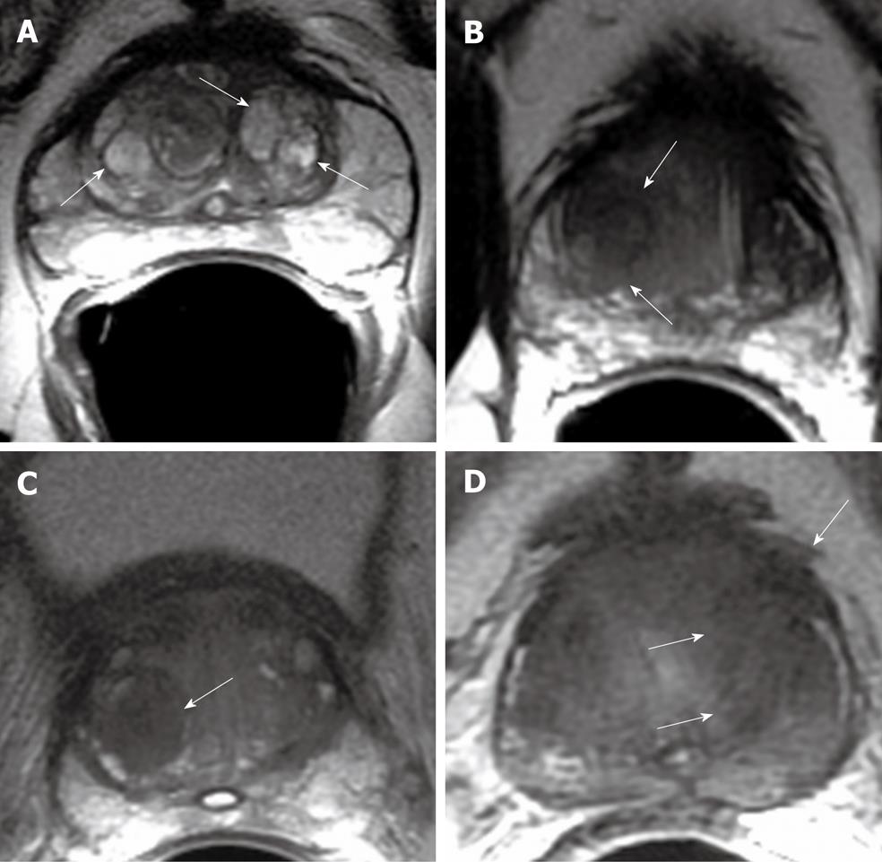
Figure 2 Axial T2W MR image.
A: Multiple, well defined hyperintense glandular benign prostatic hyperplasia (BPH) nodules in central gland (arrows); B: Well defined, amorphous, hypointense TZ tumor (arrows); C: Hypointense stromal BPH nodule in the right transition zone (TZ) (arrow); D: Hypointense TZ tumor with extracapsular extension (arrows).
Figure 3 Tumor in the left mid prostate gland demonstrated by MR.
A: Axial T2W image shows ill defined, amorphous, hypointense tumor (arrows); B: Diffusion weighted imaging (DWI) reveals focal area of bright signal consistent with tumor(arrow); C: Apparent diffusion coefficient (ADC) map reveals clear focal mass with dark signal consistent with decreased ADC (arrow).
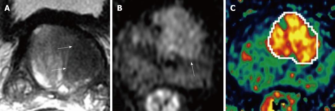
Figure 4 Left TZ tumor of prostate gland demonstrated by MR.
A: Axial T2W image depicts ill defined, round, homogenous hypointense tumor (arrows); B: DWI depicts focal area of bright signal on left mid gland (arrow); C: K-trans map in dynamic contrast-enhanced MR imaging (DCE-MRI) clearly localizes the tumor and reveals some internal heterogeneity.
- Citation: Kayhan A, Fan X, Oommen J, Oto A. Multi-parametric MR imaging of transition zone prostate cancer: Imaging features, detection and staging. World J Radiol 2010; 2(5): 180-187
- URL: https://www.wjgnet.com/1949-8470/full/v2/i5/180.htm
- DOI: https://dx.doi.org/10.4329/wjr.v2.i5.180


