Copyright
©2010 Baishideng Publishing Group Co.
World J Radiol. May 28, 2010; 2(5): 172-179
Published online May 28, 2010. doi: 10.4329/wjr.v2.i5.172
Published online May 28, 2010. doi: 10.4329/wjr.v2.i5.172
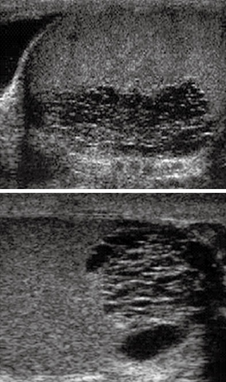
Figure 1 Ultrasound of both testes (sagittal images) demonstrates ectasia of the testes with formation of intratesticular cysts.
These finding are suggestive of a seminal tract obstructive etiology which should be managed by epididymo-vasotomy.
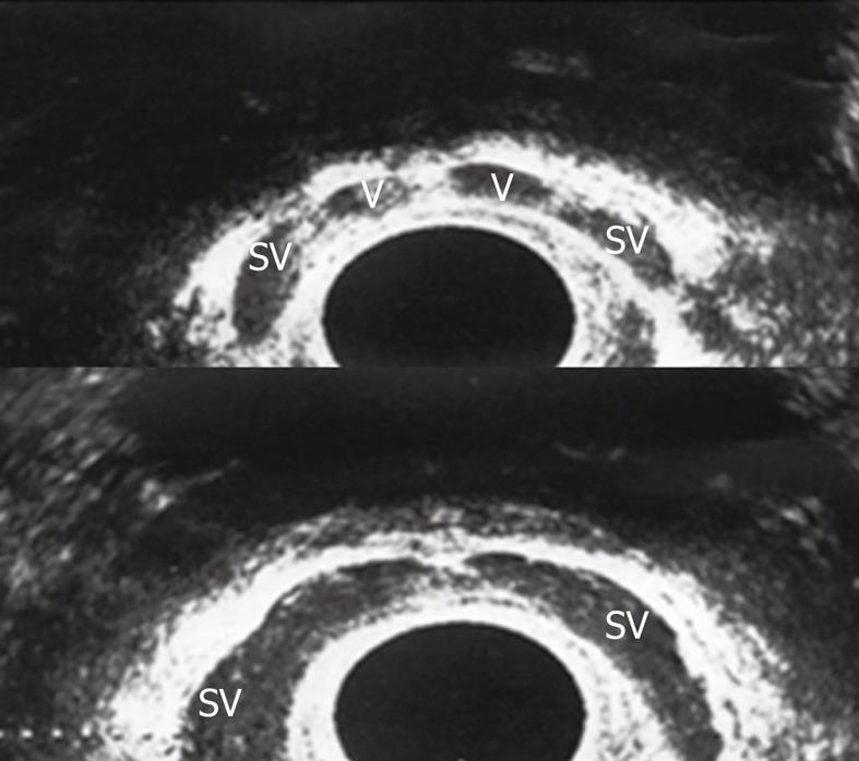
Figure 2 Transrectal ultrasound (TRUS) in the axial plane showed normal both vasal ampullae (V) and seminal vesicles (SV).
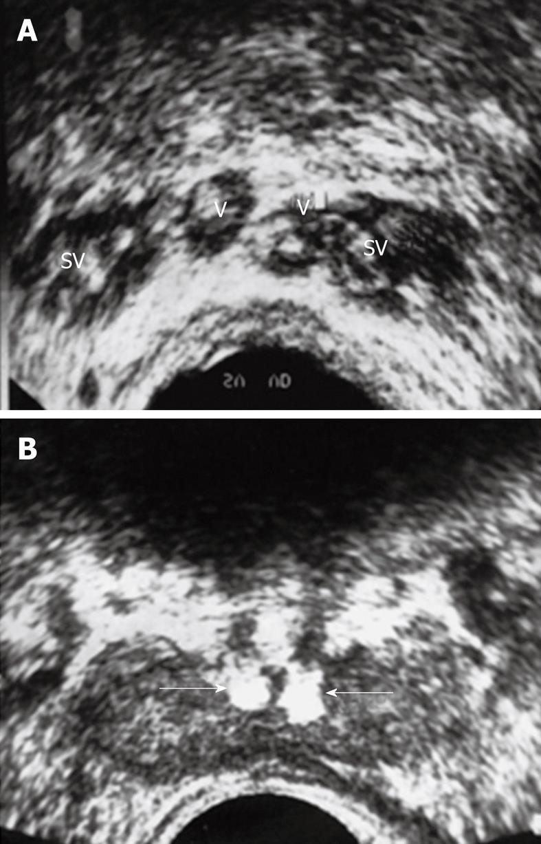
Figure 3 Twenty five years infertile man with azospermia.
A: Multiple calculi within the SV and V; B: Bilateral echogenic calculi impacted within the ejaculatory ducts (arrows).
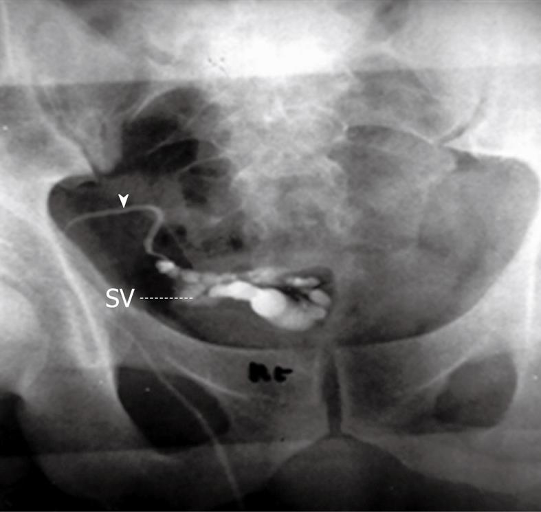
Figure 4 Percutaneous vasography shows normal right sided vasogram with opacification of the right vas (arrowhead), SV and ejaculatory duct (arrow) with retrograde opacification of the urinary bladder (UB).
Figure 5 Percutaneous right vasography in a patient with obstructed infertility shows complete obstruction of the ejaculatory duct with retention of the dye in the vas (arrowhead) and SV and non opacification of the urinary bladder.
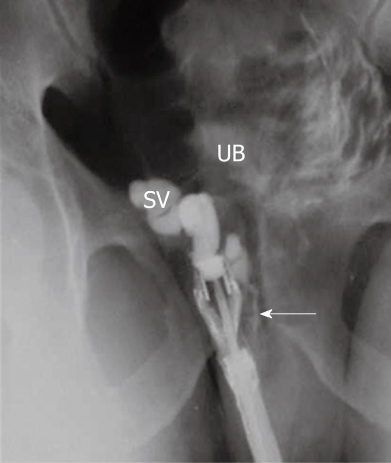
Figure 6 TRUS-right seminal vesiculography shows normal opacification of right SV and ejaculatory duct (arrow).
The injected dye is seen in the posterior urethra and UB denoting patency of the right ejaculatory duct.
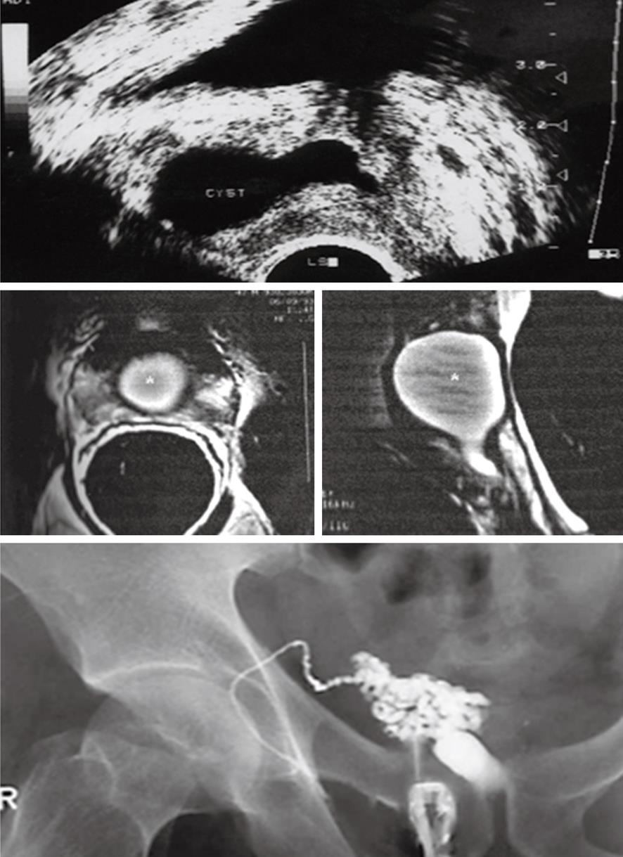
Figure 7 A 29-year-old man with primary obstructive infertility.
TRUS (upper image) and endorectal magnetic resonance imaging (middle image) show a well-defined midline urogenital cyst with intra-and extraprostatic components. TRUS-seminal vesiculography (lower image) shows the seminal vesicle is communicating with the urogenital cyst with non opacification of the urethra or urinary bladder denoting complete distal obstruction (N.B. the left vas and seminal vesicles were absent). Trans-urethral incision of the cyst lead to improvement of sperm count.
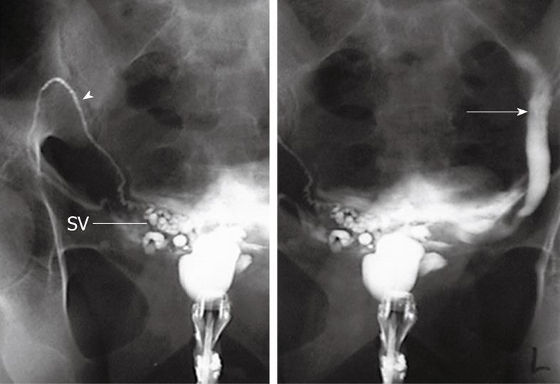
Figure 8 A 33-year-old man with primary infertility.
TRUS-guided contrast opacification of midline prostatic cyst shows the presence of a large cyst communicating on the right side with the right vas (arrowhead) and right SV. On the left side the cyst is communicating with a blind tubular structure (arrow), which proved to be an ectopic short ureter of a hypoplastic left kidney.
Figure 9 A 27-year-old with primary infertility.
A: TRUS shows a 3 cm × 2 cm thin walled midline intraprostatic urogenital cyst; B: TRUS-guided contrast opacification of the cyst revealed that the cyst was blind with no communication with the seminal tract. Semen analysis showed improvement of the sperm count 3 d after complete cyst aspiration.
- Citation: Donkol RH. Imaging in male-factor obstructive infertility. World J Radiol 2010; 2(5): 172-179
- URL: https://www.wjgnet.com/1949-8470/full/v2/i5/172.htm
- DOI: https://dx.doi.org/10.4329/wjr.v2.i5.172


