Copyright
©2010 Baishideng.
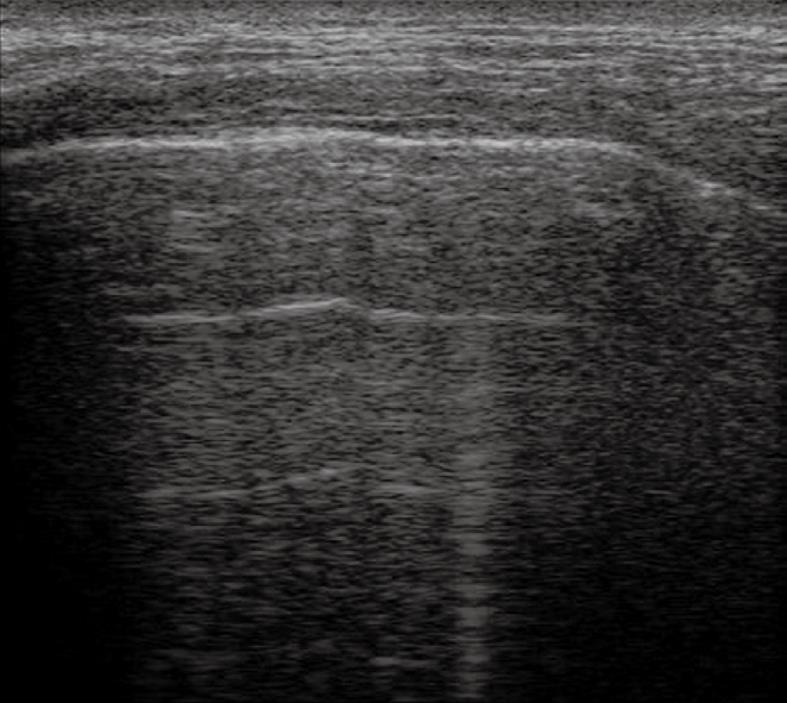
Figure 1 Normal lung.
Multiple parallel transversal echoes departing from the pleural line (reverberation artefacts) and a single vertical artefact.
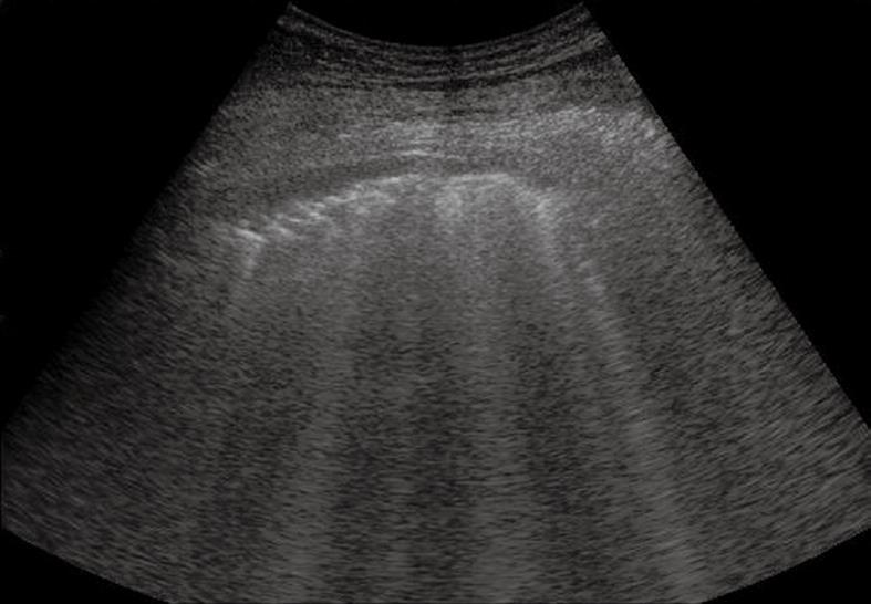
Figure 2 Pulmonary fibrosis.
Multiple vertical comet-tail artefacts and sporadic ring-down artefacts. The pleural line appears thick and irregular.
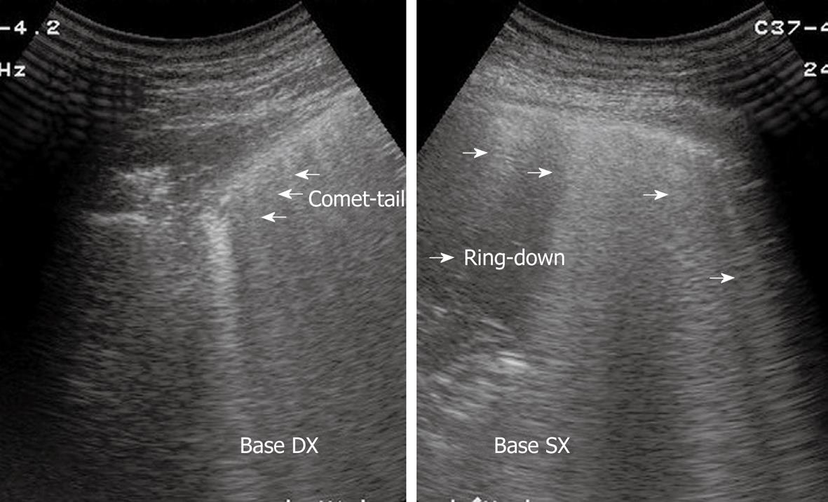
Figure 3 Right and left lung showing comet-tail and ring-down artefacts, respectively.
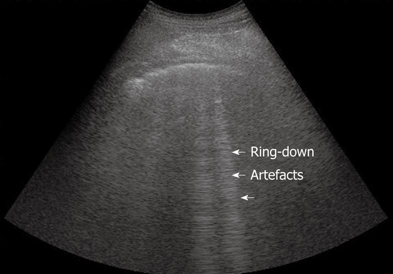
Figure 4 Normal lung.
Sporadic ring-down artefacts spreading from the pleural line into the lung surface.
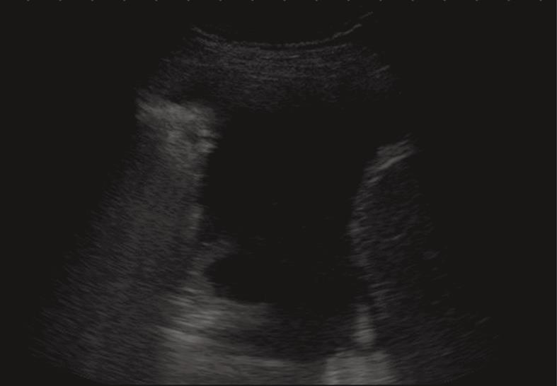
Figure 5 Anechoic echo-free pleural effusion.
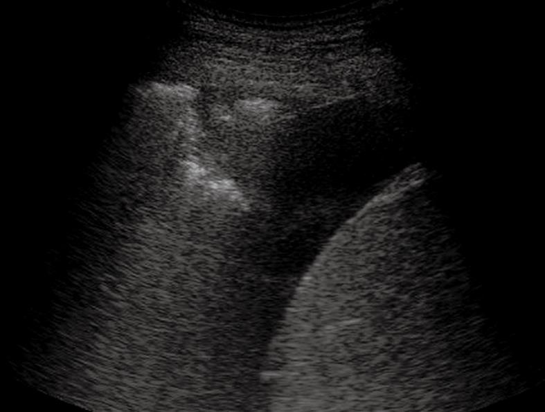
Figure 6 Heterogeneous echogenic material inside the anechoic pleural effusion.
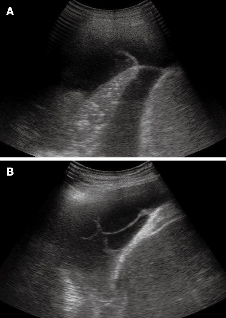
Figure 7 Sporadic (A) and multiple (B) fibrin strands or septa floating inside the anechoic pleural effusion.
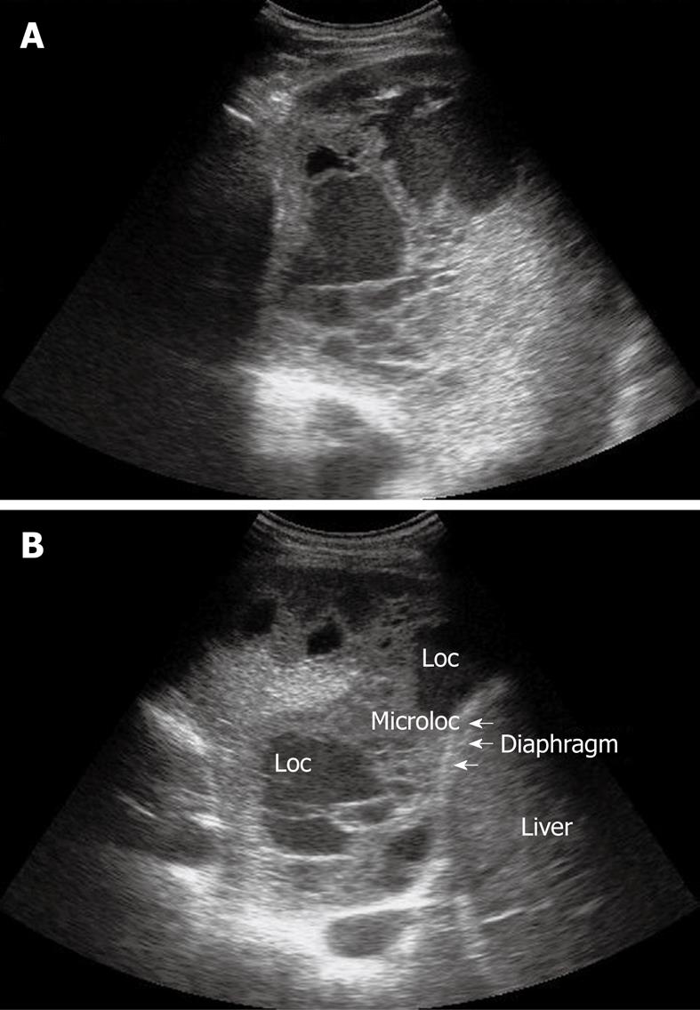
Figure 8 Homogeneous echogenic material inside the pleural space (A and B).
Loc: Loculation; Microloc: Microloculation.
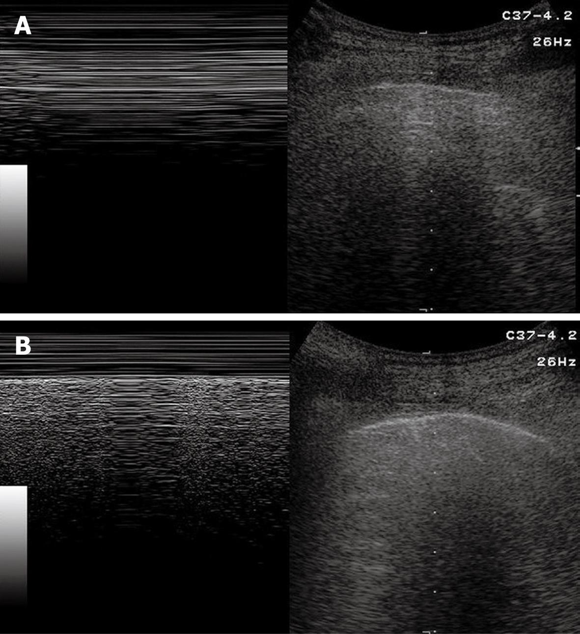
Figure 9 Pneumothorax.
A: B-mode image (right image) shows horizontal reverberation artefacts corresponding to frozen echoes in M-mode image (left image), due to loss of breathing-dependent motion of pleural line; B: After resolution of pneumothorax, B-mode image (right image) shows pleural line and a single comet-tail artefact; in M-mode image (left image), breathing-dependent movements appear as “frosted glass” artefacts, quite different from frozen echoes seen in A.
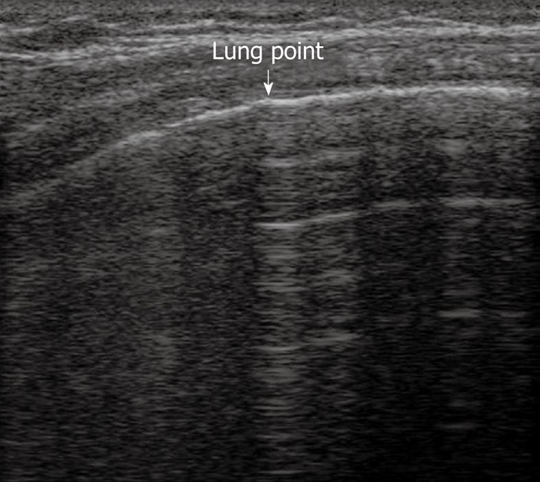
Figure 10 Lung point.
Horizontal reverberation artefacts are interrupted by reappearance of irregular, fragmented, thickened pleural line with comet-tail artefacts.
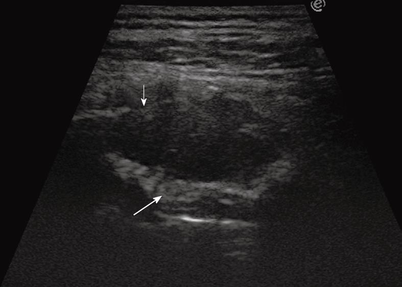
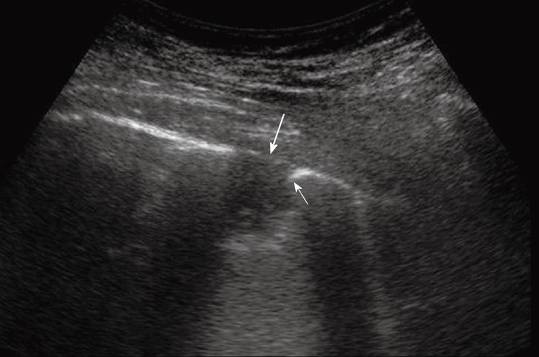
Figure 11 Extrapleural mass disrupting a rib (thin arrow) and displacing and disrupting the pleural line (large arrow).
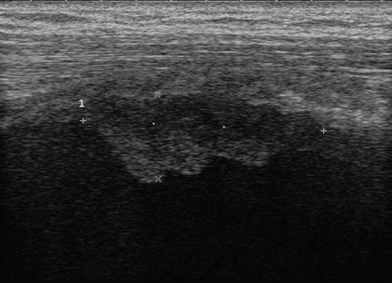
Figure 12 Pleural mesothelioma.
Solid pleural mass with irregular lobulated borders.
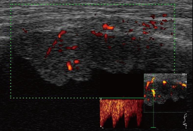
Figure 13 Color Doppler sonography shows flow signals inside the lesion, with mainly arterial vascularization.
Figure 14 Peripheral pulmonary nodule abutting and infiltrating the pleura.
Note the acute angle between the nodule and the pleural line (thin arrow) and the disruption of the pleural line (large arrow).
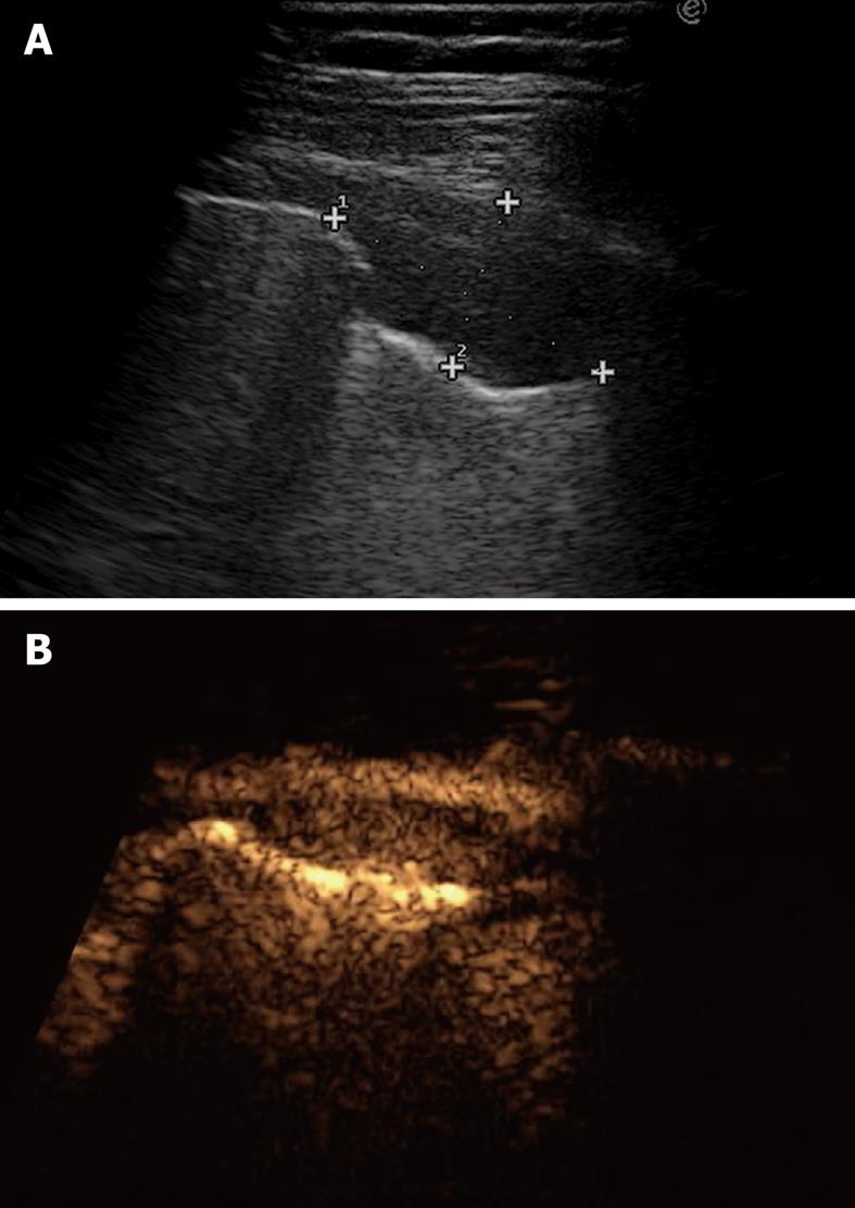
Figure 15 Pleural metastasis.
A: B-mode sonogram shows a hypoechoic lesion resembling either a solid mass or a homogeneously echoic saccate effusion; B: Contrast-enhanced sonography shows contrast enhancement of the lesion 20 s after bolus injection of ultrasound contrast agent. Sonographically-guided biopsy confirmed metastasis from breast carcinoma.
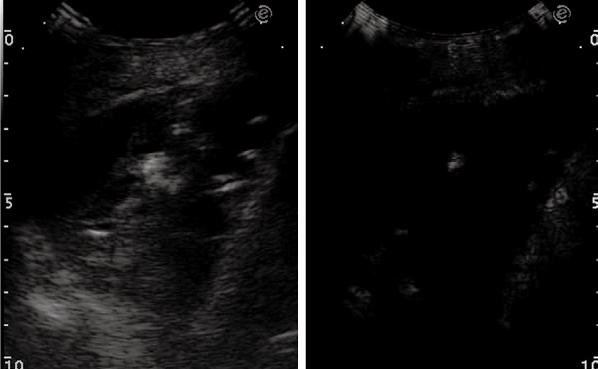
Figure 16 Pleural effusion with broad echoic debris mimicking a solid lesion (left image).
Contrast-enhanced sonography shows no enhancement within the effusion (right image).
- Citation: Sartori S, Tombesi P. Emerging roles for transthoracic ultrasonography in pleuropulmonary pathology. World J Radiol 2010; 2(2): 83-90
- URL: https://www.wjgnet.com/1949-8470/full/v2/i2/83.htm
- DOI: https://dx.doi.org/10.4329/wjr.v2.i2.83


