Copyright
©The Author(s) 2025.
World J Radiol. Jun 28, 2025; 17(6): 107205
Published online Jun 28, 2025. doi: 10.4329/wjr.v17.i6.107205
Published online Jun 28, 2025. doi: 10.4329/wjr.v17.i6.107205
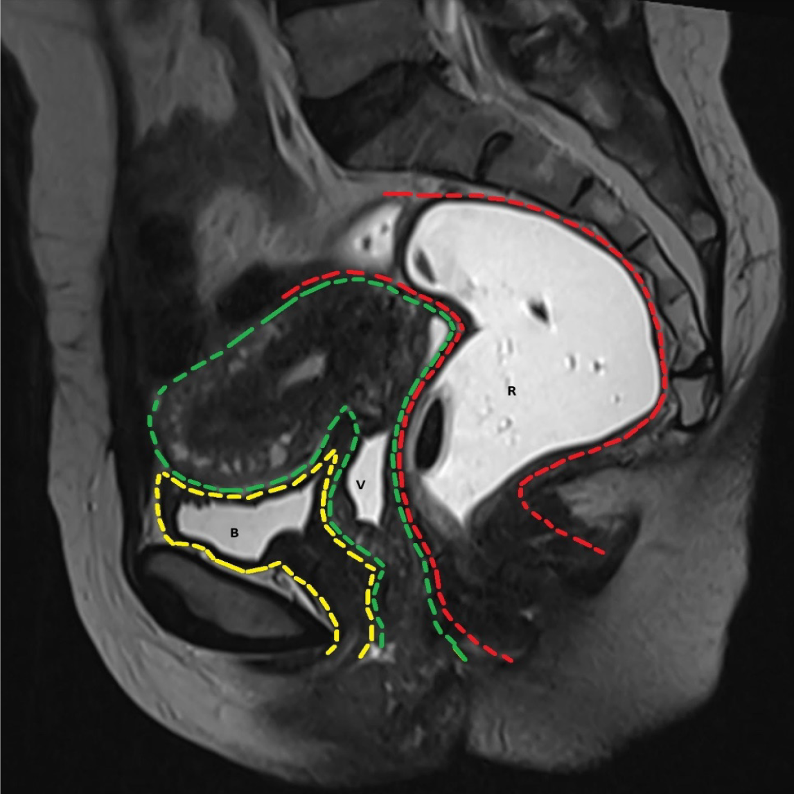
Figure 1 T2-weighted image in sagittal plane shows the three compartments of pelvis.
The anterior compartment (yellow) contains the urinary bladder (B) and urethra, middle compartment (green) contains the uterus, cervix and vagina (V) and the posterior compartment (red) contains the rectum (R) and anal canal.
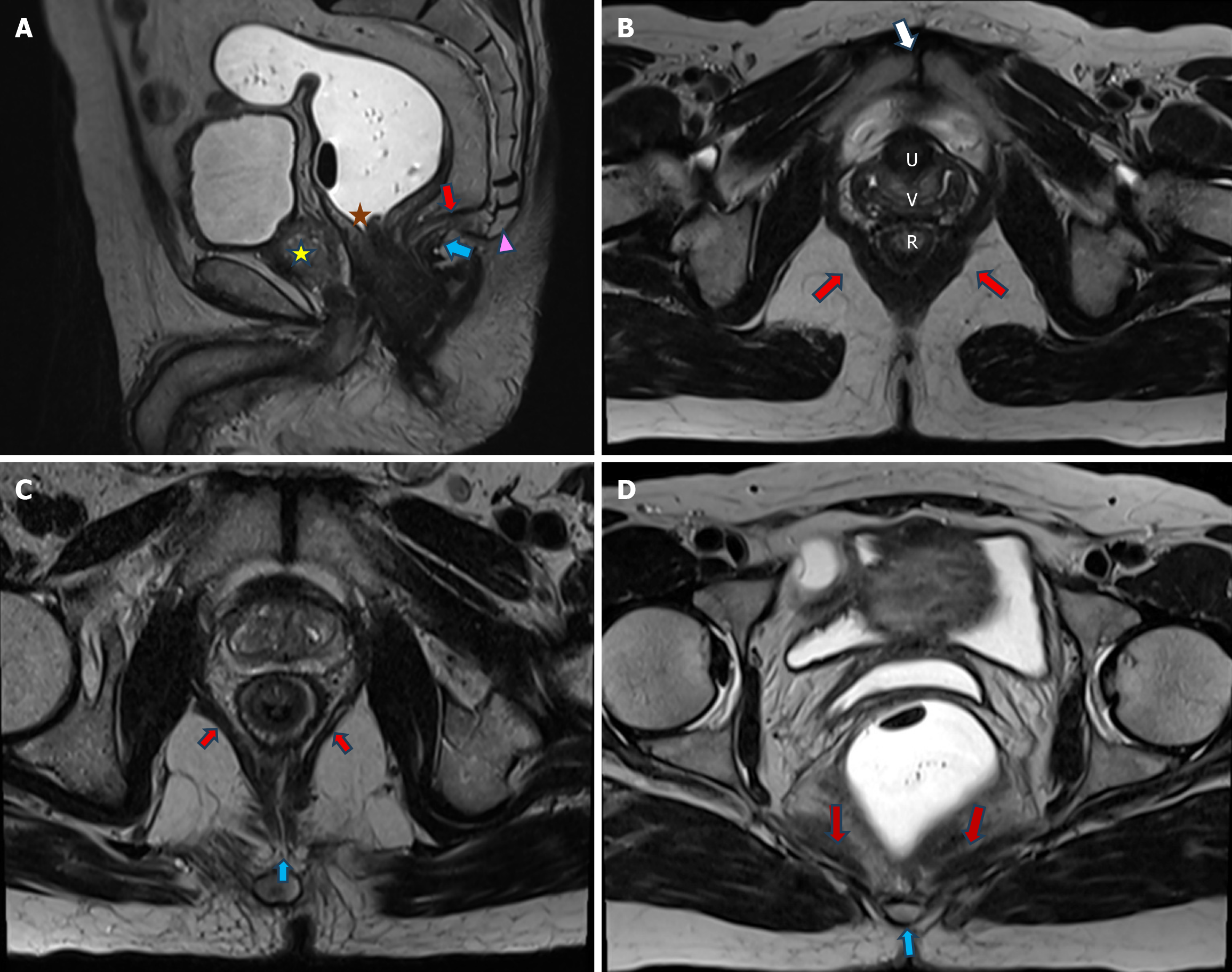
Figure 2 Normal supporting structures of pelvic floor.
A: T2-weighted image in sagittal plane shows the posterior aspect of puborectalis muscle (blue arrow) just behind the anorectal junction (orange star). Levator plate (red arrow) extends from the coccyx (pink arrowhead) to the anorectal junction (orange star). Note the normal vertical orientation of the urethra (yellow star) behind pubic bone. Bladder (B) and rectum (R) are distended; B: Axial T2-weighted image shows the sling of puborectalis muscle (red arrows) attached to the pubis (white arrow) anteriorly and encircling around the anorectal junction (R) posteriorly; C: Axial T2-weighted image cephalad to (B) shows pubococcygeus muscle (red arrows) coursing from the pubis anteriorly to the levator plate (blue arrow) posteriorly; D: Axial T2-weighted image cephalad to (C) shows iliococcygeus muscle (red arrows) extending from the obturator fascia laterally to the coccyx (blue arrow) posteriorly.
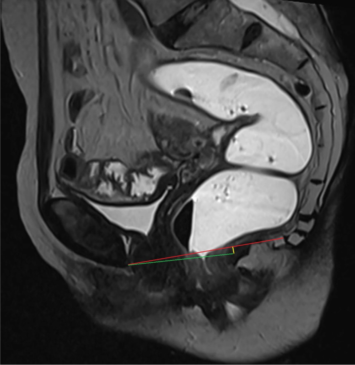
Figure 3 Sagittal T2-weighted image illustrating the placement of various reference lines.
The pubococcygeal line (PCL), denoted by the red line, spans from the inferior aspect of the pubis anteriorly to the last coccygeal articulation posteriorly. The H-line, represented by the green line, extends from the inferior aspect of the pubis anteriorly to the posterior wall of the anorectal junction. The M-line, depicted by the yellow line, is drawn perpendicularly to the PCL, originating from the posterior point of the H-line.
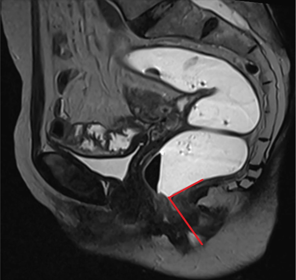
Figure 4
Anorectal angle is the angle formed at anorectal junction by the intersection of two lines, one drawn along the central axis of anal canal and another drawn along the posterior border of lower rectum.
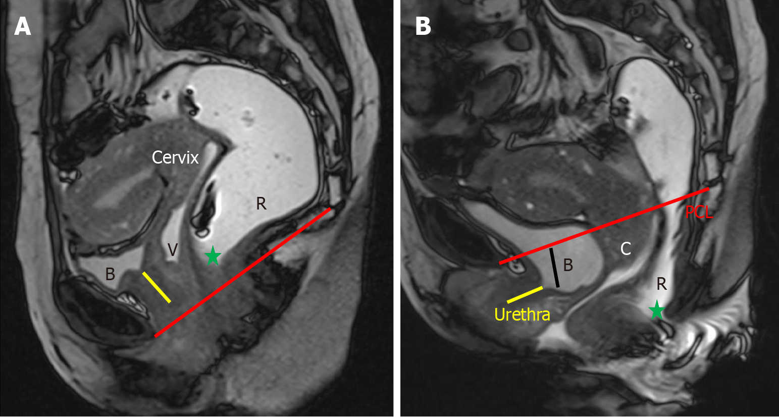
Figure 5 Urethral hypermobility and cystocele.
A: Sagittal true fast imaging with steady-state free precession (TRUFI) at rest, which shows minimal posterior angulation of the urethra (yellow line), approximately 5° relative to the vertical axis of the pubis; B: In the sagittal TRUFI image obtained during the defecation phase, the urethral axis rotates posteriorly to 162° and descends below the pubic symphysis, indicating urethral hypermobility. This is accompanied by a descent of the bladder neck below the pubococcygeal line, consistent with cystocele.
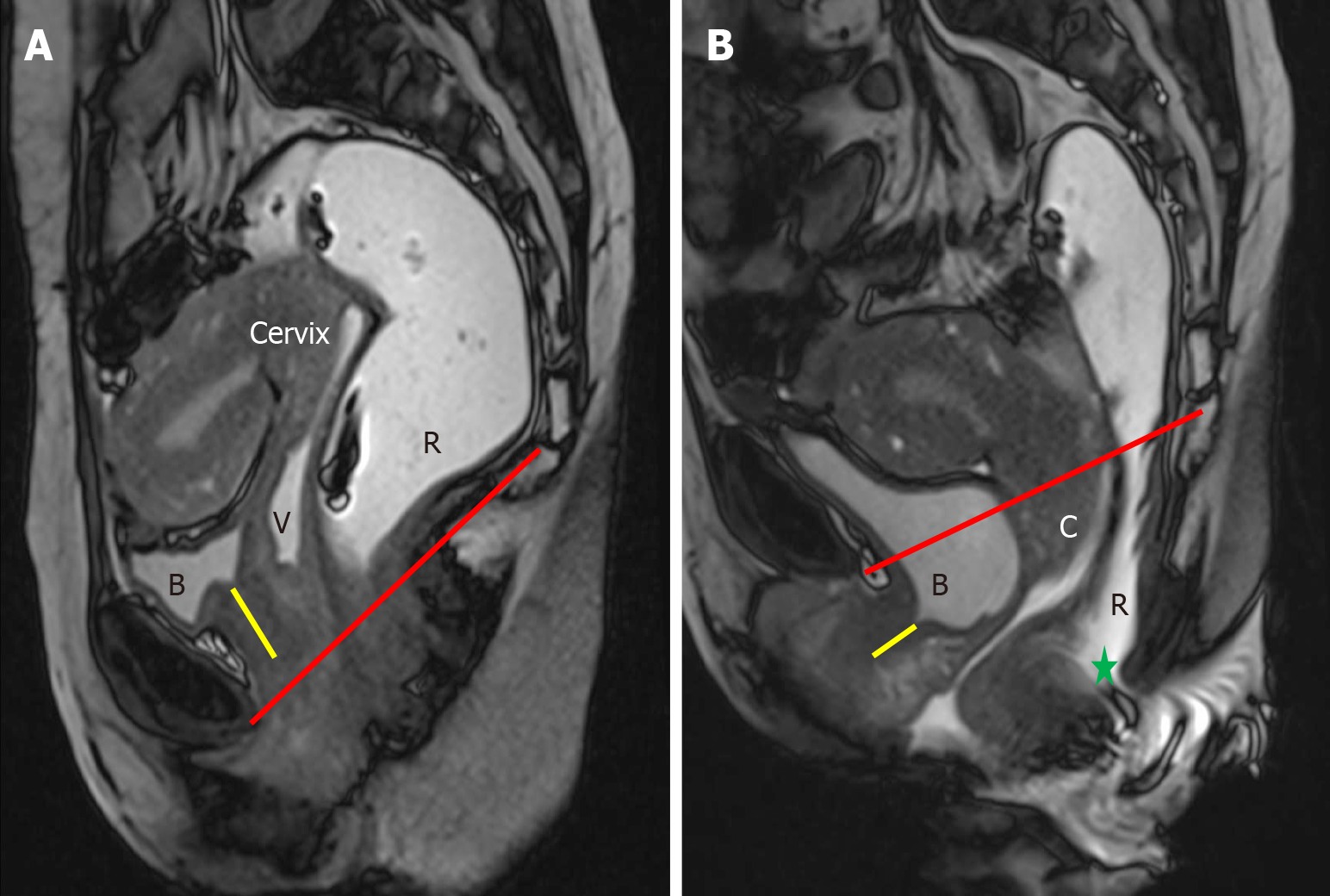
Figure 6 Cervical descent.
A: Sagittal true fast imaging with steady-state free precession image at rest, the cervix is positioned well above the pubococcygeal line (PCL); B: During defecation, the anterior lip of the cervix descends 2.1 cm below the PCL, indicating cervical descent.
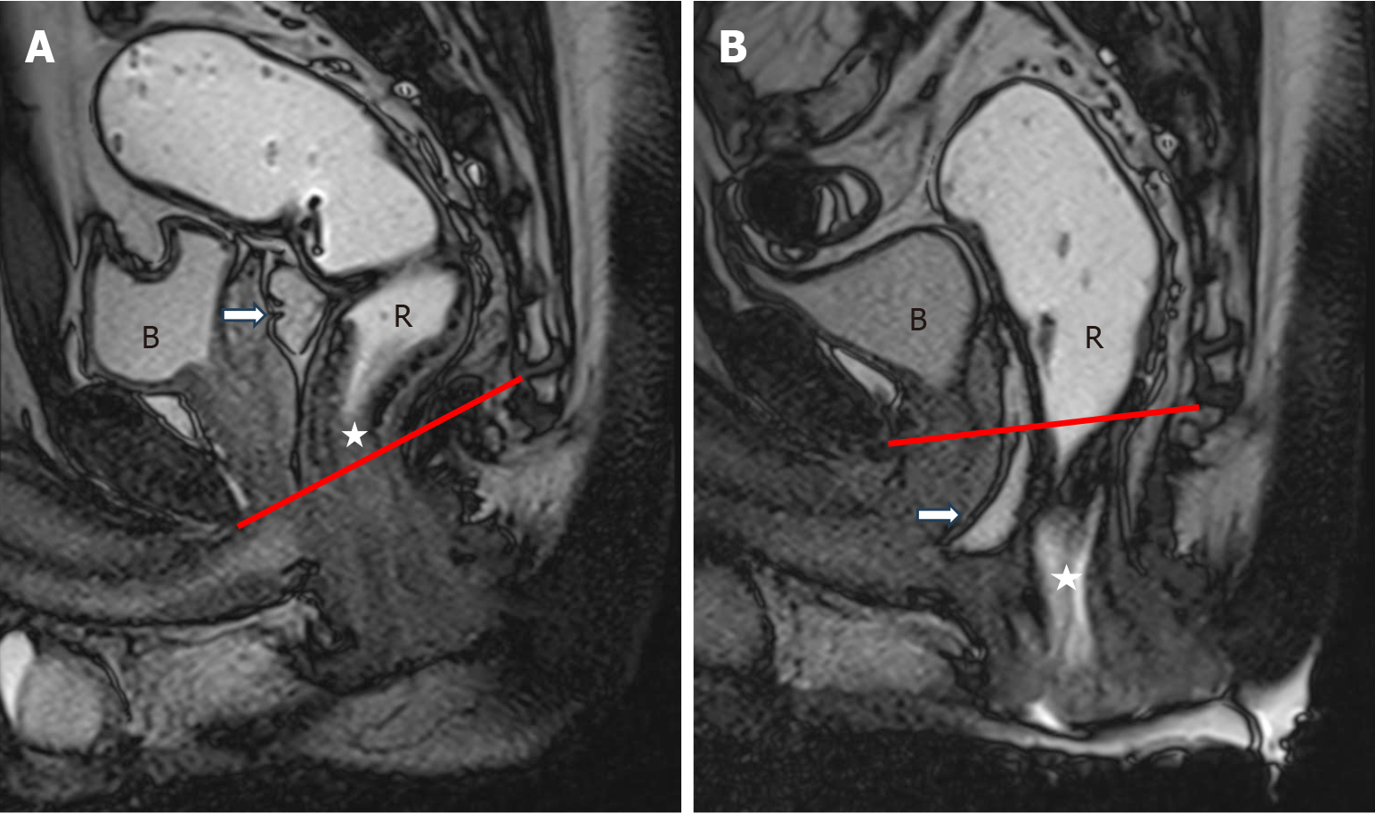
Figure 7 Peritoneocele.
A: Sagittal true fast imaging with steady-state free precession image at rest shows the normal position of cul-de-sac (white arrow) above pubococcygeal line (PCL); B: During the defecation phase there is caudal descent of the peritoneum (peritoneocele) (white arrow) below the PCL. Note descent of anorectal junction (white star).
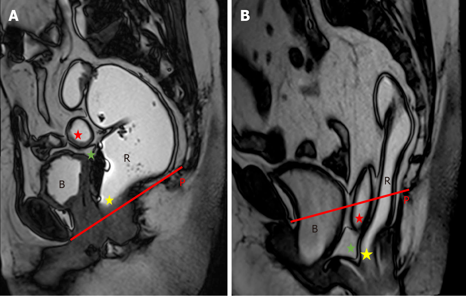
Figure 8 Enterocele.
A: Sagittal true fast imaging with steady-state free precession image at rest shows a small bowel loop (red star) well above pubococcygeal line (PCL); B: During the defecation phase there is caudal descent of this small bowel loop (red star) along with the peritoneum (green star) below the PCL. Note descent of anorectal junction (yellow star).
Figure 9 Anterior rectocele and anorectal junction descent.
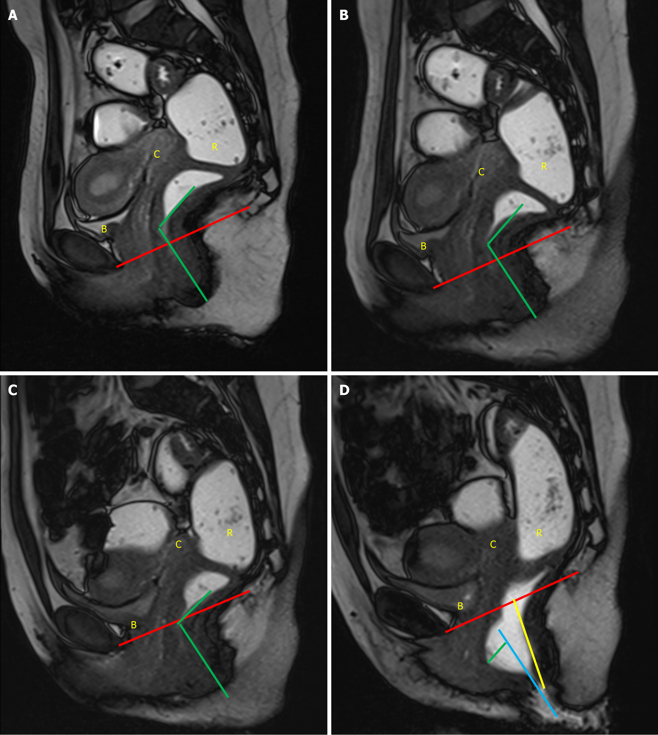
A: Sagittal true fast imaging with steady-state free precession (TRUFI) image at rest shows anorectal junction located above pubococcygeal line (PCL); B: During the squeeze phase there is reduction in anorectal angle with anorectal junction located above PCL; C: During the strain phase, anorectal junction reaches PCL; D: During the defecation phase there is caudal descent of the anorectal junction (yellow line) below the PCL. There is the formation of an anterior rectocele (green line), which is seen as a bulge of the anterior rectal wall beyond the expected anterior wall of rectum (blue line).
Figure 10 Rectal intussusception.
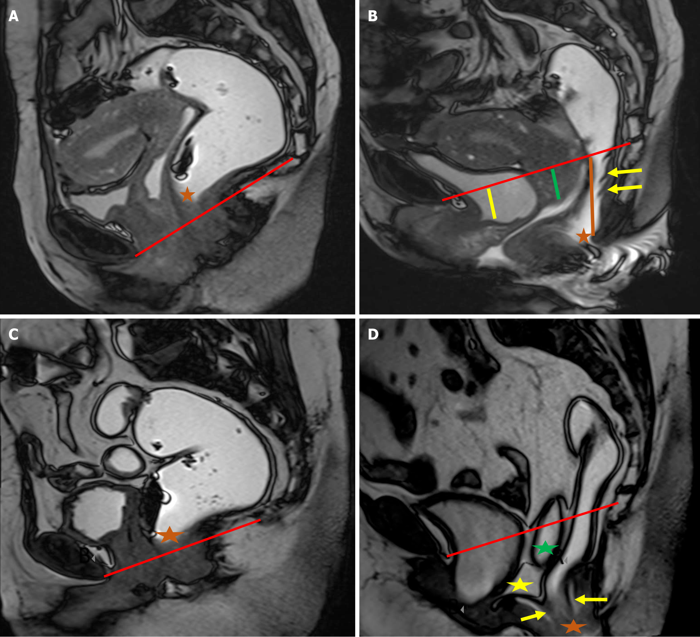
A: In the first patient, the anorectal junction (orange star) is above the pubococcygeal line (PCL) at rest; B: During the defecation phase, the anorectal junction descends caudally below the PCL (orange line). There is associated formation of three mucosal intussusceptions along the posterior wall of the rectum (yellow arrows); C: In the second patient, the anorectal junction is above the PCL at rest (orange star); D: During defecation, the anorectal junction descends below the PCL (orange star), with an associated full-thickness rectal intussusception (yellow arrows). Note the presence of an enterocele (green star) and peritoneocele (yellow star).
Figure 11 Dyssynergic defecation.
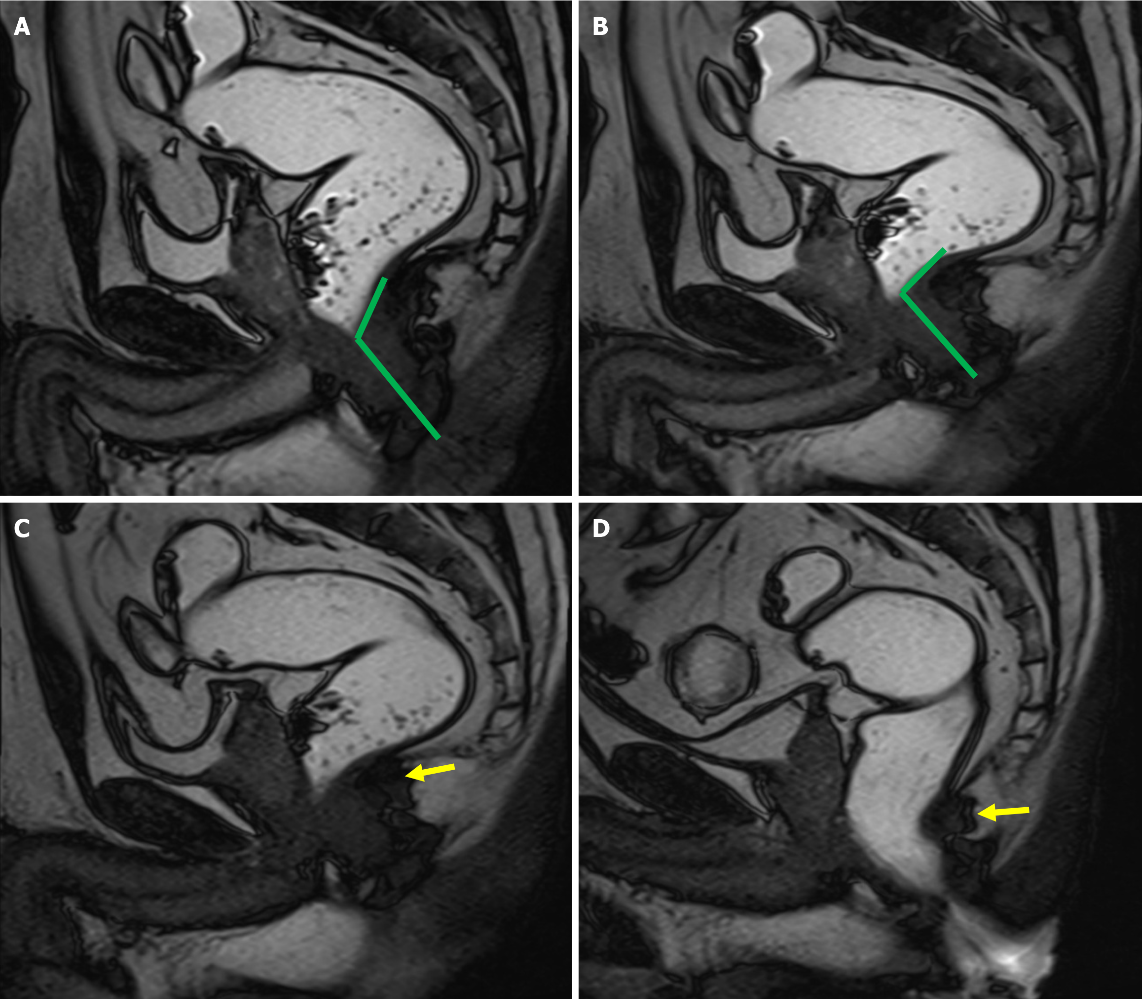
A: Sagittal true fast imaging with steady-state free precession image at rest shows an obtuse anorectal angle (ARA); B: During the squeeze maneuver the ARA reduces; C: During the strain phase, there is further reduction in the ARA with the prominent pubococcygeal muscle (yellow arrow) indenting the posterior wall of the anorectal junction; D: During defecation, the anorectal junction shows further indentation (yellow arrow) by the puborectalis muscle, which further reduces the ARA.
Figure 12 Dyssynergic defecation.
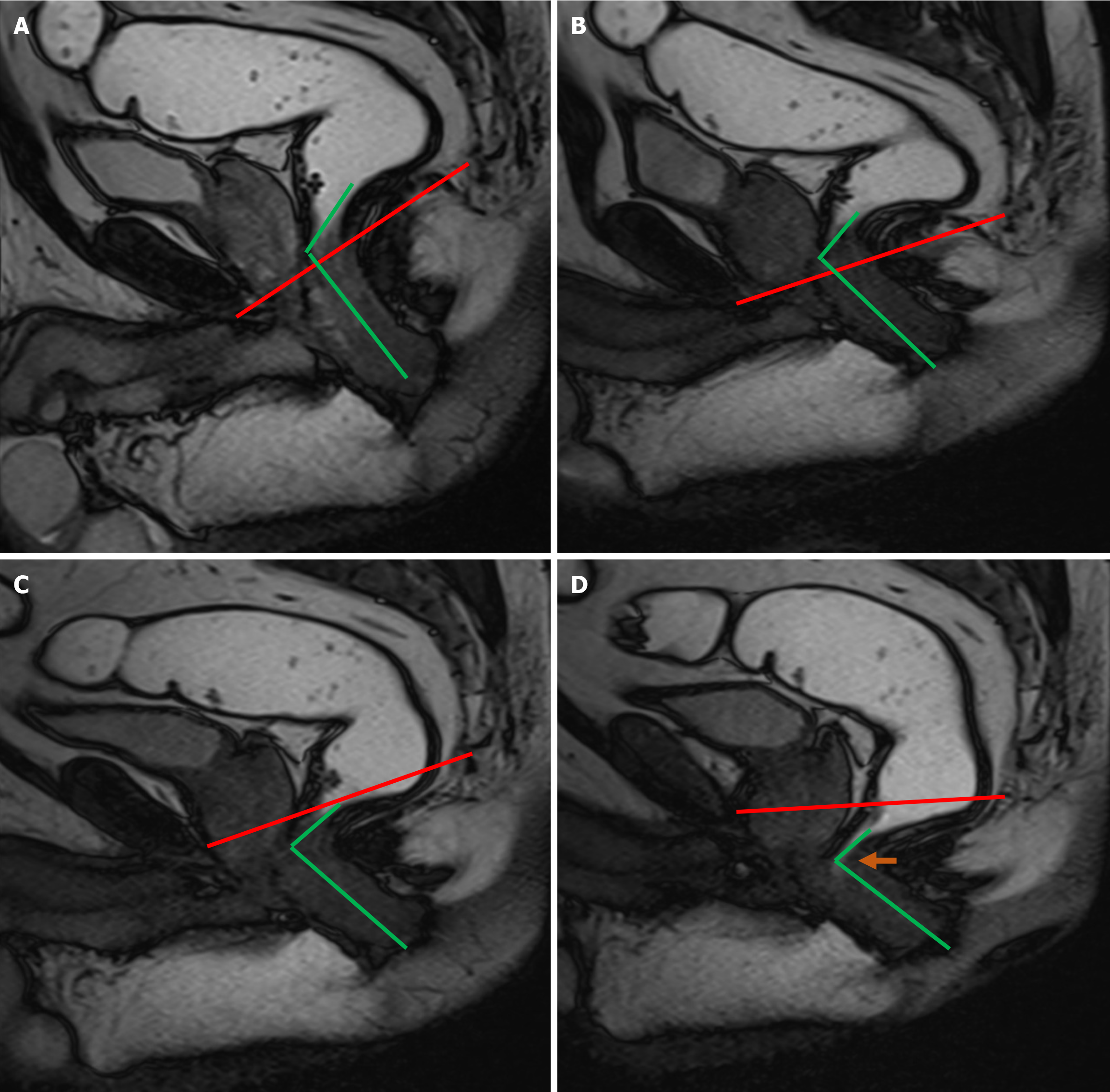
A: Sagittal true fast imaging with steady-state free precession image at rest demonstrates an anorectal angle (ARA) of greater than 90°; B: During the squeeze maneuver there is reduction in ARA; C: During the strain phase ARA reduces further; D: During the defecation phase, there is further paradoxical reduction in the ARA, with failure of the anal canal to open up.
- Citation: Parry AH, Rehaman B, Bhat SA, Wani AH, Jehangir M, Baba AA. Role of magnetic resonance defecography in the assessment of obstructed defecation syndrome. World J Radiol 2025; 17(6): 107205
- URL: https://www.wjgnet.com/1949-8470/full/v17/i6/107205.htm
- DOI: https://dx.doi.org/10.4329/wjr.v17.i6.107205


