Copyright
©The Author(s) 2025.
World J Radiol. Mar 28, 2025; 17(3): 103822
Published online Mar 28, 2025. doi: 10.4329/wjr.v17.i3.103822
Published online Mar 28, 2025. doi: 10.4329/wjr.v17.i3.103822
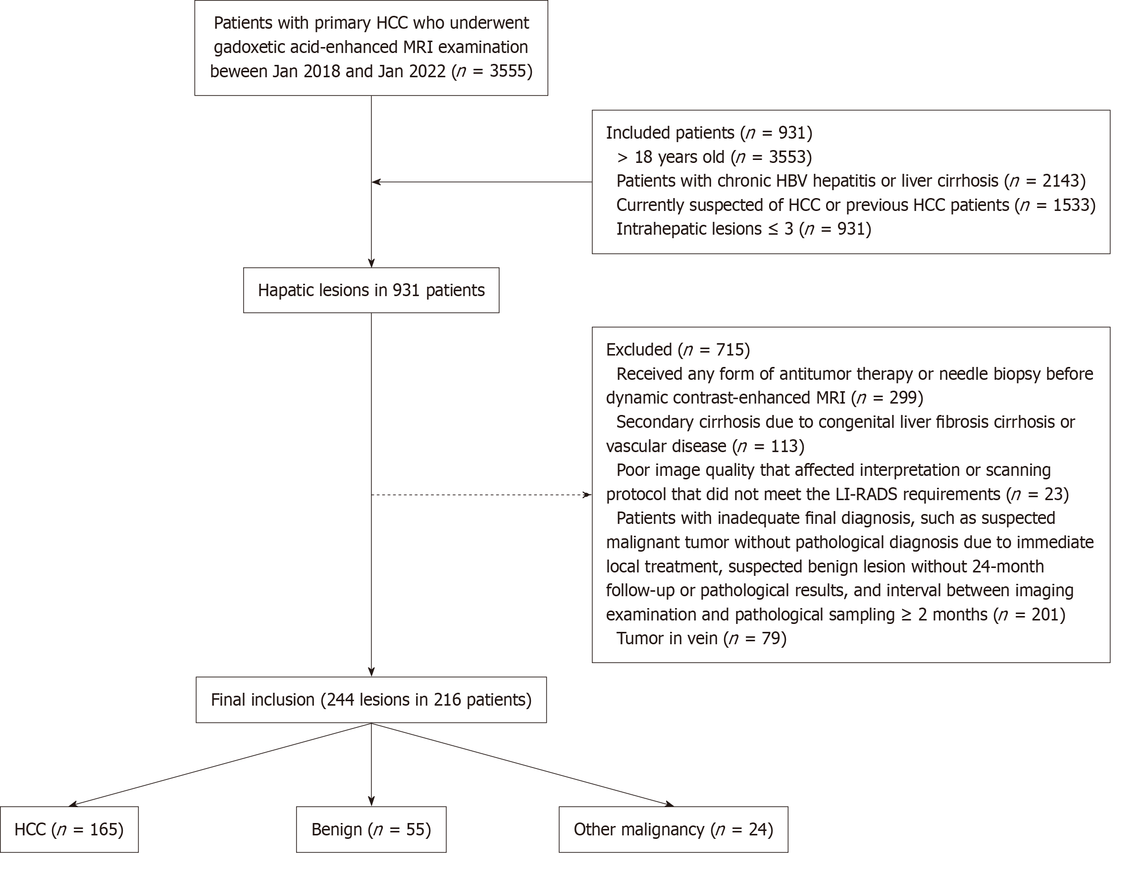
Figure 1 Flowchart.
HCC: Hepatocellular carcinoma; MRI: Magnetic resonance imaging; HBV: Hepatitis B virus; LI-RADS: Liver Imaging Reporting and Data System.
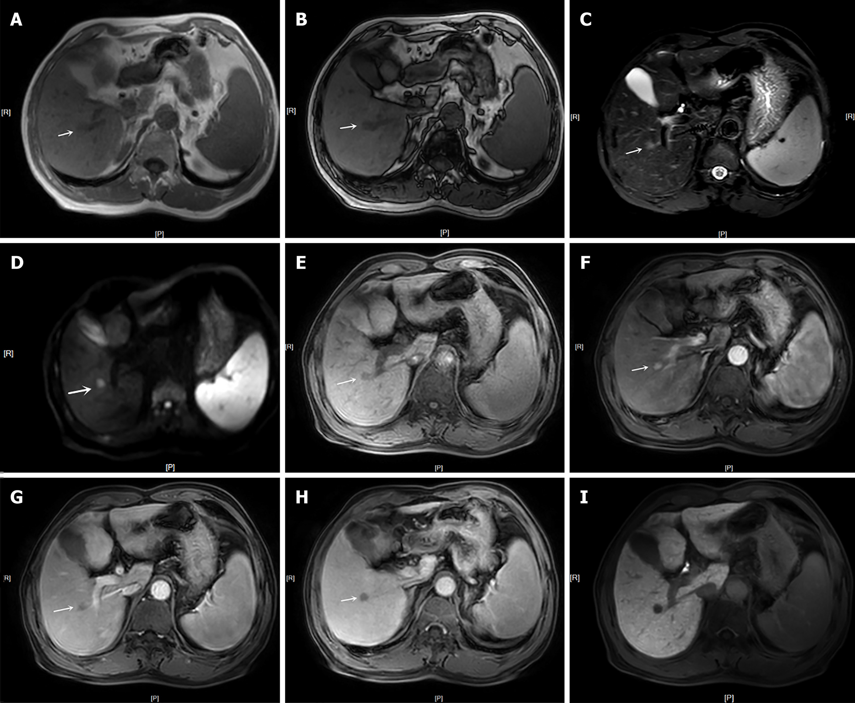
Figure 2 Axial images obtained with gadoxetic acid–enhanced magnetic resonance imaging in a 62-year-old man with cirrhosis by chronic B-viral hepatitis and surgically confirmed hepatocellular carcinoma.
This lesion was assigned as LR-4 and could not be classified as LR-5 using Liver Imaging Reporting and Data System version 2018. In contrast, our modified LR-5 criteria or alternative criteria (LR-5-M1, LR-5-M2, LR-5-M8 and LR-5-M9) by utilizing independently significant ancillary features (nonrim arterial phase hyperenhancement, nonperipheral washout, mild-moderate T2 hyperintensity and transition phase hypointensity) helped achieve the correct diagnosis of hepatocellular carcinoma. A and B: In-/out-of-phase T1-weighted image showed a 0.9 cm-diameter mass in segment VIII of the liver (white arrow); C: T2-weighted image showed mild-moderate T2 hyperintensity in the lesion; D: Diffusion-weighted imaging showed restricted diffusion in the lesion; E: Pre-enhanced phase showed hypointensity in the lesion; F: Late artery phase showed nonrim arterial phase hyperenhancement in the lesion; G: Portal venous phase showed nonperipheral washout in the lesion; H and I: The transition phase and hepatobiliary phase showed hypointensity in the lesion (white arrow).
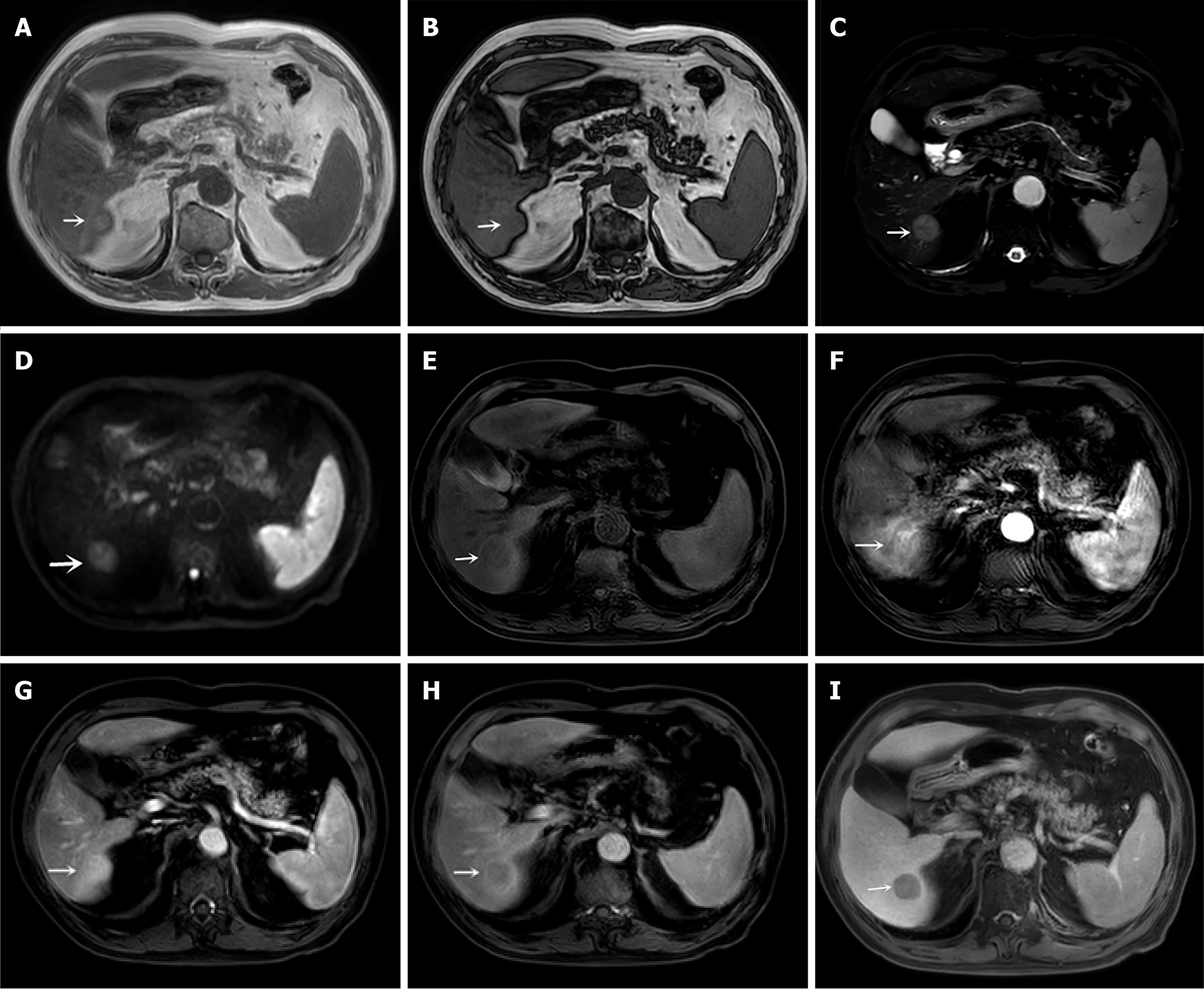
Figure 3 Axial images obtained with gadoxetic acid–enhanced magnetic resonance imaging in a 79-year-old man with cirrhosis by chronic B-viral hepatitis and surgically confirmed hepatocellular carcinoma.
This lesion was assigned as LR-4 and could not be classified as LR-5 using Liver Imaging Reporting and Data System version 2018. In contrast, our modified LR-5 criteria or alternative criteria (LR-5-M1, LR-5-M2, LR-5-M3, LR-5-M4, LR-5-M5, LR-5-M6, LR-5-M7, LR-5-M8 and LR-5-M9) by utilizing independently significant ancillary features (nonrim arterial phase hyperenhancement, mild-moderate T2 hyperintensity, transition phase hypointensity and fat in mass, more than adjacent liver) helped achieve the correct diagnosis of hepatocellular carcinoma. A and B: T1-weighted image showed a 1.9 cm-diameter mass in segment VI of the liver (white arrow), signal loss (white arrow) in the leision of out phase imaging compared to that of in phase imaging indicated fat in mass, more than adjacent liver; C: T2-weighted image showed mild-moderate T2 hyperintensity in the lesion; D: Diffusion-weighted imaging showed restricted diffusion in the lesion; E: Pre-enhanced phase showed hypointensity in the lesion; F: Late artery phase showed nonrim arterial phase hyperenhancement and corona enhancement in the lesion; G: Portal venous phase showed no washout in the lesion; H: Transition phase showed hypointensity and enhancing capsule in the lesion; I: Hepatobiliary phase showed hypointensity in the lesion (white arrow).
- Citation: Song Y, Zhang YY, Yu Q, Ma R, Xiao Y, Shen JK, Wei CG. Modified LR-5 criteria based on gadoxetic acid can improve the sensitivity in the diagnosis of hepatocellular carcinoma. World J Radiol 2025; 17(3): 103822
- URL: https://www.wjgnet.com/1949-8470/full/v17/i3/103822.htm
- DOI: https://dx.doi.org/10.4329/wjr.v17.i3.103822


