Copyright
©The Author(s) 2025.
World J Radiol. Mar 28, 2025; 17(3): 100168
Published online Mar 28, 2025. doi: 10.4329/wjr.v17.i3.100168
Published online Mar 28, 2025. doi: 10.4329/wjr.v17.i3.100168
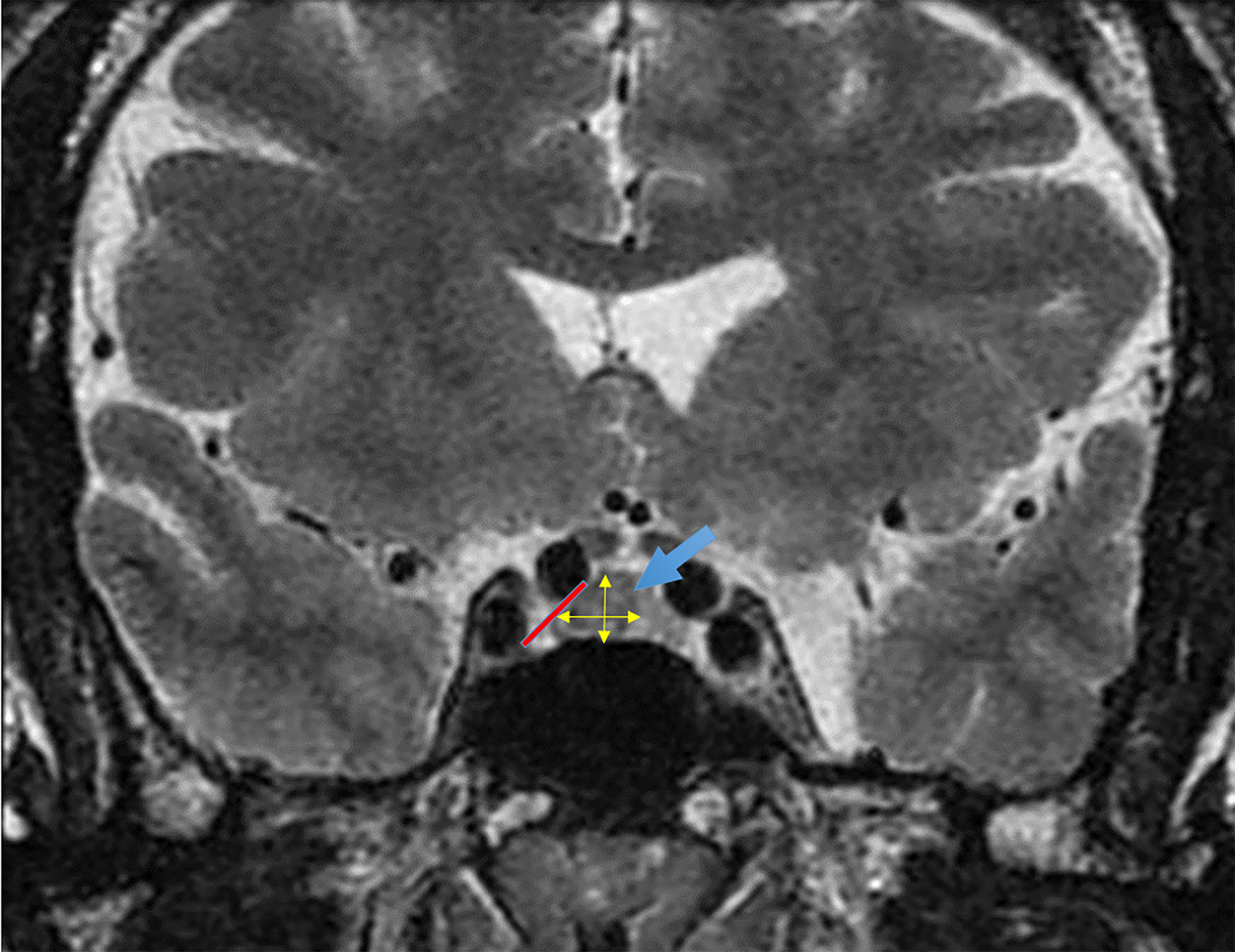
Figure 1 T2-weighted coronal magnetic resonance image.
A rounded hypo-enhancing lesion is centered in the right aspect of the pituitary gland (blue arrow). No evidence of cavernous sinus invasion is noted (red line). Measurement of the adenoma's length and height is indicated by the yellow arrows. Pituitary neuroendocrine tumor volume was calculated using the simplified ellipsoid volume formula: Volume = (length × height × width)/2.
- Citation: Alvarez M, Donato A, Rincon J, Rincon O, Lancheros N, Mancera P, Guzman I. Evaluation of pituitary tumor volume as a prognostic factor in acromegaly: A cross-sectional study in two centers. World J Radiol 2025; 17(3): 100168
- URL: https://www.wjgnet.com/1949-8470/full/v17/i3/100168.htm
- DOI: https://dx.doi.org/10.4329/wjr.v17.i3.100168


