Copyright
©The Author(s) 2024.
World J Radiol. Sep 28, 2024; 16(9): 482-488
Published online Sep 28, 2024. doi: 10.4329/wjr.v16.i9.482
Published online Sep 28, 2024. doi: 10.4329/wjr.v16.i9.482
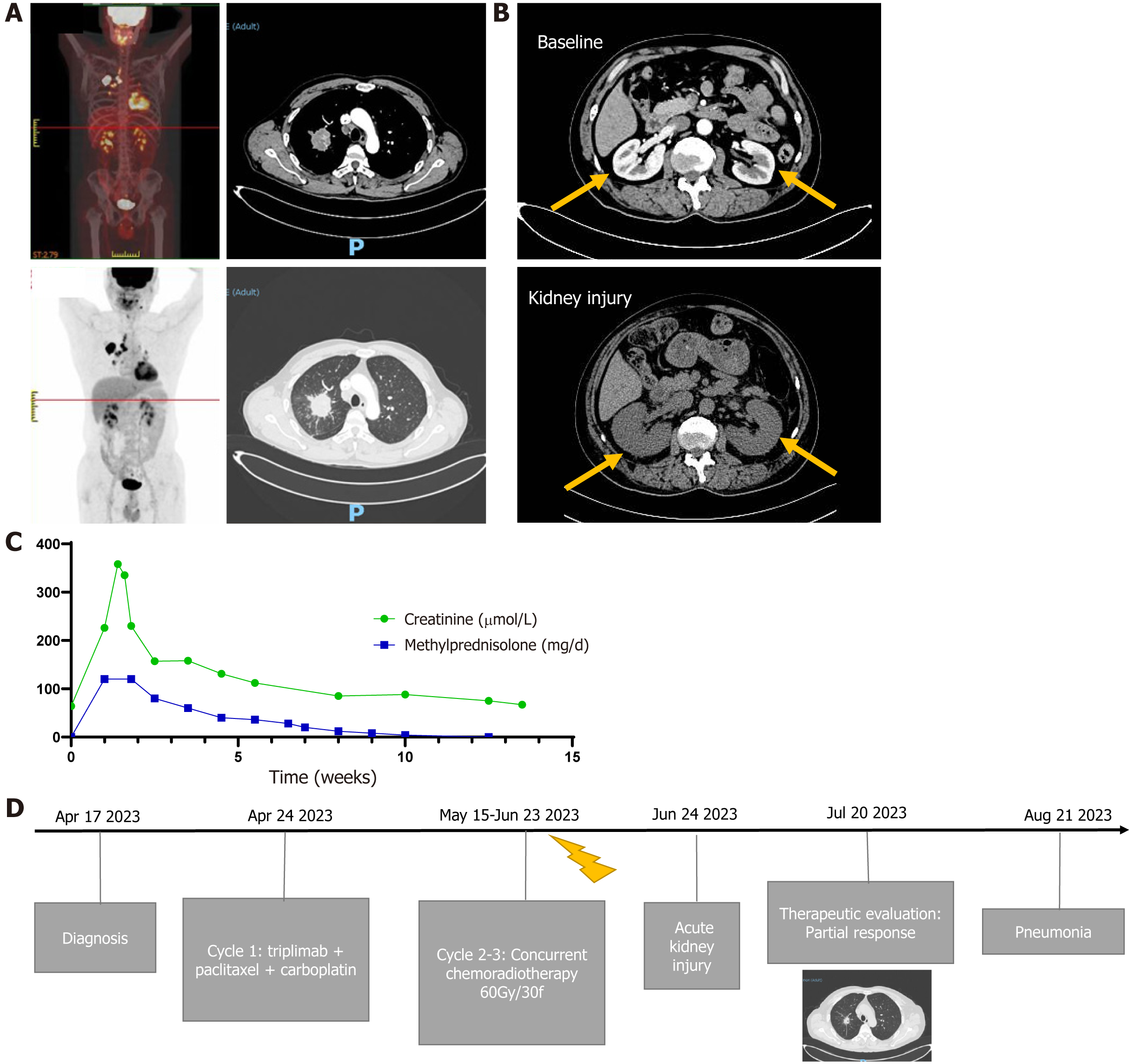
Figure 1 Time line of treatment.
A: Positron emission tomography image at diagnosis; B: Images of acute kidney injury; C: Changes in creatinine and methylprednisolone levels; D: Treatment timeline.
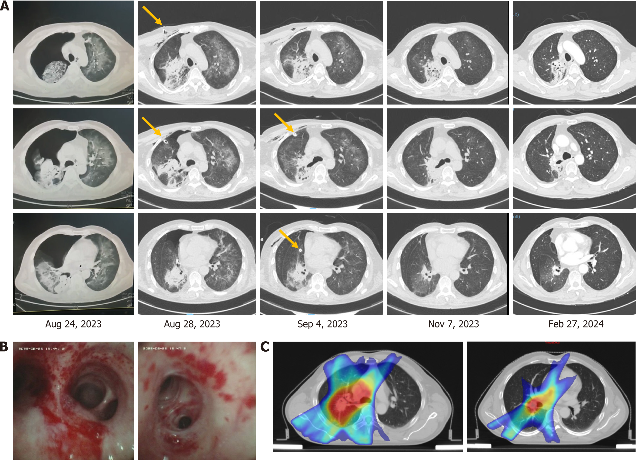
Figure 2 Radiographic and tracheoscopy findings.
A: The computed tomography image changes before and after PJP treatment. The orange arrow shows the drainage tube. B: Tracheoscopy findings. C: Tumor irradiation field.
- Citation: Zheng YW, Pan JC, Wang JF, Zhang J. Pneumocystis pneumonia in stage IIIA lung adenocarcinoma with immune-related acute kidney injury and thoracic radiotherapy: A case report. World J Radiol 2024; 16(9): 482-488
- URL: https://www.wjgnet.com/1949-8470/full/v16/i9/482.htm
- DOI: https://dx.doi.org/10.4329/wjr.v16.i9.482


