Copyright
©The Author(s) 2024.
World J Radiol. Jun 28, 2024; 16(6): 232-240
Published online Jun 28, 2024. doi: 10.4329/wjr.v16.i6.232
Published online Jun 28, 2024. doi: 10.4329/wjr.v16.i6.232
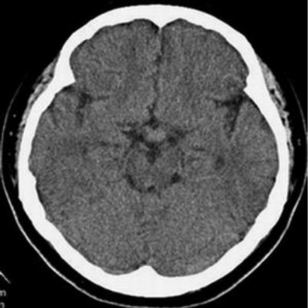
Figure 1 Cranial computed tomography showed no significant abnormalities.
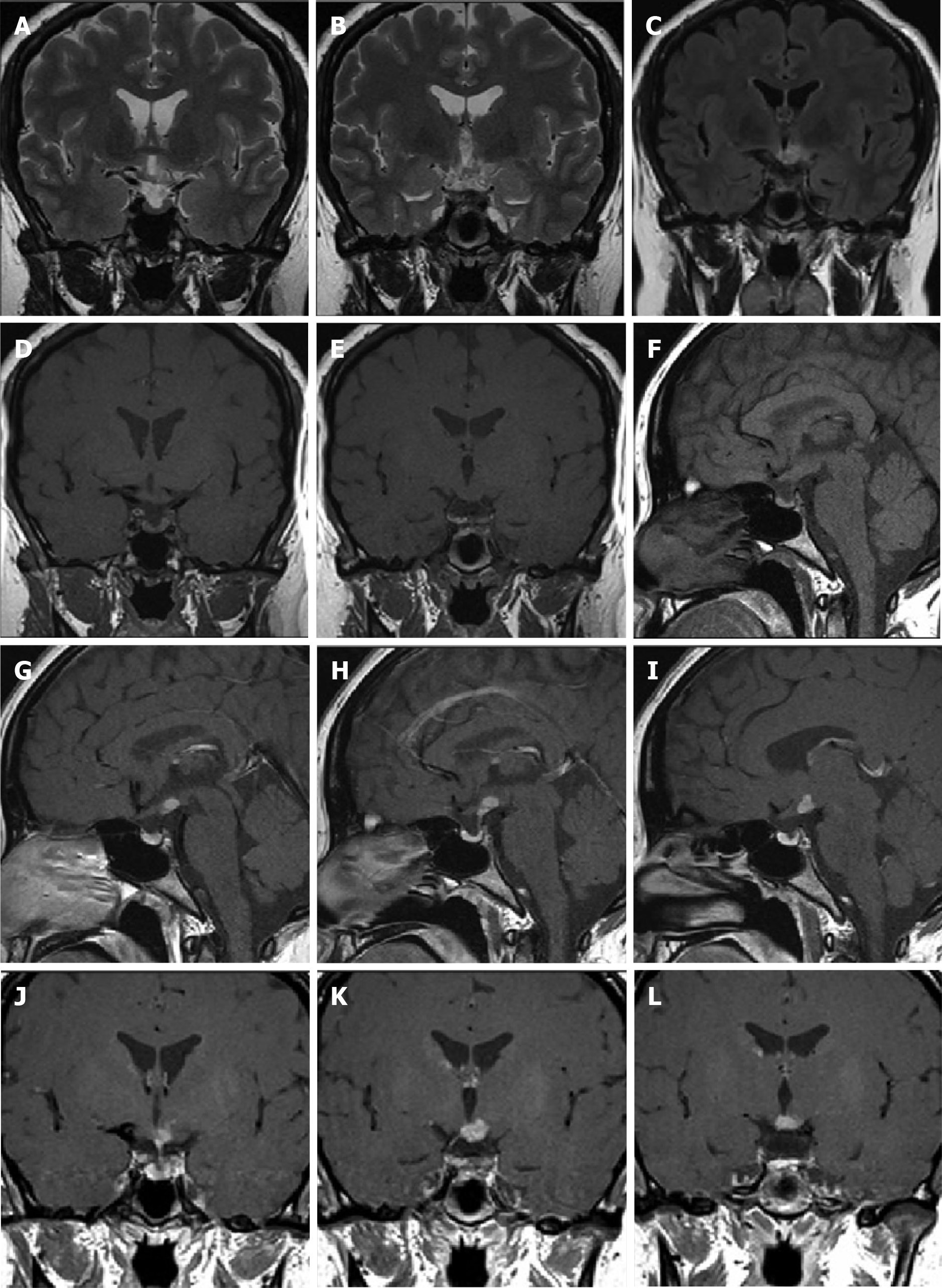
Figure 2 Cranial magnetic resonance imaging enhancement scan showed pituitary height lower than normal for the same age group.
Abnormal enhancement occurred anterior to the papillary body on the enhancement scan. A and B: Representative coronal T2-weighted images; C: Coronal fluid attenuated inversion recovery image; D: E: Representative coronal T1-weighted images; F: Sagittal T1-weighted image; G-I: Representative sagittal enhancement scans; J-L: Representative coronal enhancement scans.
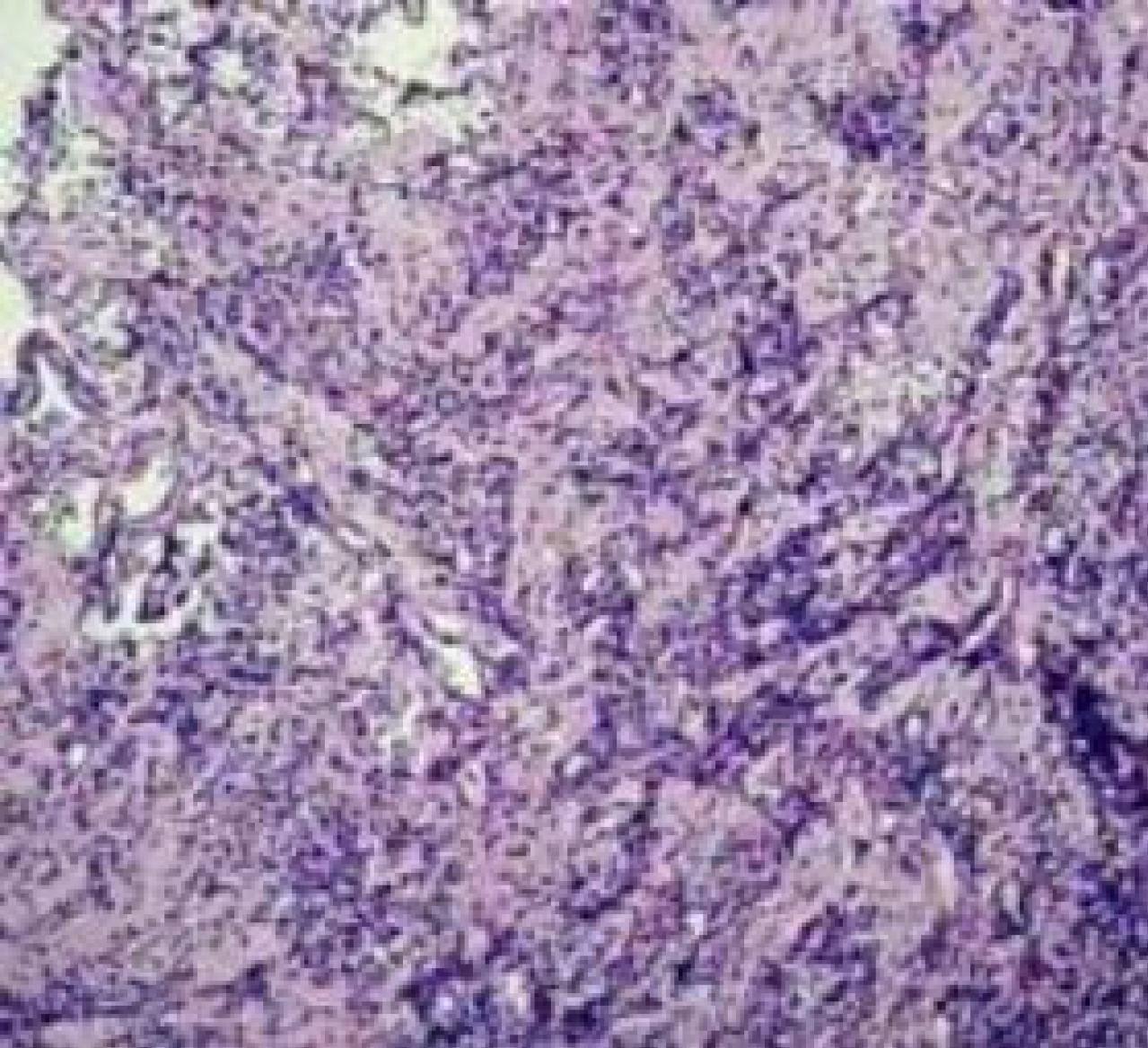
Figure 3 Postoperative pathology showed chronic inflammation.
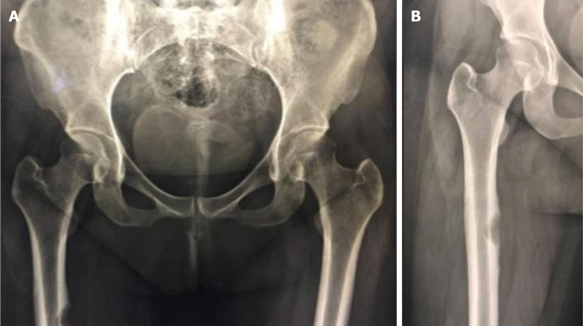
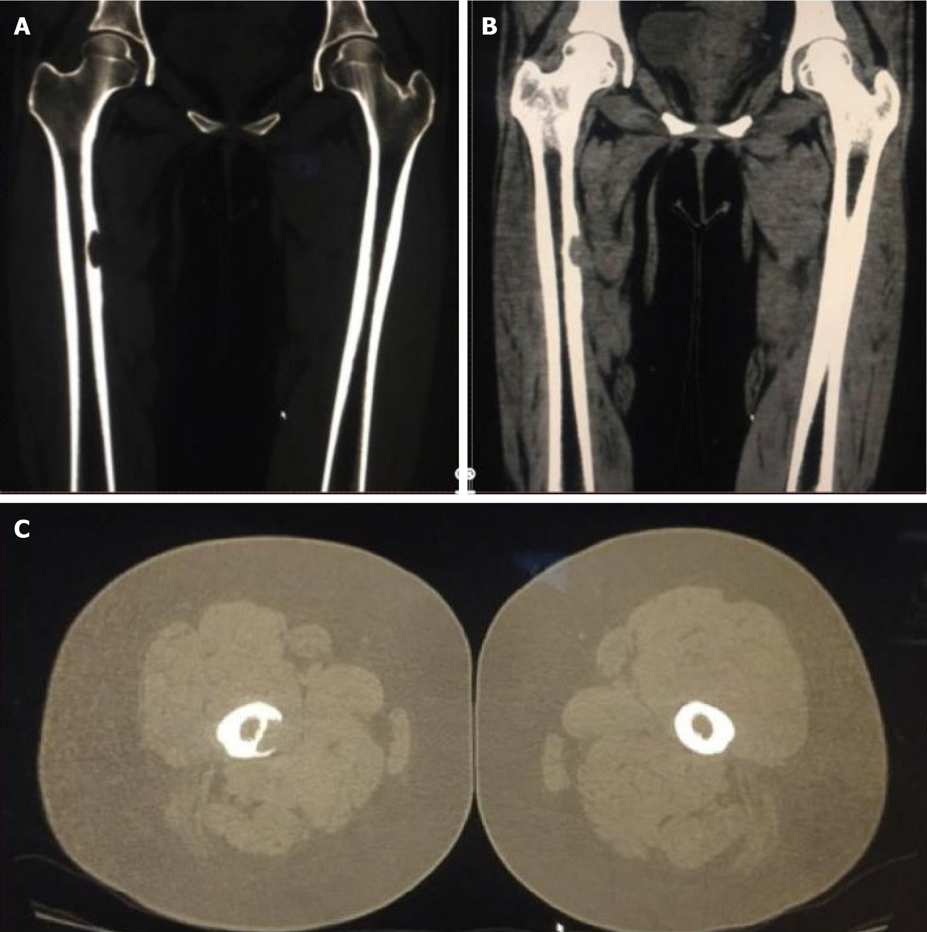
Figure 4 X-ray examination of the patient showed bone destruction and defects in the upper part of the right femur.
A: Bilateral hip; B: Right lower limb.
Figure 5 Three-dimensional computed tomography showed cortical destruction and soft tissue mass formation in the medial aspect of the right upper femur.
A: Coronal bone view; B: Coronal soft tissue view; C: Axial view.
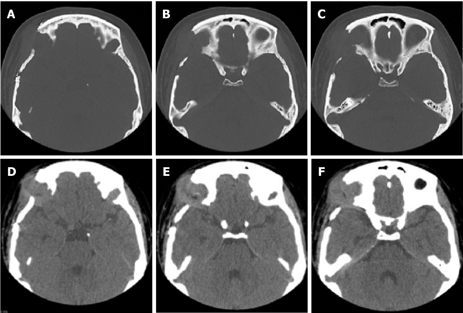
Figure 6 Orbital computed tomography showed irregular bone destruction in the upper outer wall of the right eye orbit combined with the formation of a surrounding soft tissue mass.
A-C: Representative axial bone views; D-F: Representative axial soft tissue views.
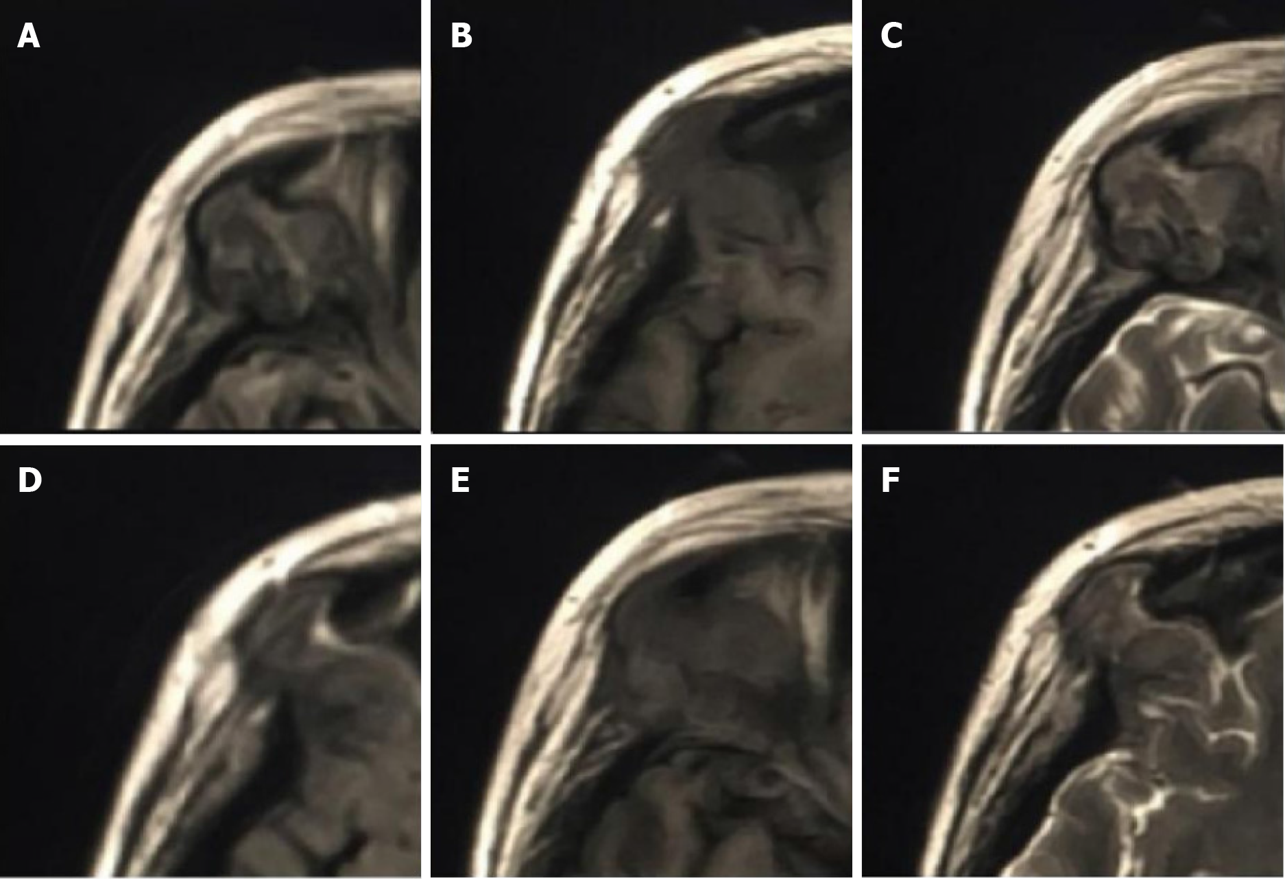
Figure 7 Orbital magnetic resonance imaging showed irregular bone destruction in the upper outer wall of the right eye orbit.
It was found with isotropic T1 and isotropic/slightly longer T2 signal shadows with isotropic signals in the fluid attenuated inversion recovery sequence. A-C: Representative the upper half of the lesion as seen on the axial fluid-attenuated inversion recovery, T2-weighted, and T1-weighted images; D-F: Representative the lower half of the lesion as seen on the axial fluid-attenuated inversion recovery, T2-weighted, and T1-weighted images.
- Citation: Zhang ZR, Chen F, Chen HJ. Multisystemic recurrent Langerhans cell histiocytosis misdiagnosed with chronic inflammation at the first diagnosis: A case report. World J Radiol 2024; 16(6): 232-240
- URL: https://www.wjgnet.com/1949-8470/full/v16/i6/232.htm
- DOI: https://dx.doi.org/10.4329/wjr.v16.i6.232


