Copyright
©The Author(s) 2024.
World J Radiol. Mar 28, 2024; 16(3): 40-48
Published online Mar 28, 2024. doi: 10.4329/wjr.v16.i3.40
Published online Mar 28, 2024. doi: 10.4329/wjr.v16.i3.40
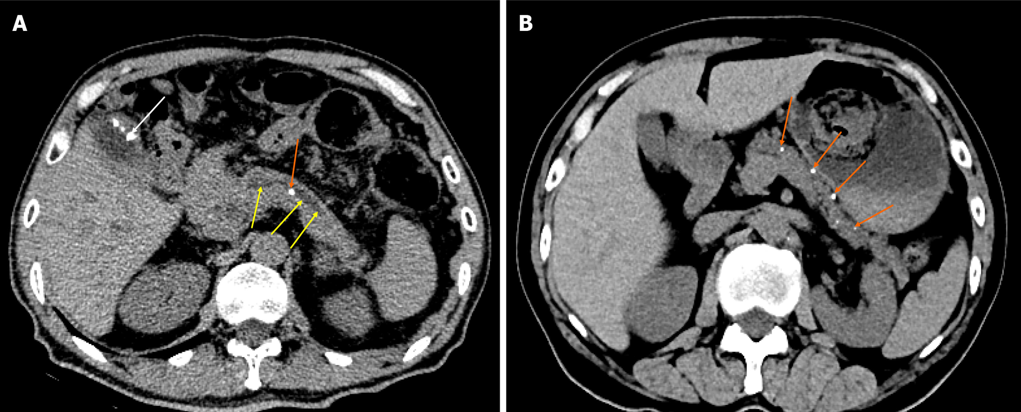
Figure 1 Chronic pancreatitis.
A: Chronic pancreatitis with main pancreatic duct stone and main pancreatic duct dilatation. A 58-year-old man with chronic pancreatitis presented with no pain. The abdominal computed tomography (CT) scan represents the main pancreatic duct stone (orange arrow), the dilated main pancreatic duct (yellow arrows) with a diameter of 0.5 cm, as well as the combined gallbladder multiple stones (white arrow); B: Chronic pancreatitis with pancreatic parenchymal atrophy and multiple pancreatic calcifications. A 69-year-old man with chronic pancreatitis. The abdominal CT scan shows a decrease in pancreatic volume, parenchymal atrophy, and multiple calcifications in the pancreatic parenchyma (orange arrows).
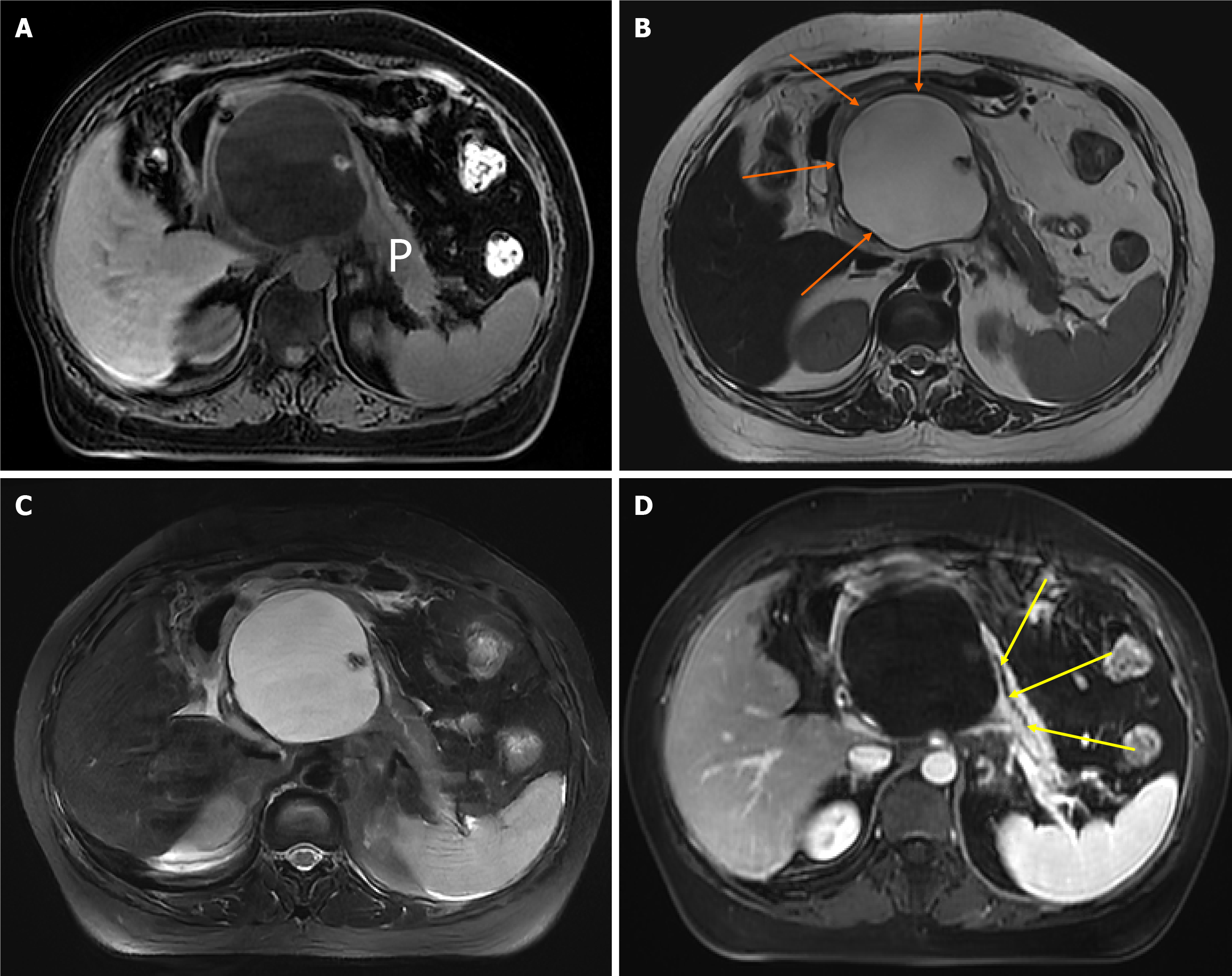
Figure 2 Chronic pancreatitis with large pancreatic cysts.
A 52-year-old woman with chronic pancreatitis presented with upper abdominal pain. The upper abdominal magnetic resonance images. A: Magnetic resonance imaging (MRI) fat-suppressed T1-weighted imaging shows a large pseudocyst of the head of the pancreas, which has a low signal; B: MRI T2-weighted imaging can show a clear boundary of pseudocyst (orange arrows) with a diameter of 7 cm × 11 cm, which has a high signal; C: MRI fat-suppressed T2-weighted imaging; D: MRI enhanced scanning venous phase shows the large pseudocyst has no enhancement as well as the displaced main pancreatic duct (yellow arrows). P: Pancreas.
- Citation: Feng Y, Song LJ, Xiao B. Chronic pancreatitis: Pain and computed tomography/magnetic resonance imaging findings. World J Radiol 2024; 16(3): 40-48
- URL: https://www.wjgnet.com/1949-8470/full/v16/i3/40.htm
- DOI: https://dx.doi.org/10.4329/wjr.v16.i3.40


