Copyright
©The Author(s) 2024.
World J Radiol. Nov 28, 2024; 16(11): 689-695
Published online Nov 28, 2024. doi: 10.4329/wjr.v16.i11.689
Published online Nov 28, 2024. doi: 10.4329/wjr.v16.i11.689
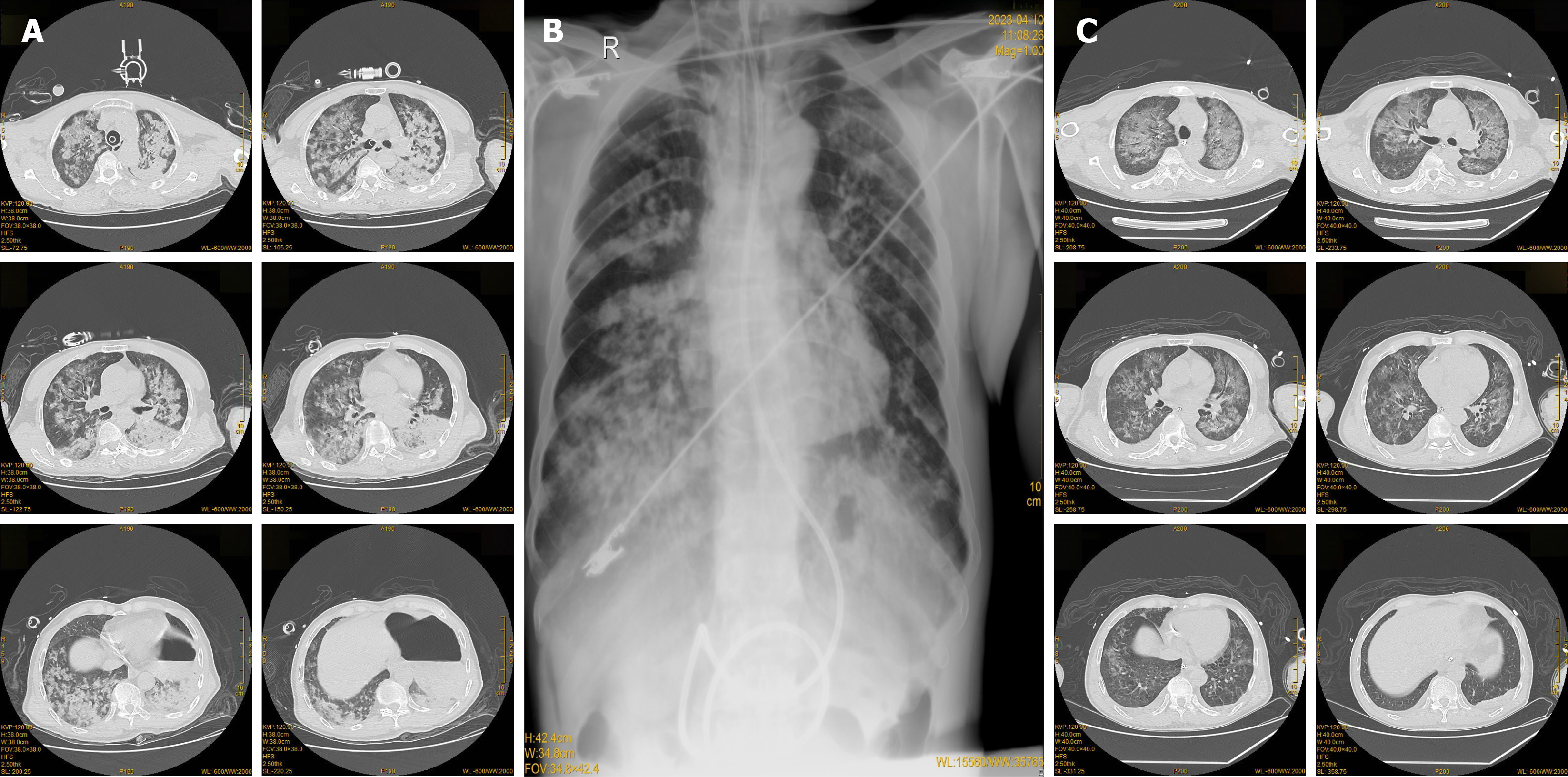
Figure 1 Imaging changes found in the course of acute respiratory distress syndrome management.
Changes on chest computed tomography (CT) before; radiography taken on April 10th; chest CT after treatment on April 13th. A: The chest CT carried out in the emergency room revealed pulmonary infiltrates in both lungs; B: A follow-up chest radiography on April 10th indicated exudative lesions in both lungs; C: After 5 days of treatment, a repeat chest CT examination showed significant resolution of the pulmonary infiltrates.
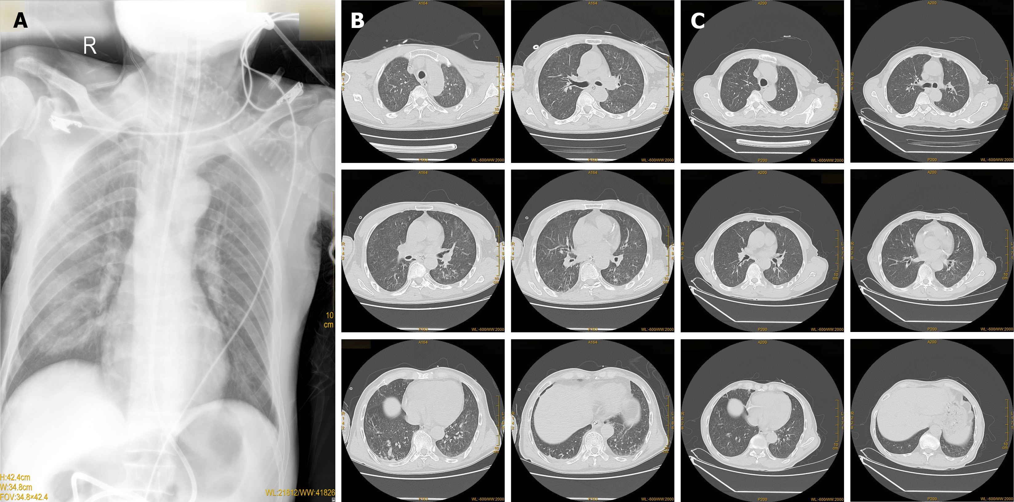
Figure 2 Gradual imaging indications of improvement post-treatment in patients.
Radiography taken on April 16th; Chest computed tomography (CT) after treatment on April 19th; Chest CT after treatment on April 27th. A: Chest radiography taken on April 16th, compared to that on April 9th, indicated a significant reduction in exudative lesions in both lungs; B: The chest CT examination on April 19th showed resolution of the exudative lesion when compared to the previous image; C: After treatment, a follow-up chest CT on April 27th indicated significant improvement in the absorption of exudative lesions in both lungs.
- Citation: Yang KY, Cui ZX. Acute respiratory distress syndrome caused by demulsifier poisoning: A case report. World J Radiol 2024; 16(11): 689-695
- URL: https://www.wjgnet.com/1949-8470/full/v16/i11/689.htm
- DOI: https://dx.doi.org/10.4329/wjr.v16.i11.689


