Copyright
©The Author(s) 2024.
World J Radiol. Nov 28, 2024; 16(11): 683-688
Published online Nov 28, 2024. doi: 10.4329/wjr.v16.i11.683
Published online Nov 28, 2024. doi: 10.4329/wjr.v16.i11.683
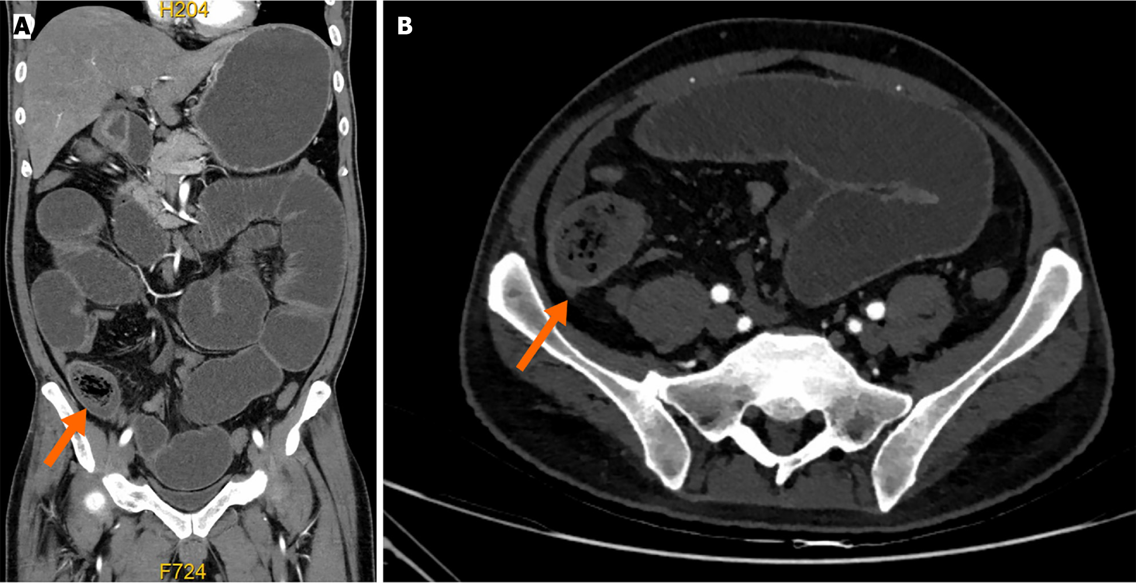
Figure 1 Computed tomography of the abdomen.
A: Coronal view demonstrating obstructing intraluminal lesion (arrowhead); B: Transverse view demonstrating obstructing intraluminal lesion (arrowhead).
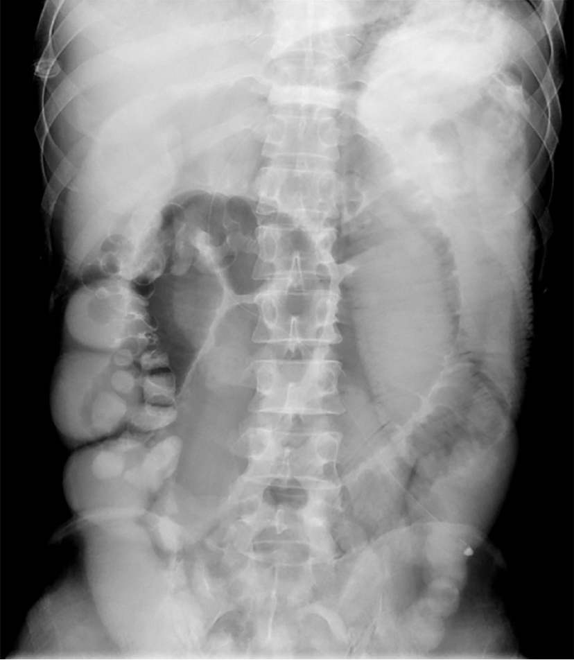
Figure 2 Full gastrointestinal radiography showing small bowel insufficiency obstruction and slow gastric emptying.
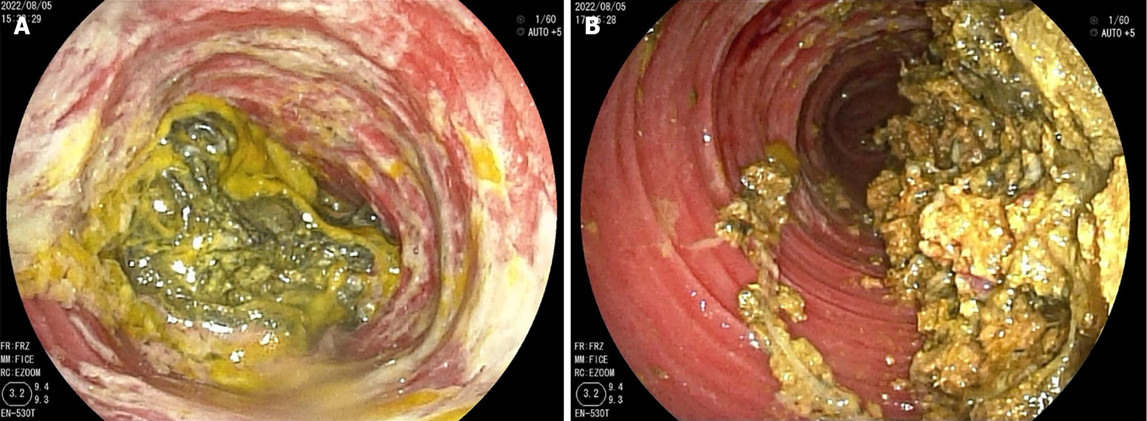
Figure 3 The result of double balloon enteroscopy.
A: Double balloon enteroscopy showing a large phytobezoar completely obstructing the bowel; B: The bezoar was smashed until obtaining small pieces with a snare, and paraffin oil was injected.
- Citation: Lu BY, Zeng ZY, Zhang DJ. Successful treatment of small bowel phytobezoar using double balloon enterolithotripsy combined with sequential catharsis: A case report. World J Radiol 2024; 16(11): 683-688
- URL: https://www.wjgnet.com/1949-8470/full/v16/i11/683.htm
- DOI: https://dx.doi.org/10.4329/wjr.v16.i11.683


