Copyright
©The Author(s) 2024.
World J Radiol. Nov 28, 2024; 16(11): 668-677
Published online Nov 28, 2024. doi: 10.4329/wjr.v16.i11.668
Published online Nov 28, 2024. doi: 10.4329/wjr.v16.i11.668
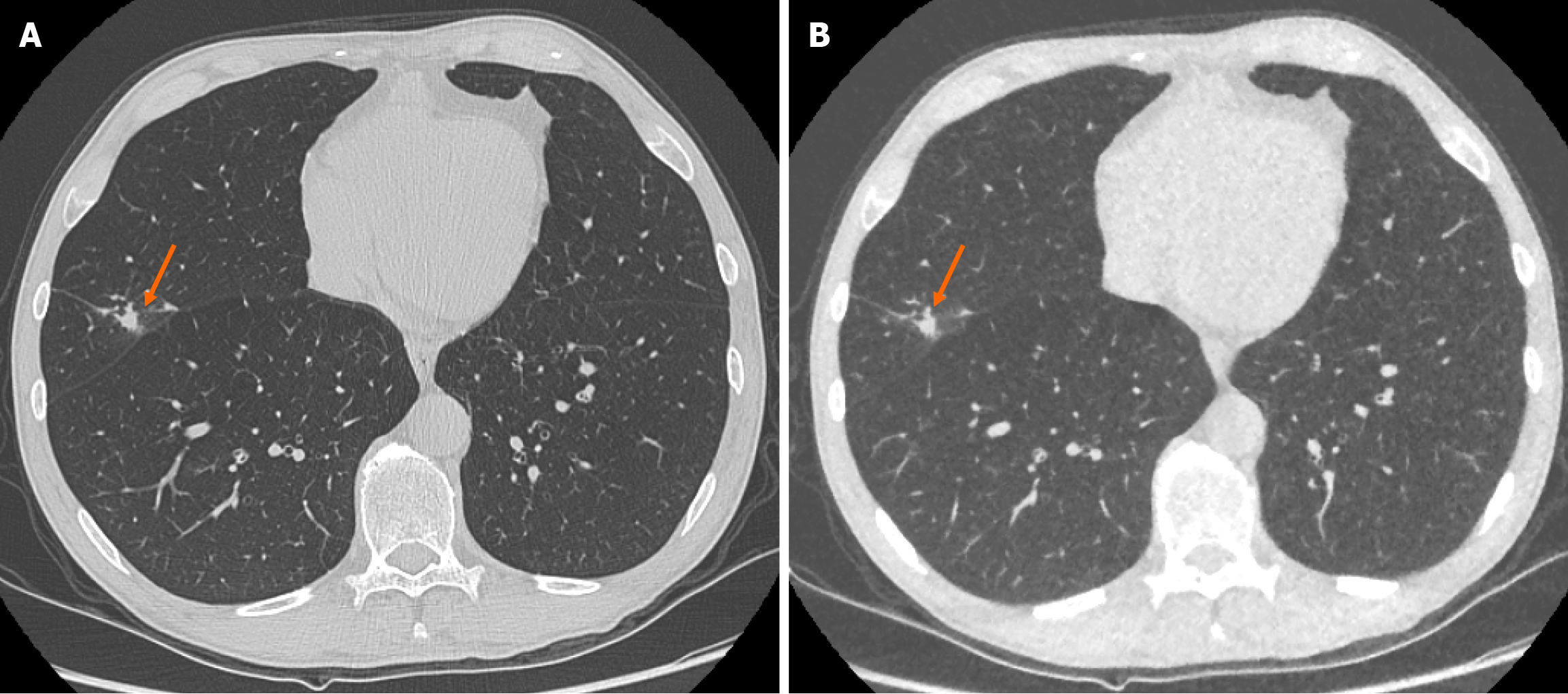
Figure 1 Representative images demonstrating image quality and ability to identify pulmonary nodules on both standard dose computed tomography chest and ultra-low-dose computed tomography chest with model-based iterative reconstruction imaging protocols.
A: Selected axial slice of a standard dose computed tomography (CT) chest presented in lung windows with a solid pulmonary nodule with spiculation and pleural tethering in the lateral segment of the middle lobe (arrow); B: Selected axial slice of an ultra-low dose CT chest in the same patient at the same level presented in lung windows with the same correctly identified pulmonary nodule in the middle lobe (arrow). These images demonstrate the ability of ultra-low-dose CT chest with model-based iterative reconstruction to adequately maintain diagnostic accuracy with regard to solid pulmonary nodules.
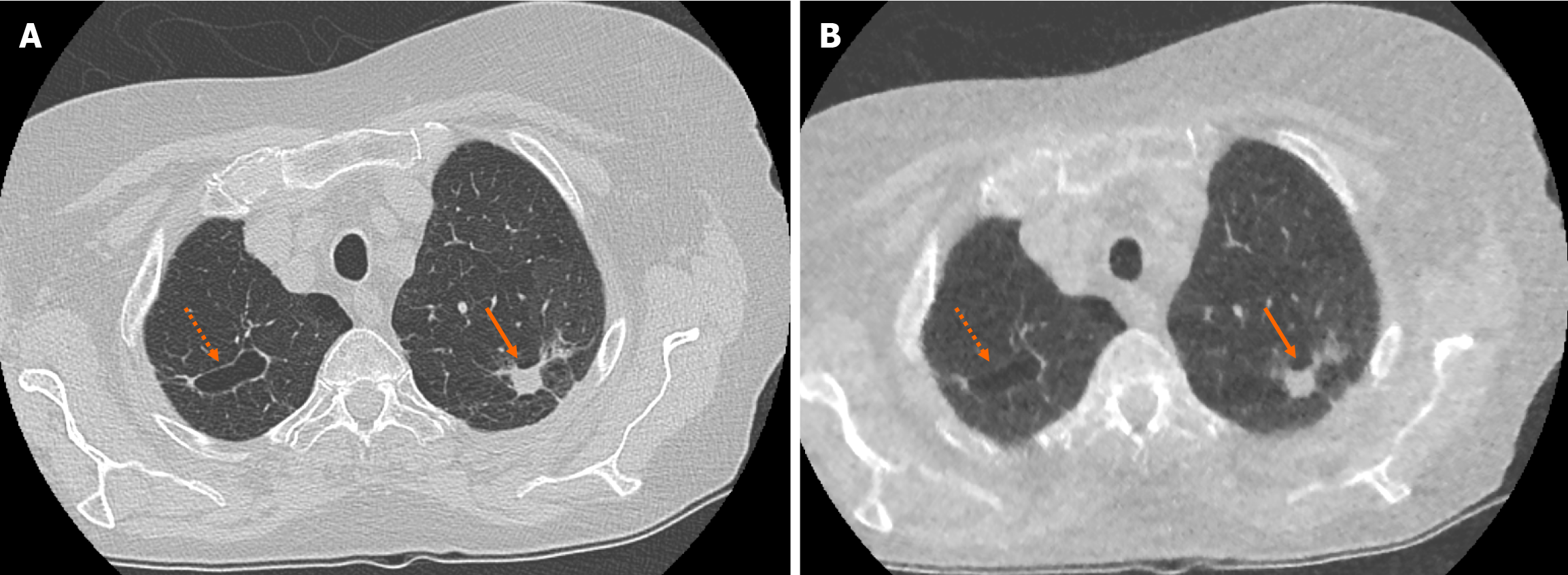
Figure 2 Example of accurate pulmonary nodule characterisation on ultra-low-dose computed tomography chest.
A: Selected axial slice of a standard dose computed tomography (CT) chest presented in lung windows with a spiculated solid pulmonary nodule with pleural tethering in the apico-posterior segment of the left upper lobe (solid arrow) and a parenchymal cyst in the apical segment of the right upper lobe (dashed arrow); B: Selected axial slice in the same patient presented in lung windows at the same level highlights the ability of ultra-low-dose CT chest with model-based iterative reconstruction to correctly characterise pulmonary nodule features such as spiculation, tethering and cavitation.
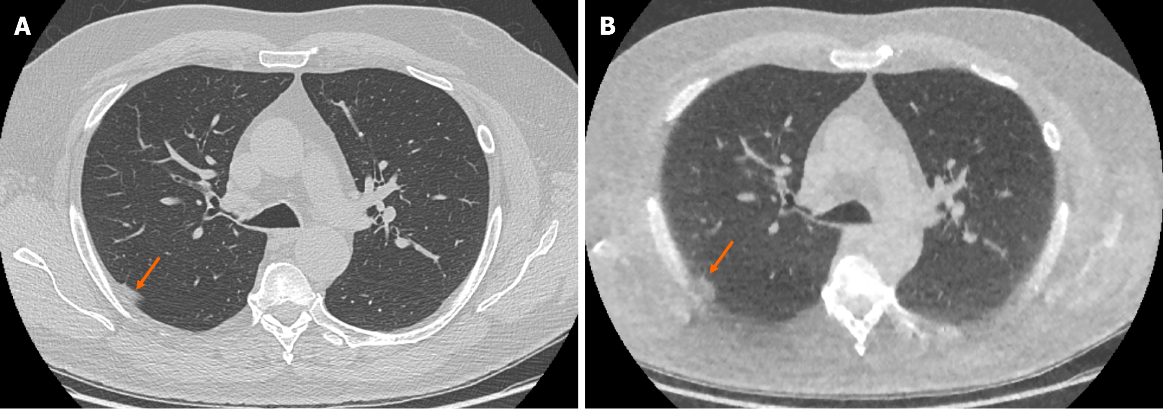
Figure 3 Example of false positive pulmonary nodule identification on ultra-low-dose computed tomography chest.
A: Selected axial slice of a standard dose computed tomography (CT) chest presented in lung windows with a small focus of peripheral atelectasis in the posterior segment of the right upper lobe (arrow); B: Selected axial slice of an ultra-low dose CT chest with model-based iterative reconstruction presented in lung windows at the same level in the same patient with the focus of soft tissue attenuation in the posterior segment of the right upper lobe incorrectly identified as a solid pulmonary nodule (arrow). The incidence of false positive solid nodule identification was minimal and did not reach statistical significance.
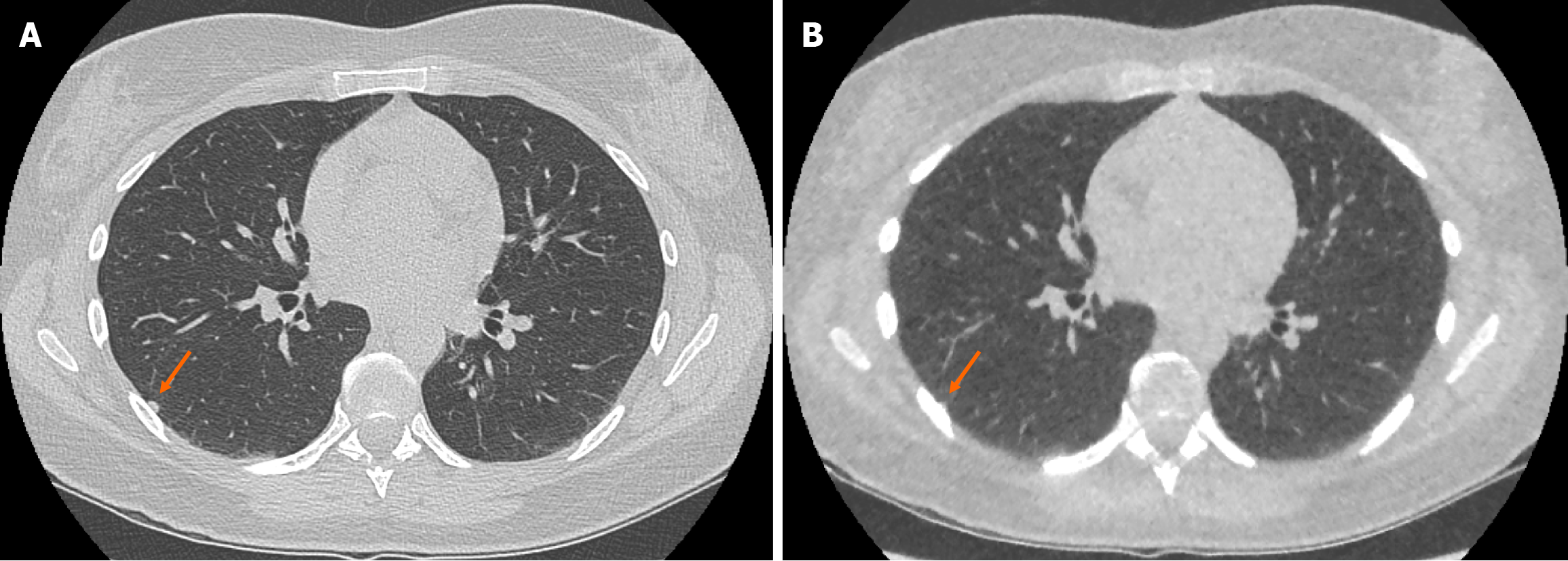
Figure 4 Example of false negative pulmonary nodule identification on ultra-low-dose computed tomography chest.
A: Selected axial slice of a standard dose computed tomography (CT) chest presented in lung windows with a solid pulmonary nodule abutting the pleura in the lateral segment of the right lower lobe (arrow); B: Selected axial slice of an ultra-low dose CT chest with model-based iterative reconstruction in the same patient at the same level presented in lung windows demonstrating the less conspicuous pulmonary nodule that was not identified (arrow). The incidence of false negative solid nodule identification was minimal and did not reach statistical significance.
- Citation: O'Regan PW, Harold-Barry A, O'Mahony AT, Crowley C, Joyce S, Moore N, O'Connor OJ, Henry MT, Ryan DJ, Maher MM. Ultra-low-dose chest computed tomography with model-based iterative reconstruction in the analysis of solid pulmonary nodules: A prospective study. World J Radiol 2024; 16(11): 668-677
- URL: https://www.wjgnet.com/1949-8470/full/v16/i11/668.htm
- DOI: https://dx.doi.org/10.4329/wjr.v16.i11.668


