Copyright
©The Author(s) 2023.
World J Radiol. Jun 28, 2023; 15(6): 191-200
Published online Jun 28, 2023. doi: 10.4329/wjr.v15.i6.191
Published online Jun 28, 2023. doi: 10.4329/wjr.v15.i6.191
Figure 1 Postoperative contrast-enhanced abdominal X-ray images following hepaticojejunostomy for the reconstruction of the biliary tract.
A: Postoperative 14th day contrast-enhanced abdominal X-ray in a 42-year-old man with major bile duct trauma (Strasberg Bismuth E1) who underwent T-tube reconstruction; the biliary tract and anastomosis was normal; B: The anastomosis and bile flow patency was normal in a 47-year-old woman who underwent hepaticojejunostomy for major bile duct trauma (Strasberg Bismuth E2); C: The stricture (arrow) in the common hepatic duct was detected in a 38-year-old woman who developed jaundice after biliary tract trauma (Strasberg Bismuth E2) and underwent percutaneous transhepatic biliary drainage.
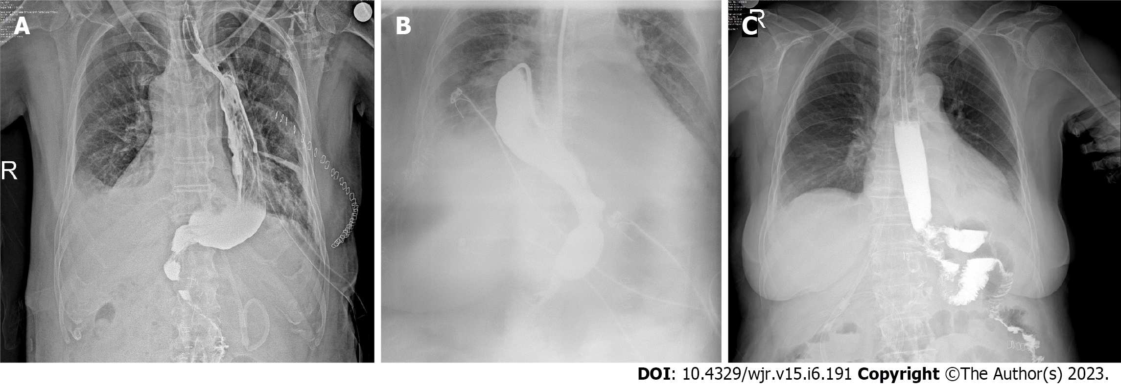
Figure 2 Contrast-enhanced abdominal X-ray images following gastrointestinal reconstruction for stomach/esophagus tumors.
A: A 67-year-old man and a 78-year-old woman who underwent esophagectomy + gastric pull-up for distal esophageal tumor. The esophagogastric passage was normal without any findings of fistula or leakage at the sites of anastomoses on the postoperative 4th day; C: The position of the jejunal loop/stump and the passage was normal in a 72-year-old woman who underwent total gastrectomy and Roux-n-Y anastomosis for cardia tumor.
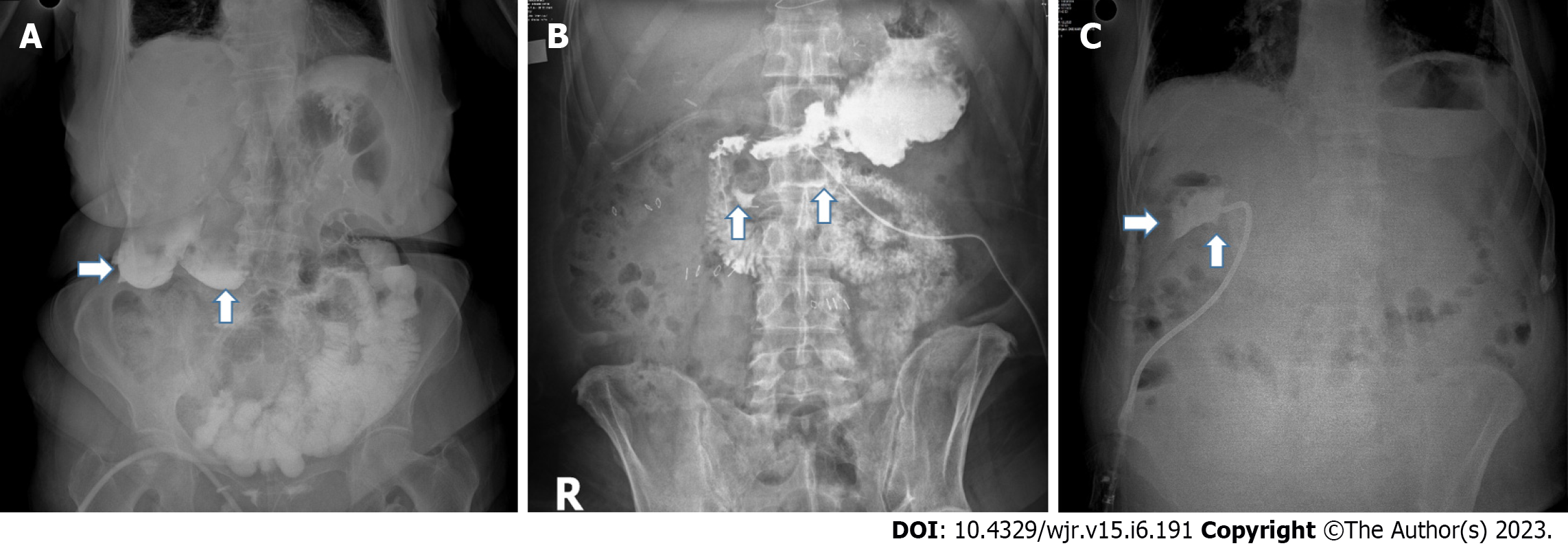
Figure 3 Contrast-enhanced abdominal X-ray images showing anastomotic leakage.
A: Anastomotic leakage in a 77-year-old man with total gastric resection and Roux-n-Y anastomosis (arrows) for gastric cancer on the 6th postoperative day; B: Anastomotic leakage detected in a 63-year-old man on the postoperative 4th day who underwent esophagectomy + gastric pull-up for distal esophagus tumor; C: Repeated control contrast-enhanced abdominal X-ray showed healing in the same patient on postoperative 30th day.
Figure 4 Functionality of the gastrointestinal passage and patency of anastomosis on contrast-enhanced abdominal X-rays.
A: A 57-year-old woman whose esophagojejunal anastomosis functionality and passage continuity were investigated and evaluated as normal on the 4th postoperative day; B: The gastric remnant, position, and functionality were normal in a 37-year-old woman (BMI-47.2) on the 3rd postoperative day after sleeve gastrectomy; C: Zenker’s diverticulum that was unexpectedly detected in a 44-year-old man with gastric resection for gastric cancer, who presented with dysphagia in the postoperative follow-up period.
Figure 5 Leakage and perforation on contrast-enhanced abdominal X-rays.
A: Contrast material leakage into the abdominal cavity (arrows) due to the peptic ulcer perforation seen on contrast-enhanced abdominal X-ray (CE-AXR) in a 76-year-old woman who cannot be diagnosed by clinical findings; B: CE-AXR in a 57-year-old woman who underwent laparoscopic stromal tumor resection and developed unexpected abdominal pain on the 5th postoperative day showed an opening at the suture line and contrast material leakage (arrows); C: In a 68-year-old woman who underwent hilar resection and portoenterostomy for Klatskin tumor, CE-AXR obtained by administering contrast material through the drain showed the biliary fistula tract and subhepatic pouch (arrows).
Figure 6 Contrast-enhanced abdominal X-rays showing intestinal obstruction.
A: Lateral contrast-enhanced abdominal X-ray (CE-AXR) showed gastric dilation due to gastric outlet obstruction (Arrow) in a 48-year-old woman with persistent nausea and vomiting after pylorus-preserving pancreatoduodenectomy; B: Abdominal cocoon was detected on CE-AXR in a 67-year-old woman who suffered from acute abdomen; C: CE-AXR showed a 74-year-old man who was thought to develop intestinal obstruction due to adhesions (arrow).
Figure 7 Non-surgical patients’ contrast-enhanced abdominal X-rays for evaluations of intestinal passage continuity.
A: A 69-year-old man underwent pancreatoduodenectomy for a pancreas tumor in the 6th postoperative year and presented with persistent nausea and vomiting due to incarcerated intestinal loops collected in a giant incisional hernia; B: It was seen in the right lower quadrant of the abdomen and the intestinal passage was evaluated as normal; C: Normal passage of the contrast material was seen on contrast-enhanced abdominal X-ray in a 59-year-old woman who was treated with chemotherapy and presented with persistent nausea and vomiting.
- Citation: Dilek ON, Atay A, Gunes O, Karahan F, Karasu Ş. Role of contrast-enhanced serial/spot abdominal X-rays in perioperative follow-up of patients undergoing abdominal surgery: An observational clinical study. World J Radiol 2023; 15(6): 191-200
- URL: https://www.wjgnet.com/1949-8470/full/v15/i6/191.htm
- DOI: https://dx.doi.org/10.4329/wjr.v15.i6.191


