Copyright
©The Author(s) 2023.
World J Radiol. Jun 28, 2023; 15(6): 182-190
Published online Jun 28, 2023. doi: 10.4329/wjr.v15.i6.182
Published online Jun 28, 2023. doi: 10.4329/wjr.v15.i6.182
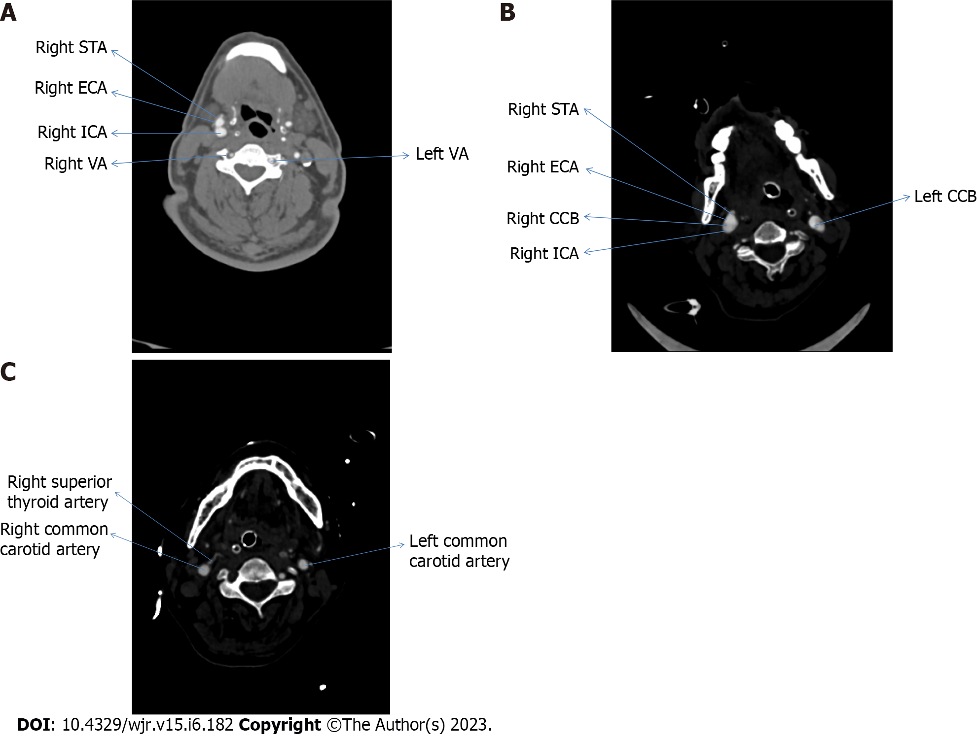
Figure 1 Computed tomography angiographic image (axial section).
A: Showing the origin of the right superior thyroid artery from the external carotid artery; B: Bifurcation of the common carotid artery; C: Common carotid artery. STA: Superior thyroid artery; ECA: External carotid artery; ICA: Internal carotid artery; VA: Vertebral artery; CCB: Bifurcation of common carotid artery.
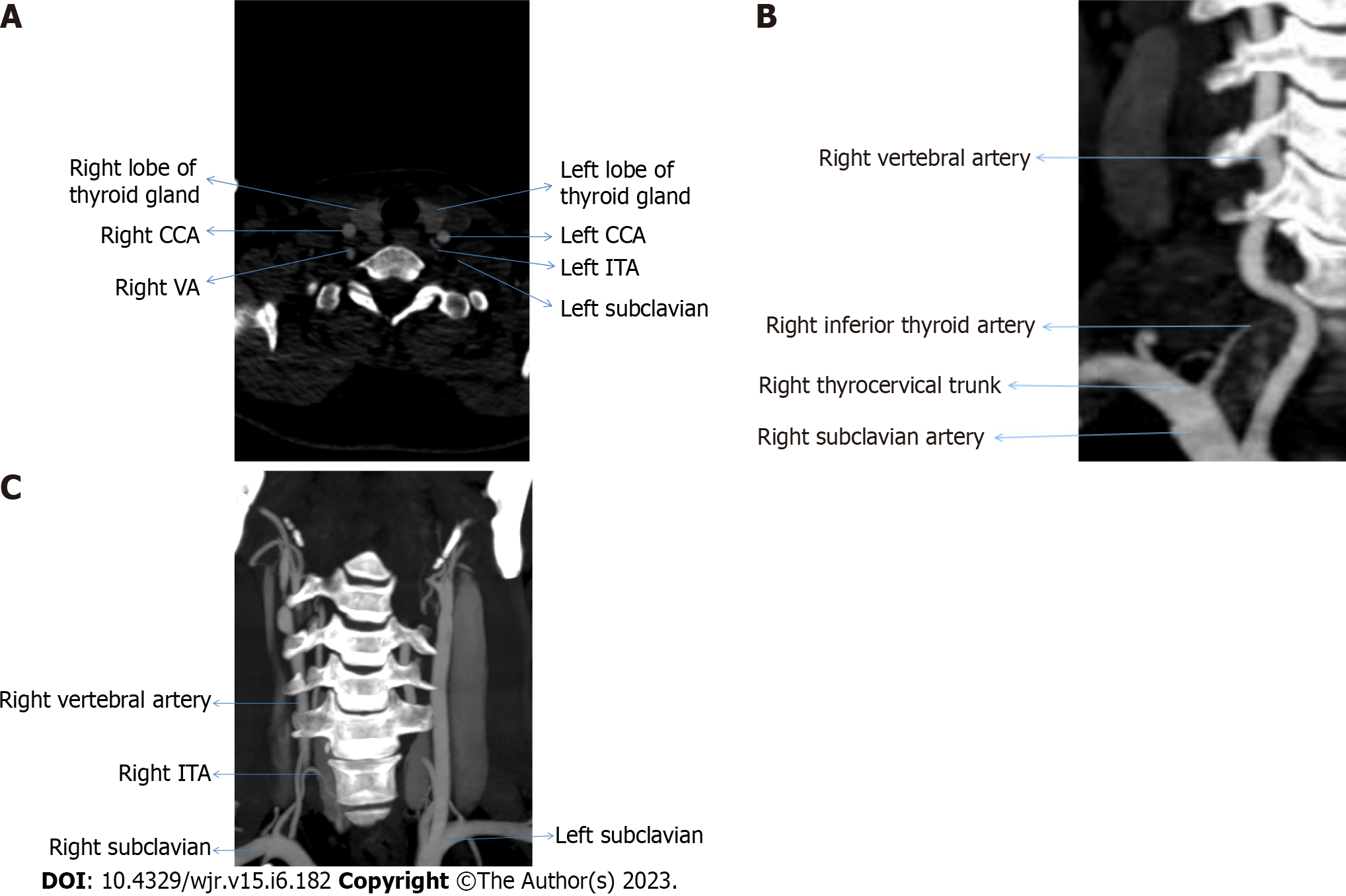
Figure 2 Computed tomography.
A: Origin of inferior thyroid artery from left subclavian artery; B: Right subclavian artery; C: Right thyrocervical trunk. ITA: Inferior thyroid artery; VA: Vertebral artery; CCA: Common carotid artery.
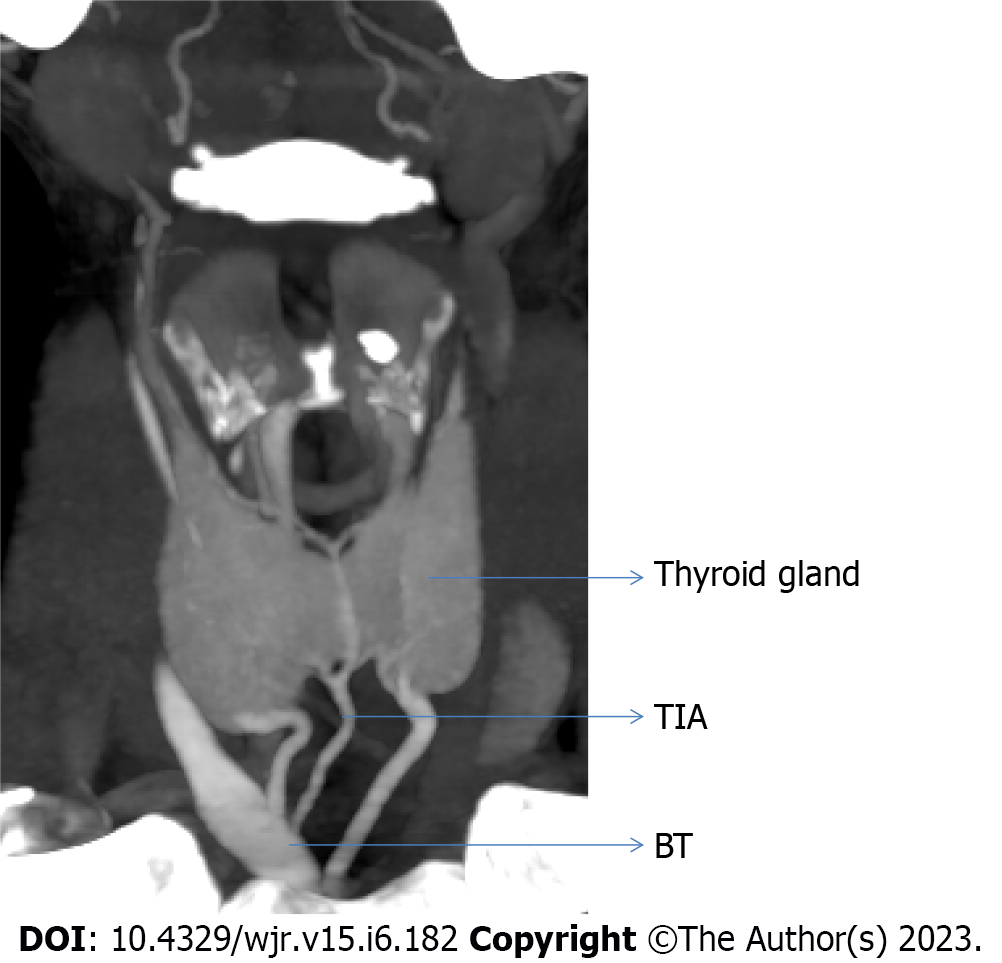
Figure 3 Showing origin of thyroid ima artery from the brachiocephalic trunk.
TIA: Thyroid ima artery; BT: Brachiocephalic trunk.
- Citation: Bhardwaj Y, Singh B, Bhadoria P, Malhotra R, Tarafdar S, Bisht K. Computed tomography angiographic study of surgical anatomy of thyroid arteries: Clinical implications in neck dissection. World J Radiol 2023; 15(6): 182-190
- URL: https://www.wjgnet.com/1949-8470/full/v15/i6/182.htm
- DOI: https://dx.doi.org/10.4329/wjr.v15.i6.182


