Copyright
©The Author(s) 2023.
World J Radiol. Apr 28, 2023; 15(4): 118-126
Published online Apr 28, 2023. doi: 10.4329/wjr.v15.i4.118
Published online Apr 28, 2023. doi: 10.4329/wjr.v15.i4.118
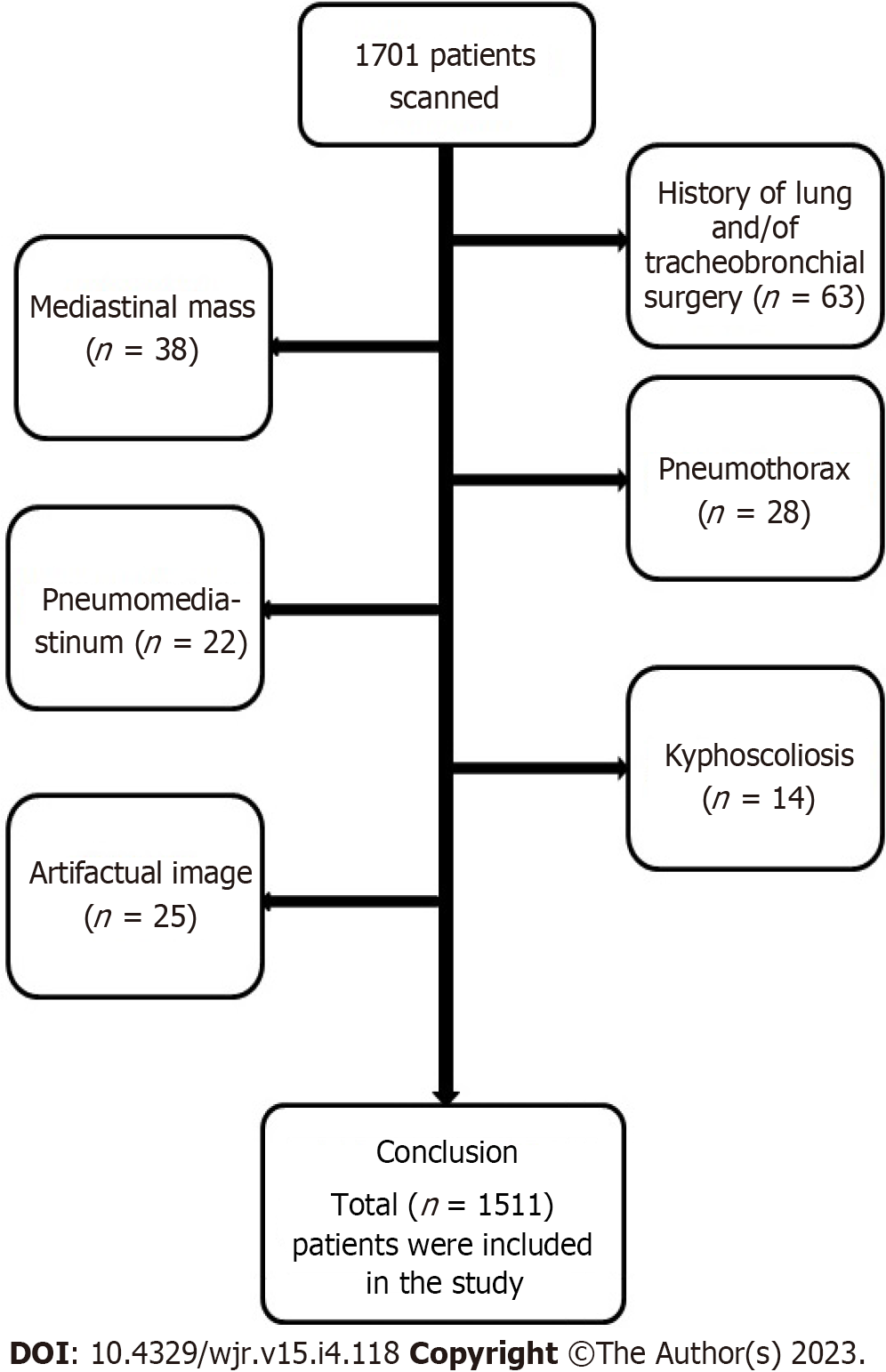
Figure 1 Diagram showing the study population.
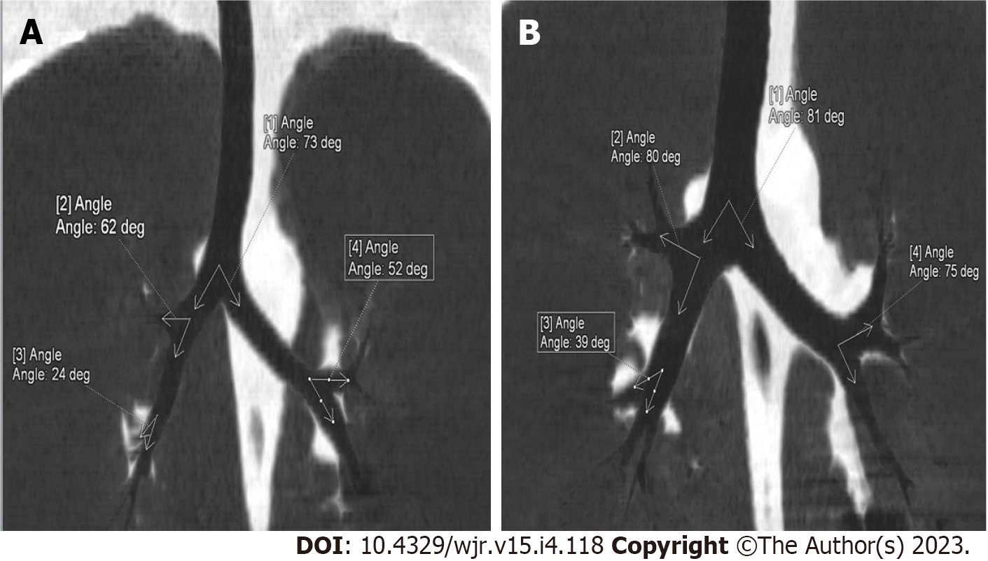
Figure 2 Measurement of tracheobronchial angles on coronal plane thoracic images in pediatric and adult patients with MinIP technique.
A: Tracheobronchial angle measurements of a 9-year-old male pediatric patient are shown, B: Tracheobronchial angle measurements of a 25-year-old adult male patient are shown: (1) Angle of right and left main bronchus in coronal plane; (2) R upper-intermedius (coronal): The angle of the right upper lobe bronchus and the right intermedius bronchus in the coronal plane; (3) R middle-lower (coronal): The angle of the right middle lobe bronchus and the right lower lobe bronchus in the coronal plane; (4) L upper-lower (coronal): The angle of left upper lobe bronchus and left upper lobe bronchus in the coronal plane.
- Citation: Kahraman Ş, Yazar MF, Aydemir H, Kantarci M, Aydin S. Detection of tracheal branching with computerized tomography: The relationship between the angles and age-gender. World J Radiol 2023; 15(4): 118-126
- URL: https://www.wjgnet.com/1949-8470/full/v15/i4/118.htm
- DOI: https://dx.doi.org/10.4329/wjr.v15.i4.118


