Copyright
©The Author(s) 2023.
World J Radiol. Feb 28, 2023; 15(2): 42-55
Published online Feb 28, 2023. doi: 10.4329/wjr.v15.i2.42
Published online Feb 28, 2023. doi: 10.4329/wjr.v15.i2.42
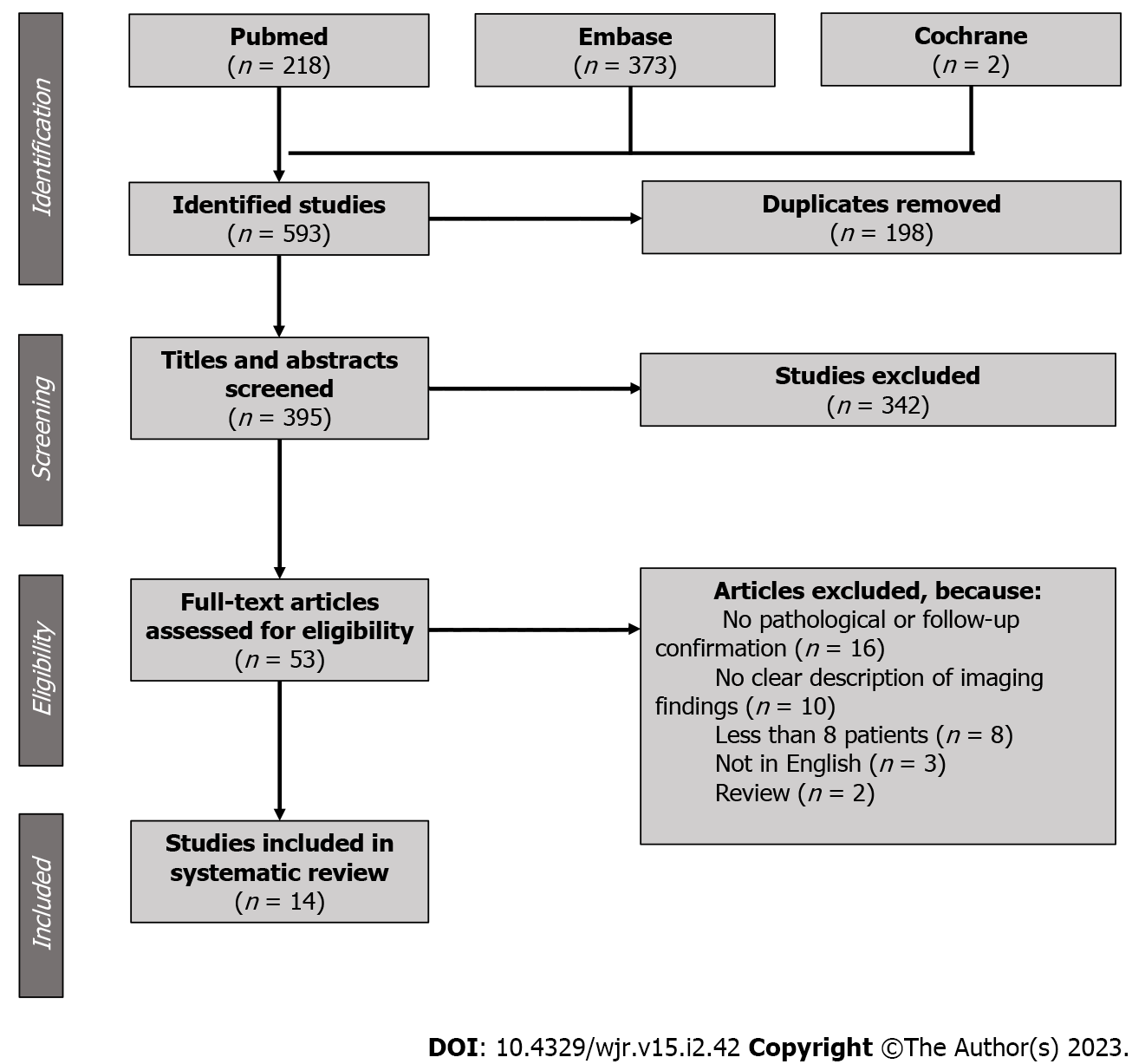
Figure 1 PRISMA flowchart showing studies inclusion and exclusion criteria.
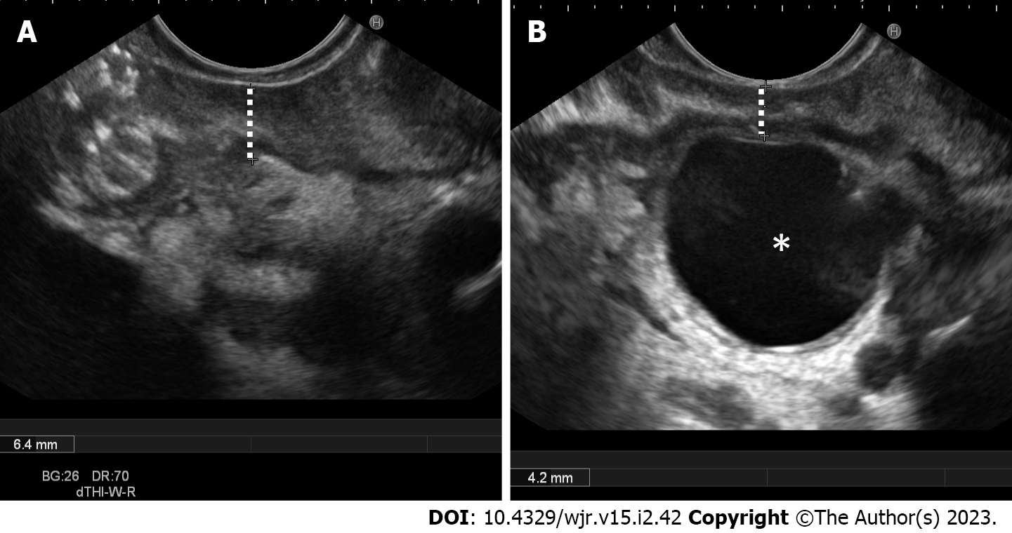
Figure 2 Endoscopic ultrasound.
A: Endoscopic ultrasound (EUS) clearly shows second duodenal portion wall thickening in a patient with chronic alcohol abuse history and abdominal pain, findings suggestive of solid subtype of paraduodenal pancreatitis; B EUS shows mild second duodenal portion wall thickening and a large duodenal wall cyst (star), findings pathognomonic for paraduodenal pancreatitis.
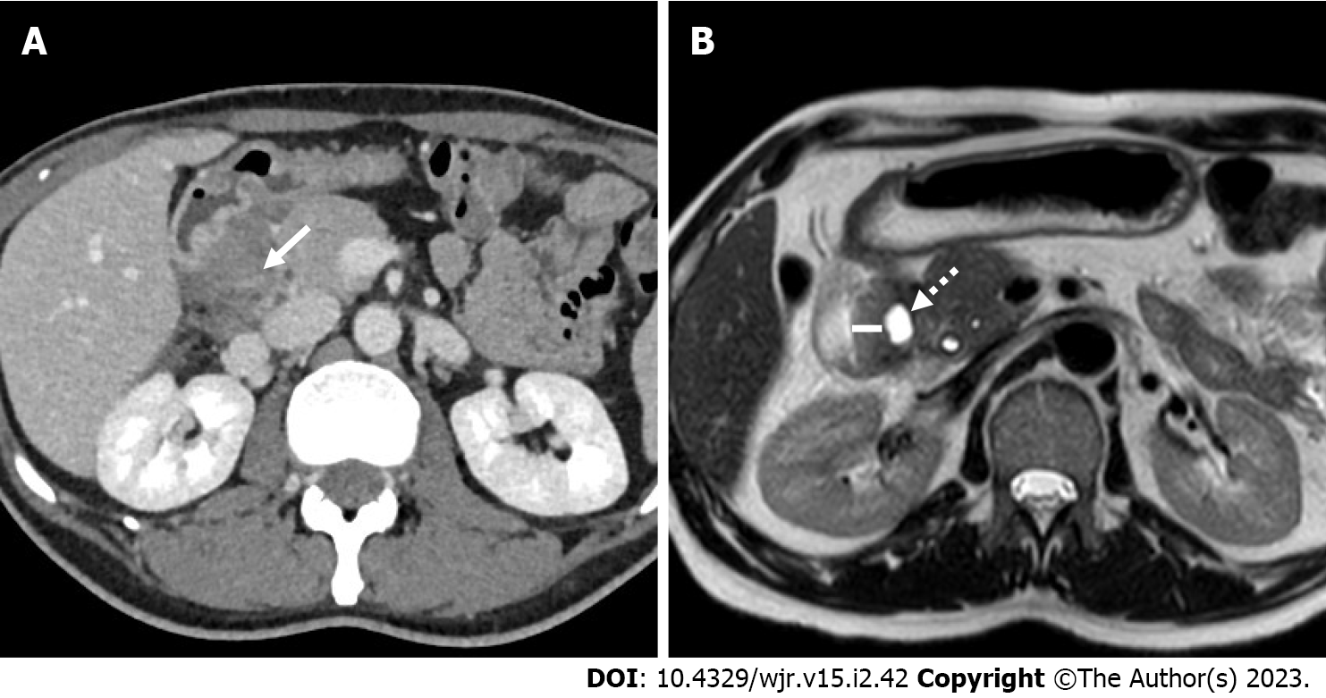
Figure 3 A 49-year-old male patient with weight loss and abdominal pain.
A: Axial 3 mm thick multiplanar reconstruction of portal venous phase computed tomography acquisition shows a hypodense mass in the groove region with patchy enhancement (arrow); B: Axial T2-weighted magnetic resonance imaging image acquired 2 mo later clearly shows eccentric second duodenal portion wall thickening (line) with cystic component (dotted arrow).
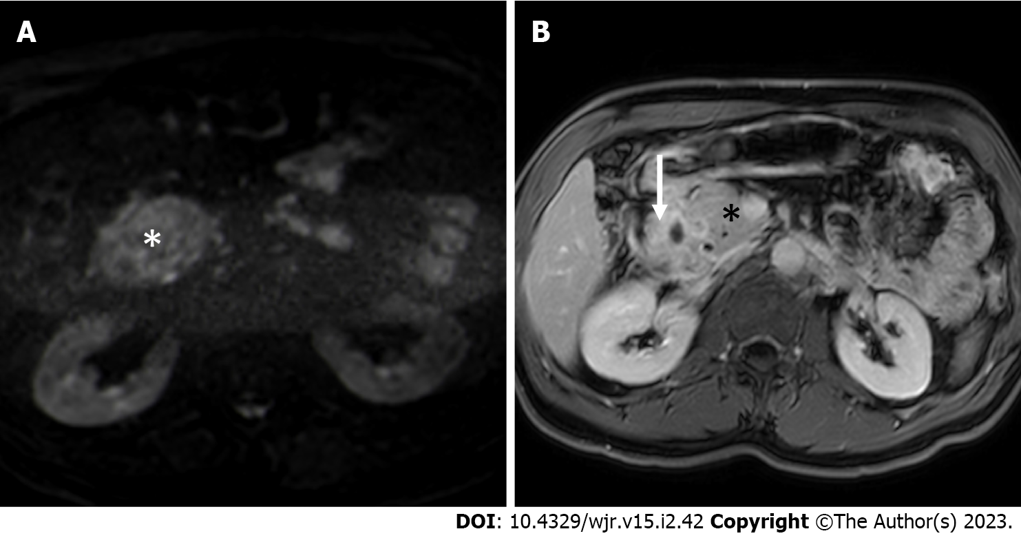
Figure 4 Magnetic resonance imaging.
A: Axial high b value (b = 800 s/mm2) diffusion weighted imaging image shows absence of increased diffusivity restriction in the thickened groove area (star) in comparison to adjacent “normal” pancreas, finding associated with paraduodenal pancreatitis and uncommon in pancreatic cancer; B: Axial delayed phase T1-weighted magnetic resonance imaging acquisition shows increased enhancement of the duodenal walls and of the groove region (arrow) in comparison to “normal” pancreas (star), finding often associated with paraduodenal pancreatitis.
- Citation: Bonatti M, De Pretis N, Zamboni GA, Brillo A, Crinò SF, Valletta R, Lombardo F, Mansueto G, Frulloni L. Imaging of paraduodenal pancreatitis: A systematic review. World J Radiol 2023; 15(2): 42-55
- URL: https://www.wjgnet.com/1949-8470/full/v15/i2/42.htm
- DOI: https://dx.doi.org/10.4329/wjr.v15.i2.42


