Copyright
©The Author(s) 2023.
World J Radiol. Oct 28, 2023; 15(10): 281-292
Published online Oct 28, 2023. doi: 10.4329/wjr.v15.i10.281
Published online Oct 28, 2023. doi: 10.4329/wjr.v15.i10.281
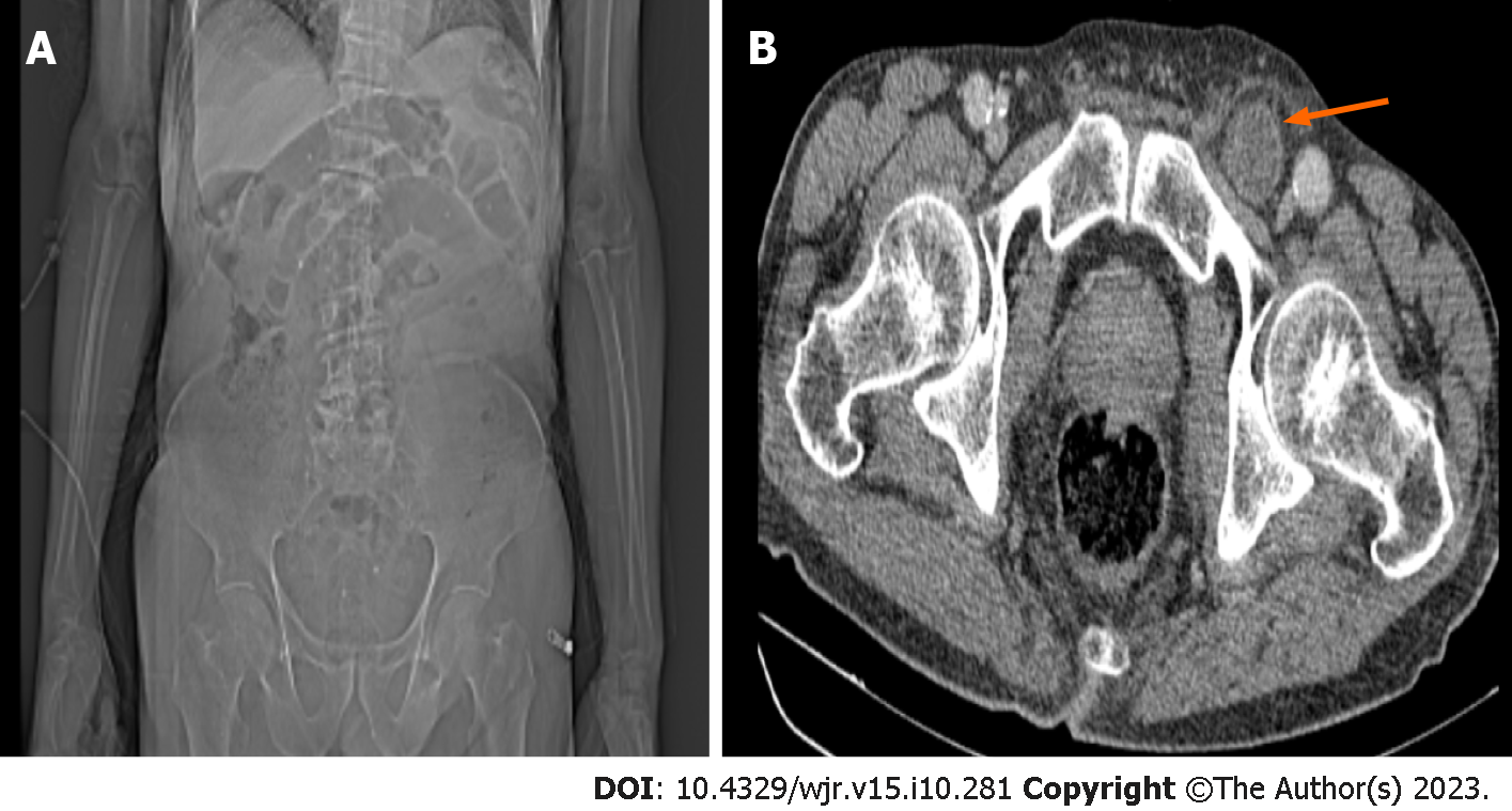
Figure 1 A 79-year-old male patient admitted to the emergency department with a complaint of abdominal pain that was more pro
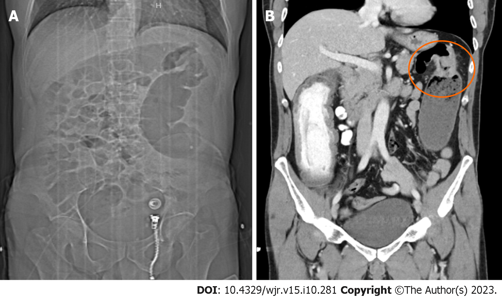
Figure 2 A 49-year-old male patient admitted to the emergency room with complaints of abdominal pain and vomiting.
A: In the computed tomography (CT) scenogram, sudden narrowing at the level of the splenic flexure and distension in the proximal and distal colon loops are seen; B: In the coronal CT image, a mass lesion causing a circular increase in wall thickness is seen at the level of the splenic flexure. All three observers reported a possible obstruction at the level of the large intestine.
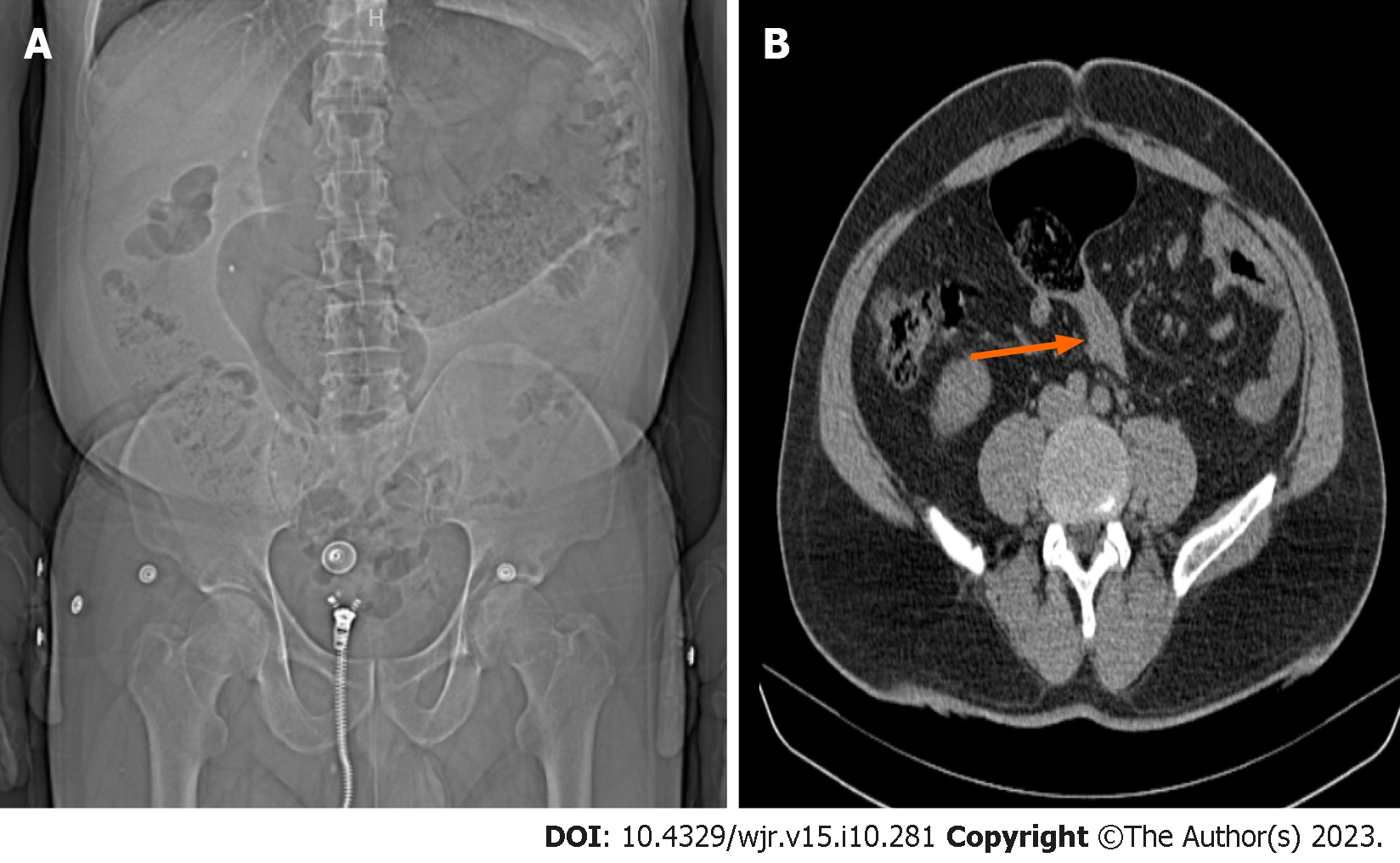
Figure 3 A 56-year-old male patient admitted to the emergency department with complaints of abdominal pain, gas and inability to pass stool.
A: In the computed tomography (CT) scanogram, swelling in the midline of the abdomen and folds in the colon loops are seen; B: In the coronal CT image, narrowing in the lower transition zones secondary to sigmoid volvulus is observed (arrow). All three observers reported a possible obstruction at the level of the large intestine.
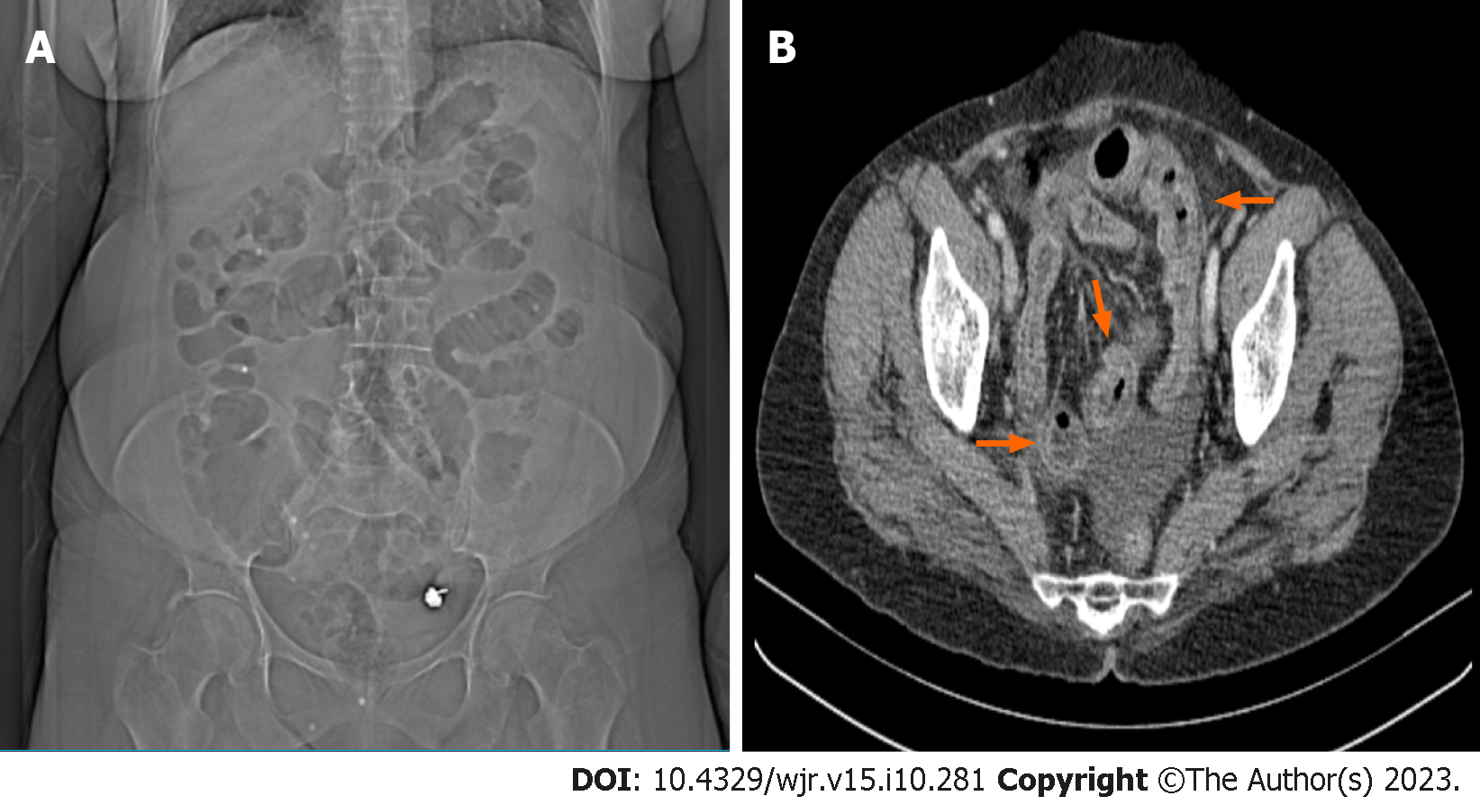
Figure 4 A 61-year-old female patient admitted to the emergency department complaining of abdominal pain.
A: Radiolucent air values in the large and small intestine segments are seen in the computed tomography (CT) scenogram image; B: Axial CT image shows trilaminar thickness increase (red arrows) in the small intestine wall compatible with ileitis, dilated appearance in the mesenteric vascular structures and free fluid in the pelvic area. Following observation of the scenogram images, the first observer reported a possible obstruction at the small intestine level, the second observer reported a possible obstruction at the large intestine level, and the third observer reported that no mechanical obstruction was detected.
- Citation: Kadirhan O, Kızılgoz V, Aydin S, Bilici E, Bayat E, Kantarci M. Does the use of computed tomography scenogram alone enable diagnosis in cases of bowel obstruction? World J Radiol 2023; 15(10): 281-292
- URL: https://www.wjgnet.com/1949-8470/full/v15/i10/281.htm
- DOI: https://dx.doi.org/10.4329/wjr.v15.i10.281


