Copyright
©The Author(s) 2022.
World J Radiol. Aug 28, 2022; 14(8): 311-318
Published online Aug 28, 2022. doi: 10.4329/wjr.v14.i8.311
Published online Aug 28, 2022. doi: 10.4329/wjr.v14.i8.311
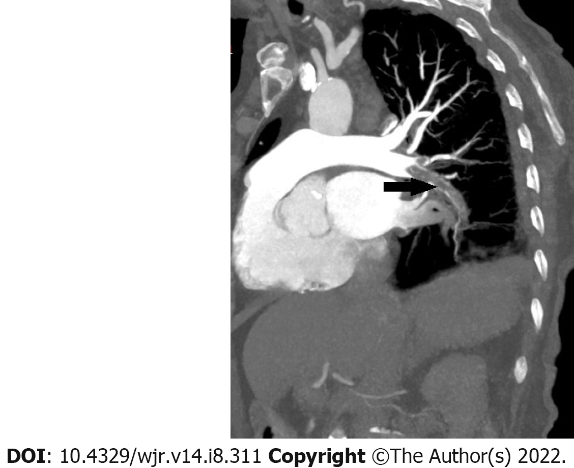
Figure 1 A 77-year-old male patient.
Curved multiplanar reformatted image shows embolus in the right lung lower lobe artery (arrow).
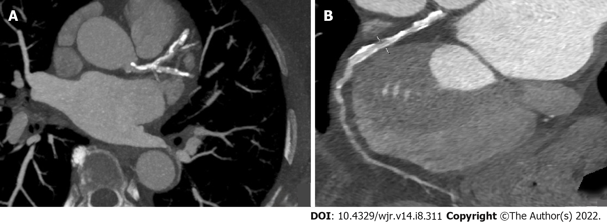
Figure 2 A 73-year-old male patient.
A: Diffuse calcific and soft plaque formations are seen in the left main, left anterior descending (LAD), and left circumflex arteries on axial maximum intensity projection image; B: Moderate stenosis (50% to 69%) is present (linear marker) in the proximal segment of LAD on curved multiplanar reformatted image.
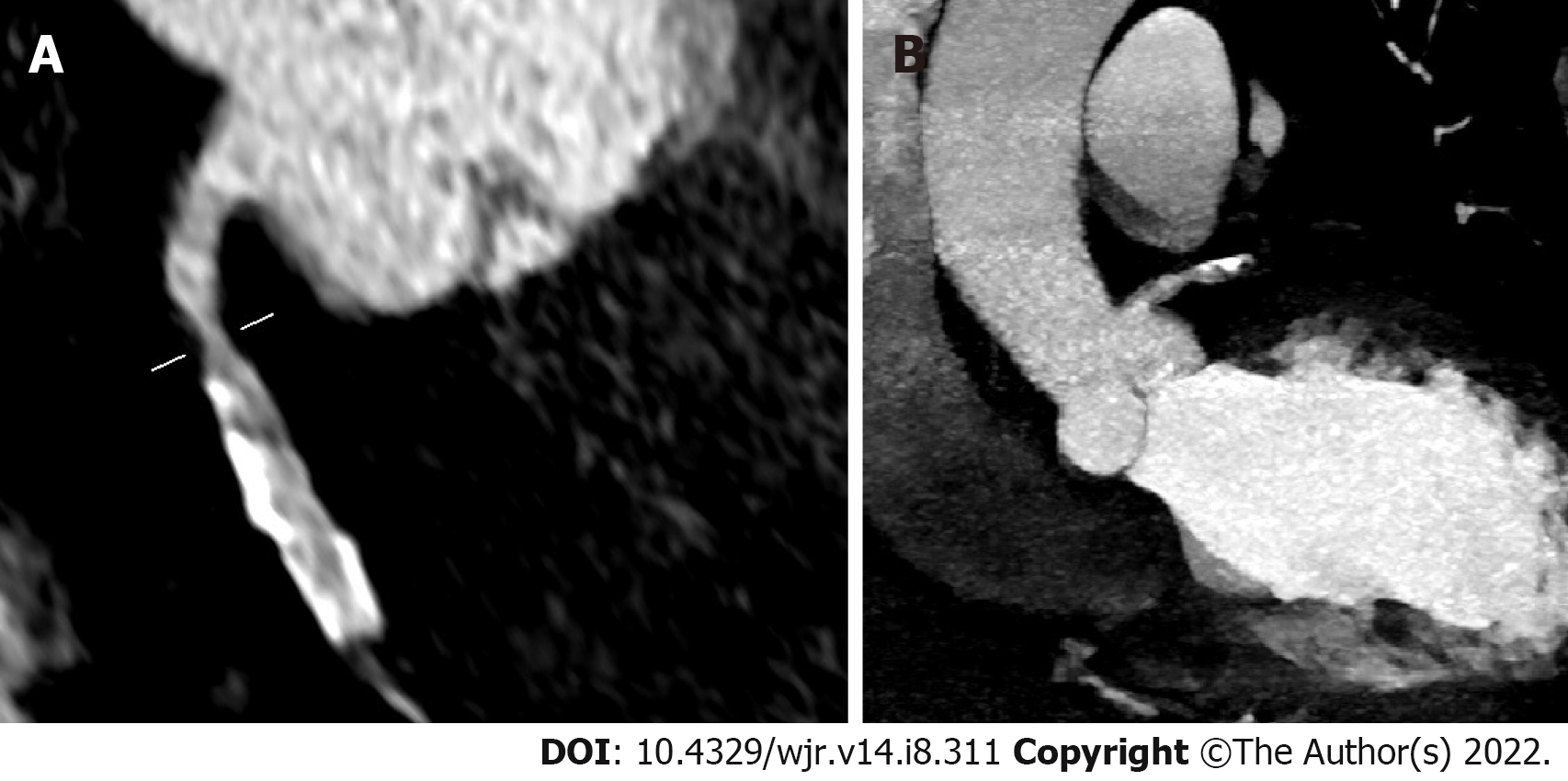
Figure 3 A 63-year-old female patient.
A: Moderate stenosis (50% to 69%; linear marker); B: Calcific plaque formations are seen in the right coronary artery on coronal maximum intensity projection images.
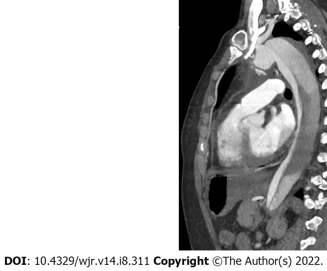
Figure 4 A 78-year-old male patient.
Sagittal maximum intensity projection image depicts Stanford type B dissection.
- Citation: Bahadir S, Aydın S, Kantarci M, Unver E, Karavas E, Şenbil DC. Triple rule-out computed tomography angiography: Evaluation of acute chest pain in COVID-19 patients in the emergency department. World J Radiol 2022; 14(8): 311-318
- URL: https://www.wjgnet.com/1949-8470/full/v14/i8/311.htm
- DOI: https://dx.doi.org/10.4329/wjr.v14.i8.311


