Copyright
©The Author(s) 2021.
World J Radiol. Jul 28, 2021; 13(7): 233-242
Published online Jul 28, 2021. doi: 10.4329/wjr.v13.i7.233
Published online Jul 28, 2021. doi: 10.4329/wjr.v13.i7.233
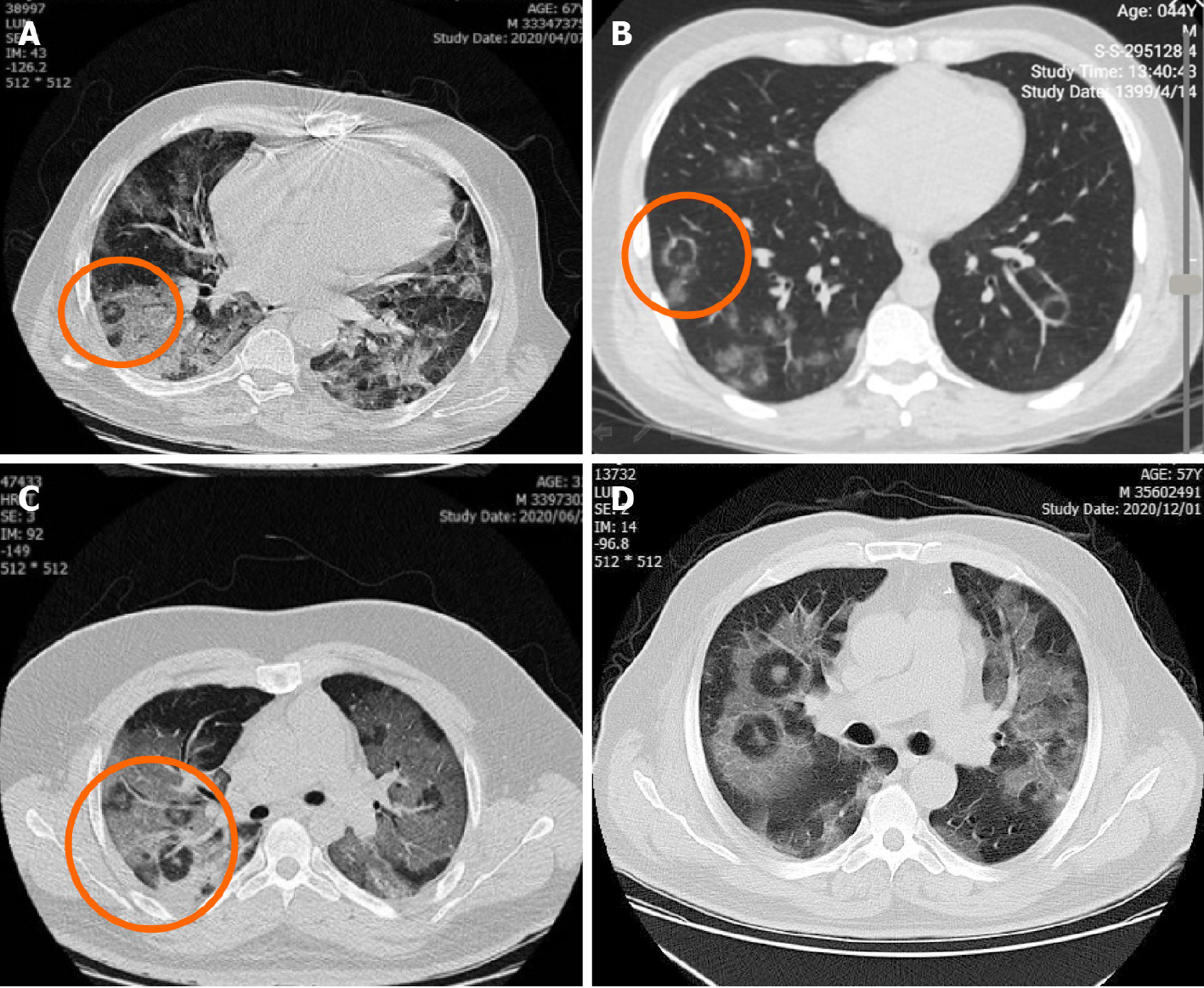
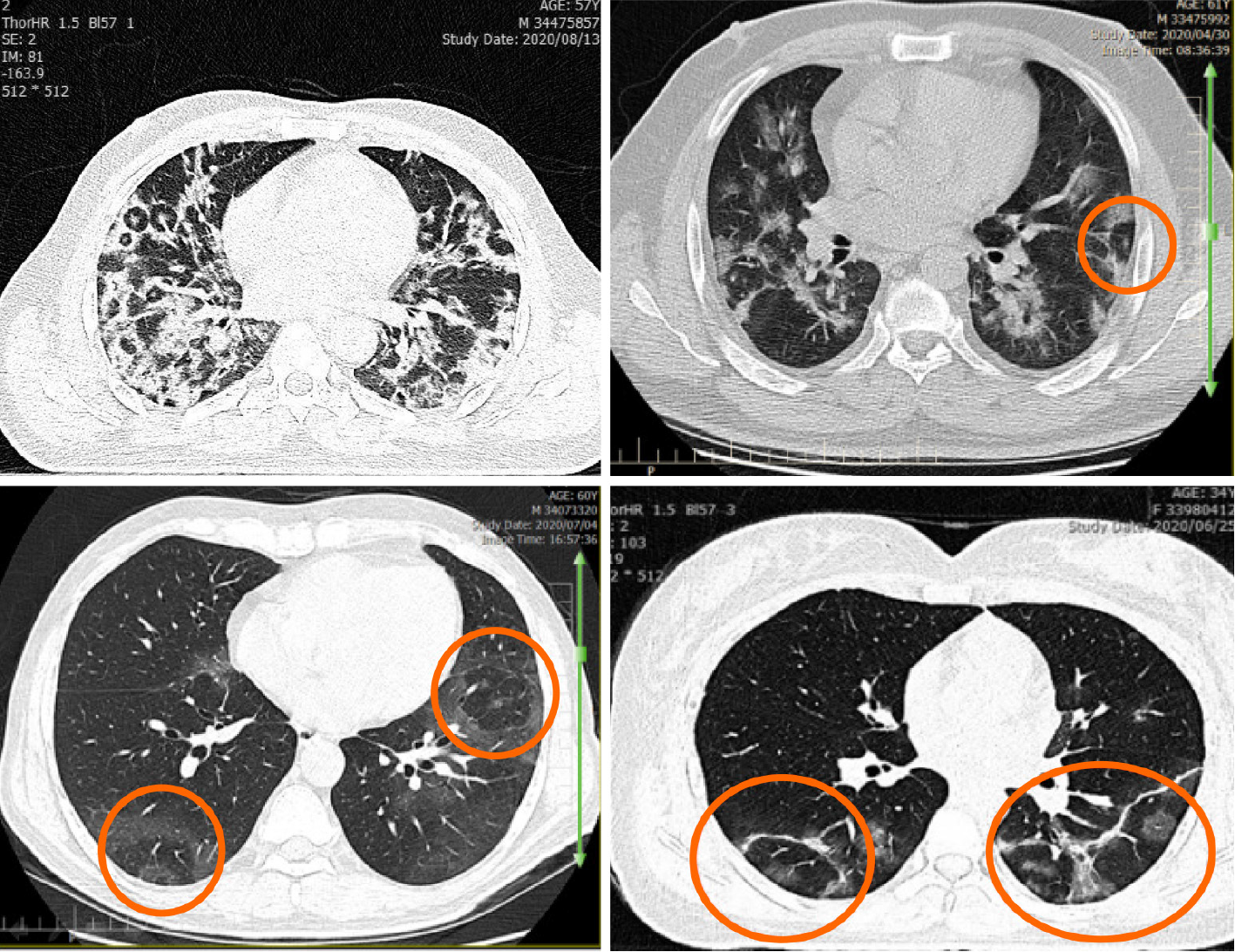
Figure 1 “Pulmonary target sign” in 4 different cases varies according to the location of the lesions.
A and B: Peripheral location; C and D: Central location.
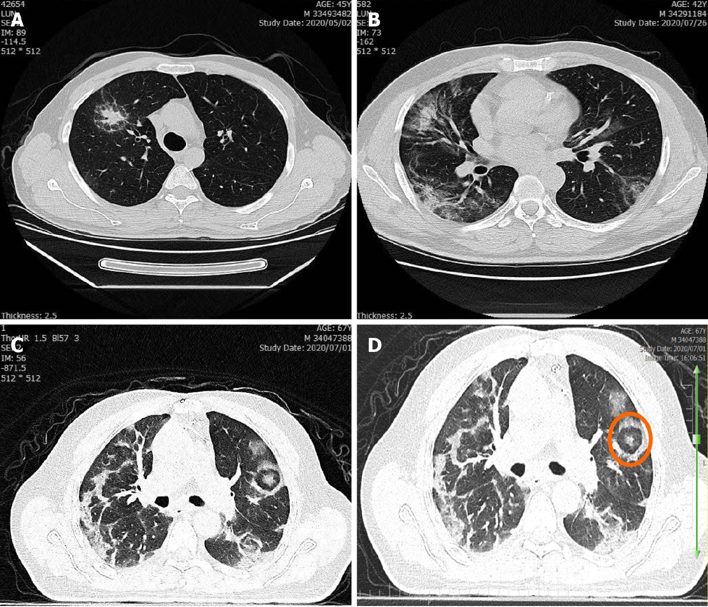
Figure 2 Variants of “pulmonary target sign” in 4 different cases.
A: “Pulmonary target sign” (PTS) similar to a solitary pulmonary nodule; B: “Rings of Saturn” as a variant of PTS; C and D: PTS with parallel pleural sign.
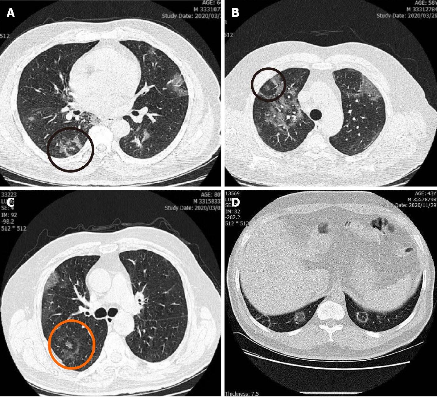
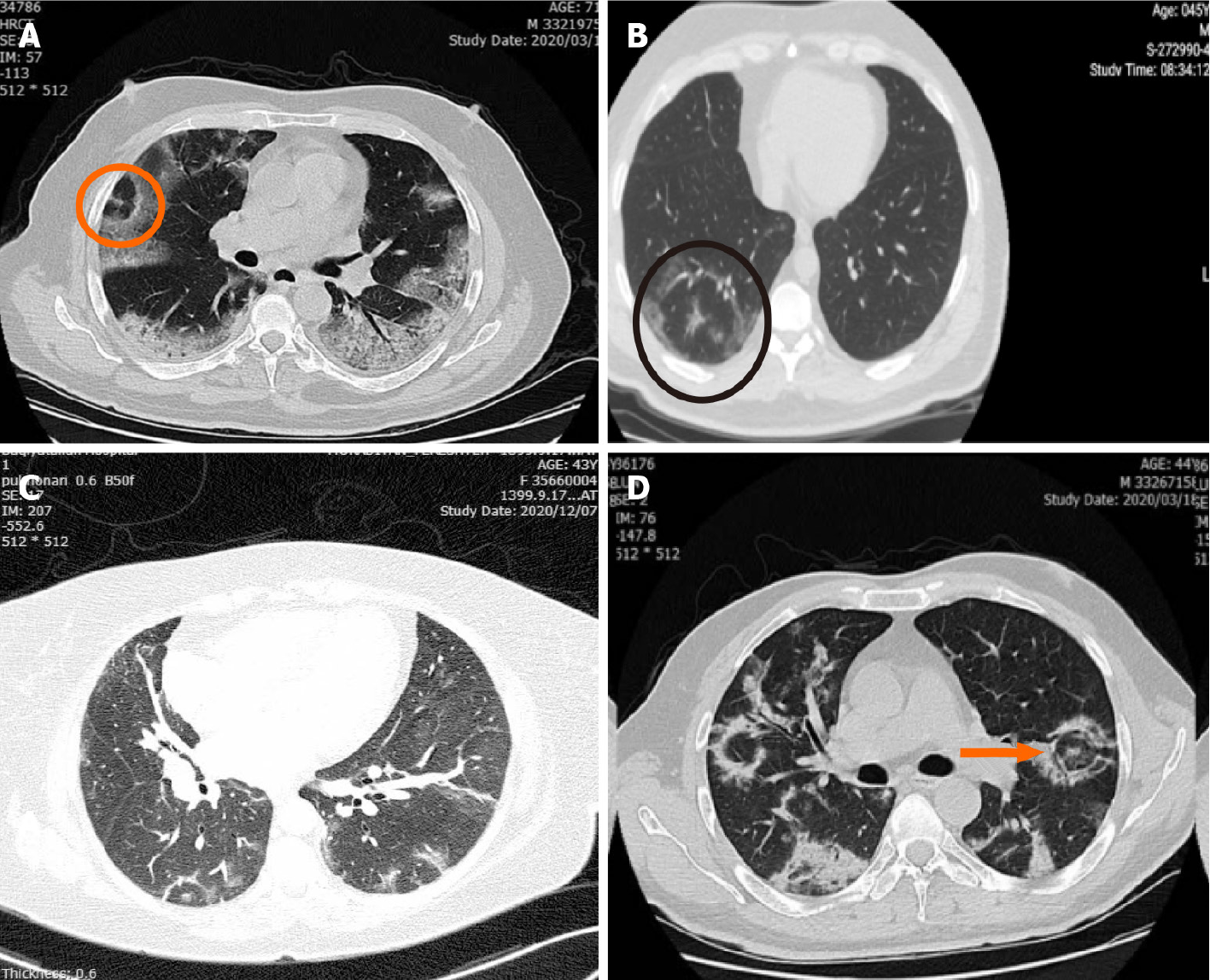
Figure 3 “Pulmonary target sign” in 4 different patients.
A and B: “Pulmonary target sign” (PTS) as a pleural based lesion; C: PTS with incomplete peripheral ring; D: Complete peripheral ring.
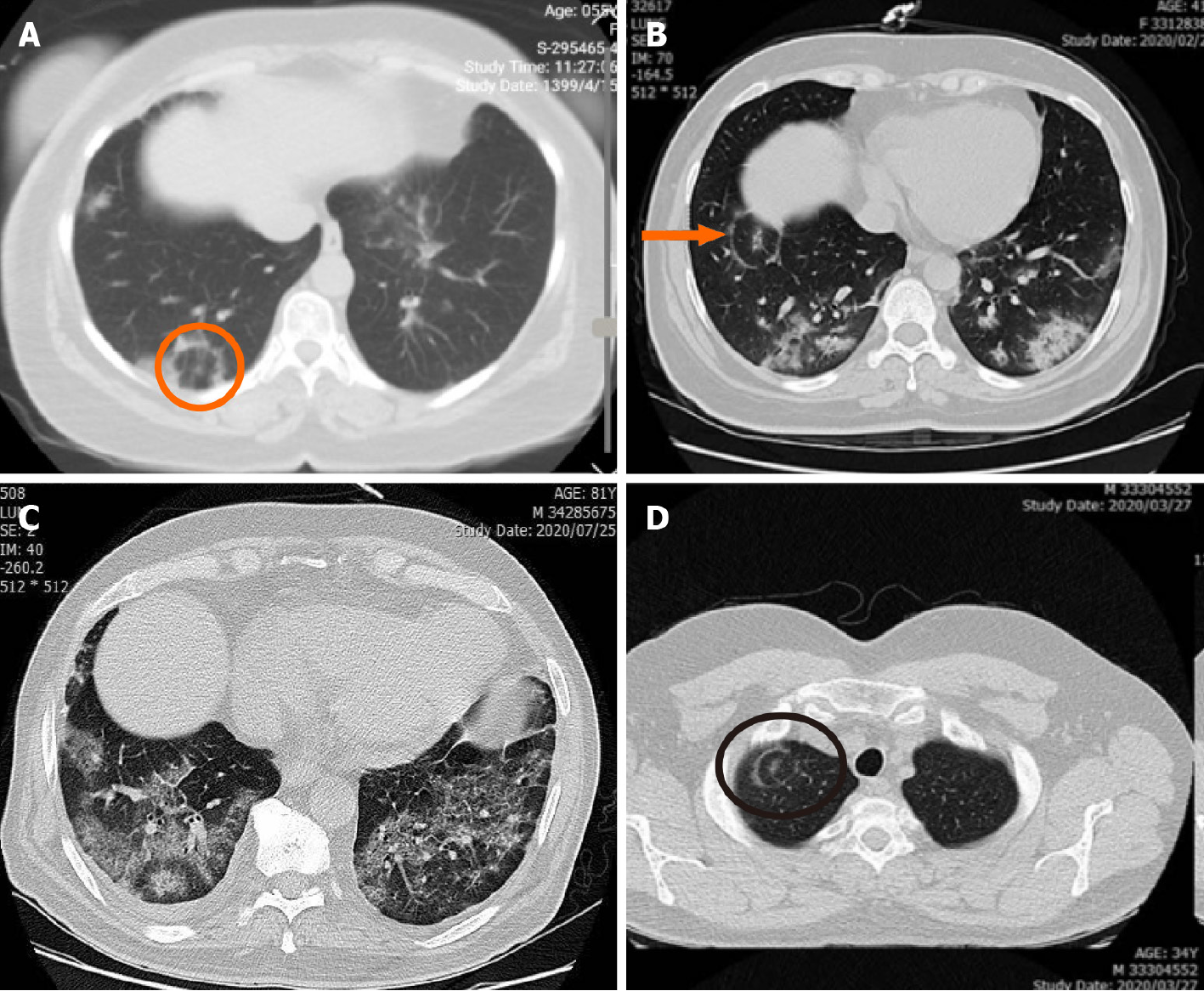
Figure 4 “Pulmonary target sign” in 4 different individuals.
A, B and C: Basal location of “pulmonary target sign”; D: Apical location.
Figure 5 “Pulmonary target sign” in 4 different cases.
“Pulmonary target sign” along with a broncho-vascular bundle.
Figure 6 Laterality of “pulmonary target sign” in 4 different cases.
A and B: Multiple unilateral “pulmonary target sign”; C: bilateral lesions; D: Solitary lesion.
Figure 7 Correlation of “pulmonary target sign” with adjacent ground-glass opacities or consolidation.
A: Circular adjacent ground-glass opaci
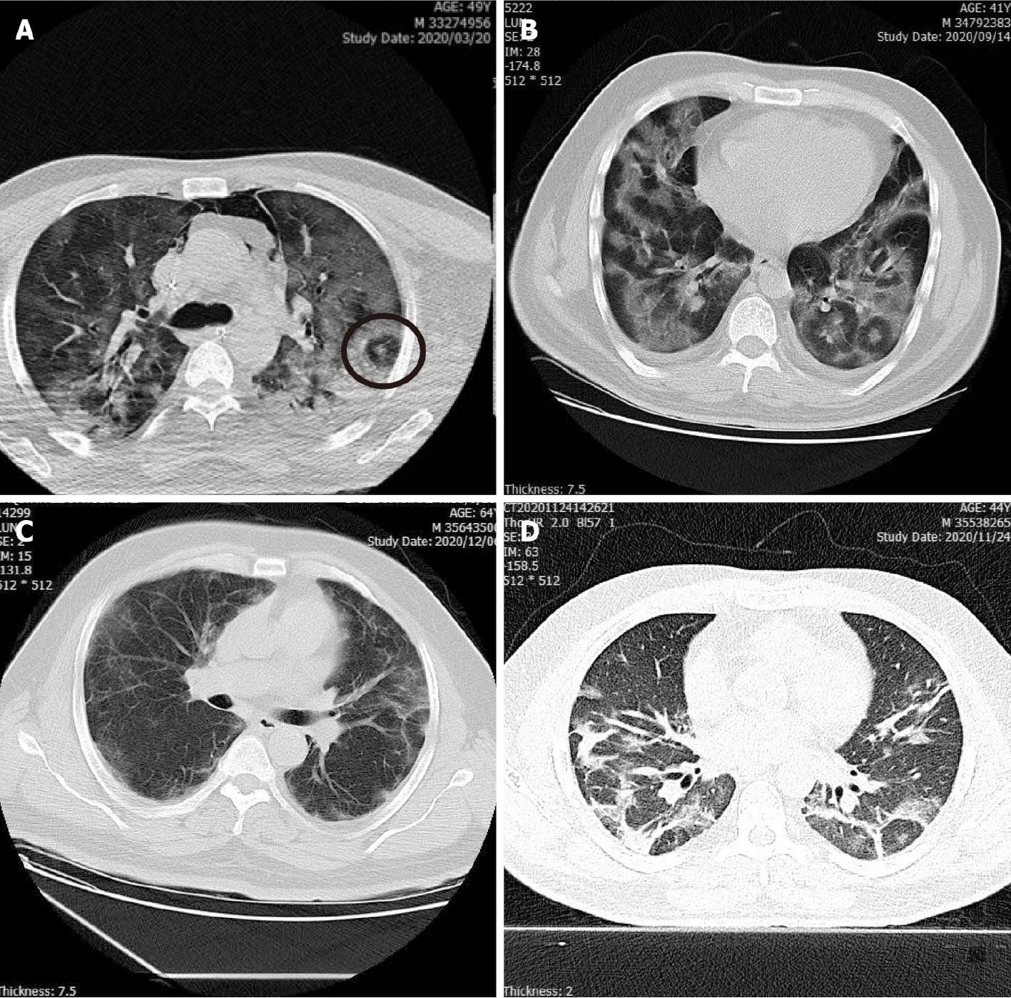
Figure 8 “Pulmonary target sign” with coronavirus disease 2019 complications.
A: Pneumothorax and pneumomediastinum; B: Pleural effusion; C: Pleural thickening; D: Fibrotic band.
- Citation: Jafari R, Jonaidi-Jafari N, Maghsoudi H, Dehghanpoor F, Schoepf UJ, Ulversoy KA, Saburi A. “Pulmonary target sign” as a diagnostic feature in chest computed tomography of COVID-19. World J Radiol 2021; 13(7): 233-242
- URL: https://www.wjgnet.com/1949-8470/full/v13/i7/233.htm
- DOI: https://dx.doi.org/10.4329/wjr.v13.i7.233


