Copyright
©The Author(s) 2021.
World J Radiol. Jun 28, 2021; 13(6): 157-170
Published online Jun 28, 2021. doi: 10.4329/wjr.v13.i6.157
Published online Jun 28, 2021. doi: 10.4329/wjr.v13.i6.157
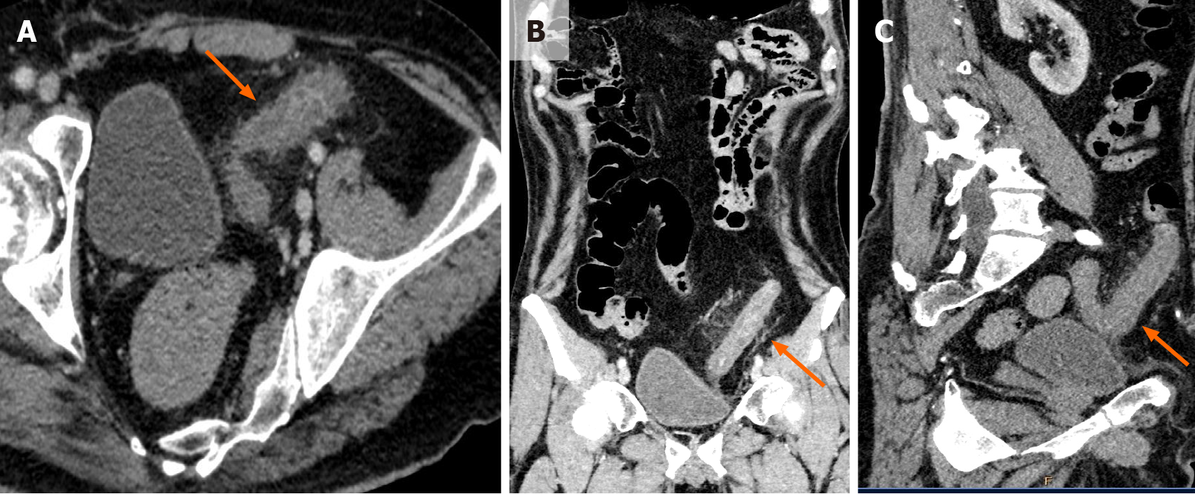
Figure 1 Contrast enhanced computed tomography imaging in a patient with coronavirus disease 2019 presenting with bloody diarrhea.
A: Axial; B: Coronal; C: Sagittal. Contrast enhanced computed tomography images of the abdomen and pelvis show diffuse edematous wall thickening involving the sigmoid colon (orange arrows in A, B, and C). Associated pericolic fat stranding is seen. In the absence of other causes of bowel wall thickening, a diagnosis of viral colitis was considered.
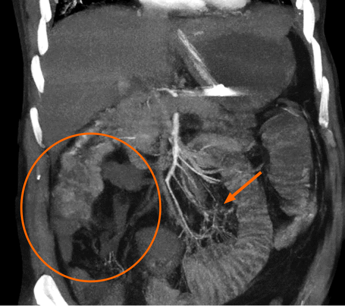
Figure 2 Acute mesenteric ischemia diagnosed on computed tomography mesenteric angiography performed in a critical patient with coronavirus disease 2019 presenting with severe abdominal pain.
The coronal maximum intensity projection image reveals a lack of opacification of the right sided branches of the superior mesenteric artery (SMA), namely, the ileo-colic and right colic branches (orange circle). The distal branches of the SMA also appear hypoperfused (orange arrow). Imaging features are consistent with SMA thrombosis.
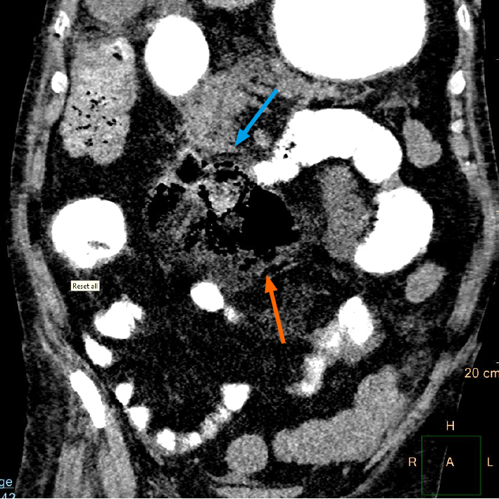
Figure 3 Bowel perforation detected on a computed tomography scan of the abdomen and pelvis performed for evaluation of severe abdominal pain and abdominal distension in a critical patient with coronavirus disease 2019.
Positive oral contrast is seen to opacify small and large bowel loops. There is evidence of jejunal perforation with a localized air collection in the mesentery (orange arrow) at the site and adjacent inflammation (blue arrow). The proximal jejunal loops appear dilated. The patient underwent emergency laparotomy with resection and anastomosis.
Figure 4 Small bowel ischemia detected on computed tomography mesenteric angiography study performed in a coronavirus disease 2019 patient presenting with severe abdominal pain and abdominal distension.
A and B: Axial contrast enhanced computed tomography (CT) images of the abdomen and pelvis; C: Sagittal contrast enhanced CT images of the abdomen and pelvis, showing dilated distal ileal loops in the right iliac fossa with non-enhancing, barely discernible walls suggestive of bowel gangrene (blue arrows in A, B, and C). The proximal small bowel loops appear dilated with air-fluid levels within, suggestive of stasis (orange arrow in B).
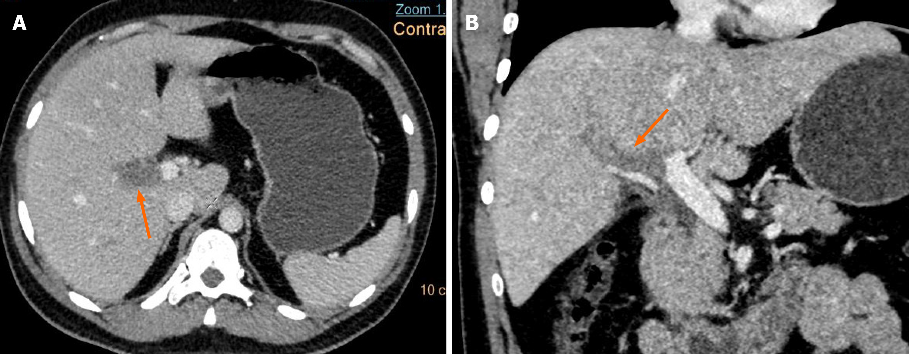
Figure 5 Venous thrombosis diagnosed in a coronavirus disease 2019 patient presenting with abdominal pain.
A: Axial; B: Coronal. Contrast enhanced computed tomography (CT) images of the abdomen show a hypodense filling defect in the main portal vein at the hilum extending into the right branch (orange arrows in A and B), suggestive of portal vein thrombosis. There were no features of mesenteric ischemia seen on the CT.
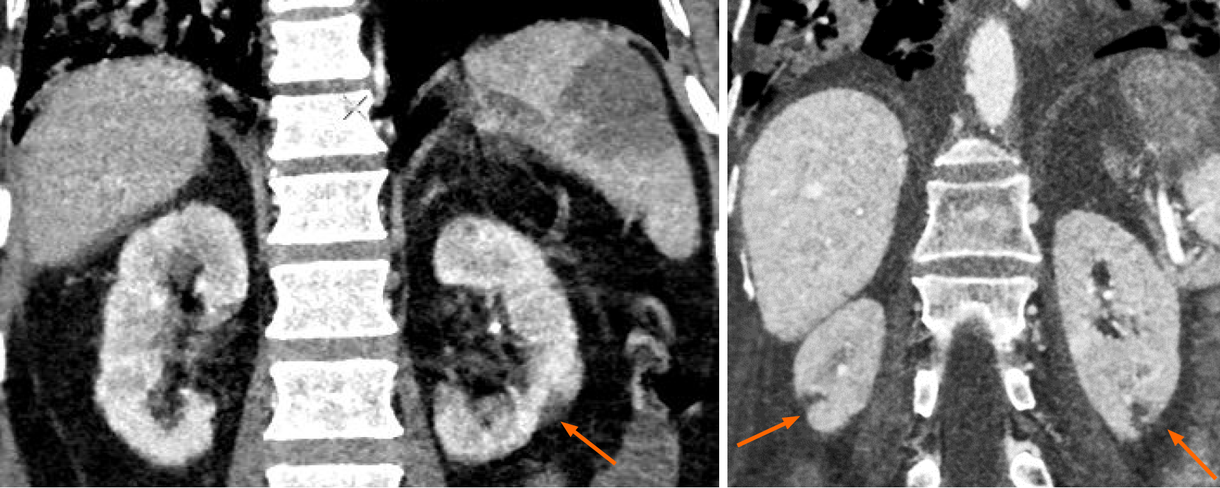
Figure 6 Solid organ infarction in a patient with coronavirus disease 2019.
Coronal contrast enhanced computed tomography (CT) images of the abdomen show discrete, wedge-shaped non-enhancing renal parenchymal defects, with their apex pointing towards the medulla and base parallel to the subcapsular region (orange arrows), suggestive of renal infarcts. These were incidentally detected during CT pulmonary angiography.
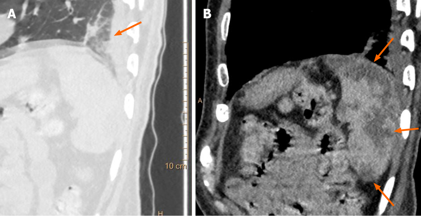
Figure 7 Incidentally detected splenic infarction in a patient with coronavirus disease 2019.
A: Sagittal chest computed tomography (CT) image shows subpleural ground glass opacities in the lung fields in a case of coronavirus disease 2019 (orange arrow); B: Sagittal contrast enhanced CT image of the abdomen in the same patient show incidentally detected, sharply marginated, wedge shaped, hypodense areas in the spleen, suggestive of splenic infarcts (orange arrows).
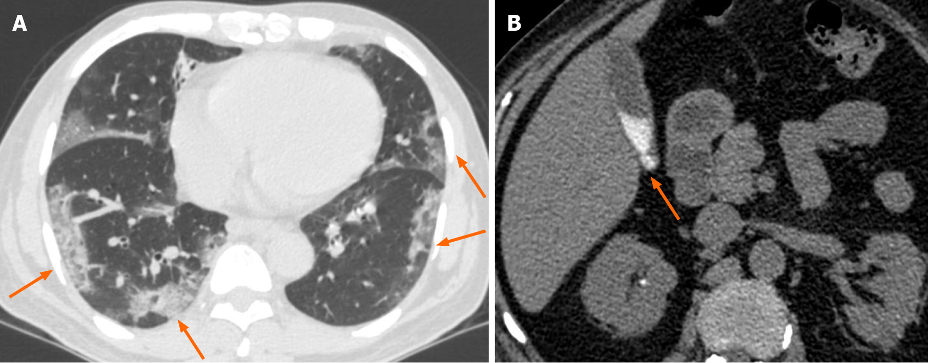
Figure 8 Evidence of cholestasis in a critically ill coronavirus disease 2019 patient.
A: Axial chest computed tomography (CT) image shows multiple peripheral ground glass opacities associated with interstitial thickening in both lung fields (orange arrows in A); B: Axial non-contrast CT image of the abdomen shows incidentally detected hyperdense sludge within the gall bladder lumen (orange arrow in B), suggesting cholestasis.
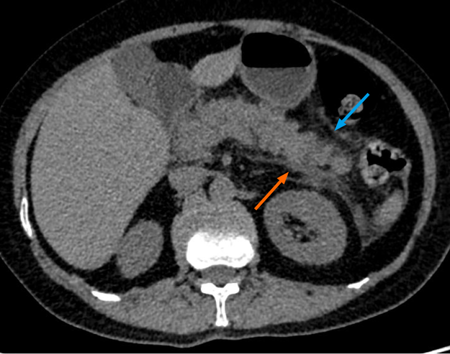
Figure 9 Acute viral pancreatitis in a coronavirus disease 2019 patient presenting with abdominal pain.
Non-contrast axial computed tomography image of the abdomen in a case of suspected viral pancreatitis (intravenous contrast could not be administered due to a history of renal parenchymal disease with elevated creatinine) is shown. The distal body and tail of pancreas reveal fuzzy margins with peri-pancreatic fat stranding (blue arrow). Thickening of the left anterior conal fascia is noted with a streak of fluid in the left retro-mesenteric plane (orange arrow). Elevated serum amylase and lipase levels, in conjunction with these imaging findings, were highly suggestive of a diagnosis of acute viral pancreatitis in a patient with coronavirus disease 2019 presenting with abdominal pain.
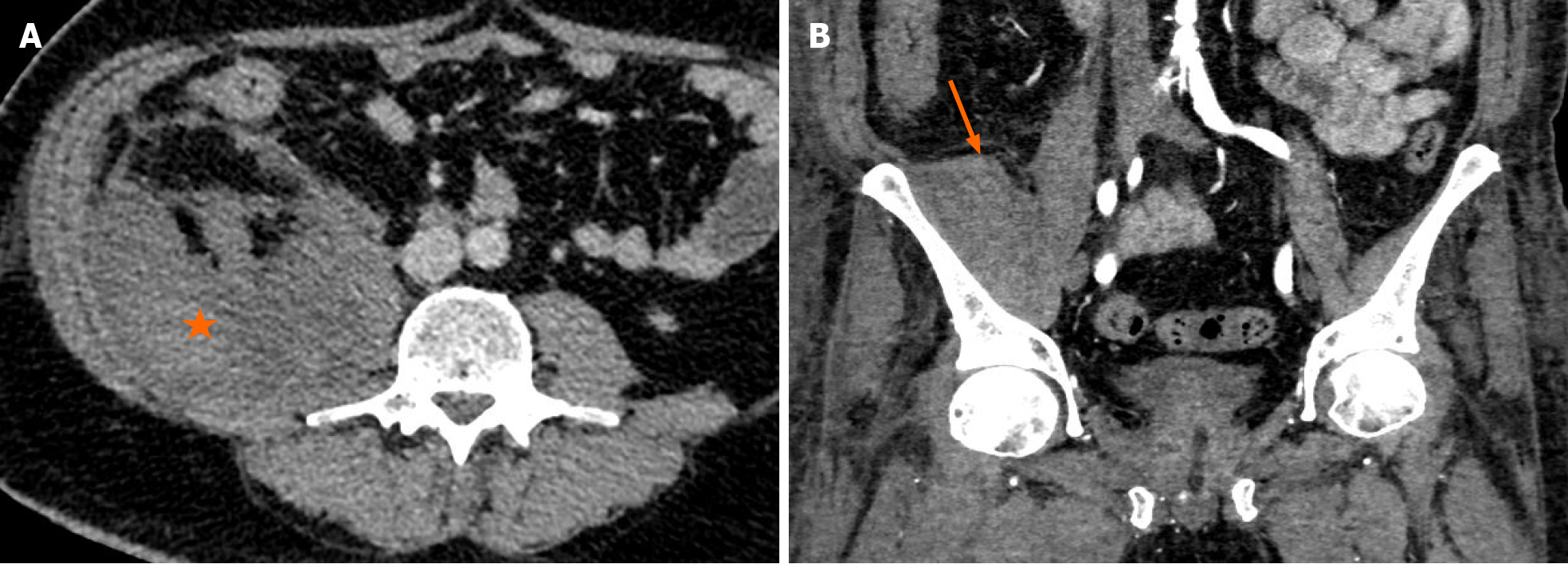
Figure 10 Bleeding manifestations encountered in treated cases of coronavirus disease 2019.
A: Contrast enhanced axial computed tomography (CT) image of the abdomen shows a hyperdense collection in the right ilio-psoas compartment (orange asterisk in A), suggestive of a hematoma; B: Contrast enhanced coronal CT image of the abdomen shows a hyperdense collection in the right iliacus muscle (orange arrow in B) in another patient. Both patients were being treated with anticoagulants for coronavirus disease 2019 associated hypercoagulable state evidenced by elevated D-dimer levels. CT imaging was performed to evaluate the cause of a sudden fall in hemoglobin.
- Citation: Vaidya T, Nanivadekar A, Patel R. Imaging spectrum of abdominal manifestations of COVID-19. World J Radiol 2021; 13(6): 157-170
- URL: https://www.wjgnet.com/1949-8470/full/v13/i6/157.htm
- DOI: https://dx.doi.org/10.4329/wjr.v13.i6.157


