Copyright
©The Author(s) 2021.
World J Radiol. May 28, 2021; 13(5): 122-136
Published online May 28, 2021. doi: 10.4329/wjr.v13.i5.122
Published online May 28, 2021. doi: 10.4329/wjr.v13.i5.122
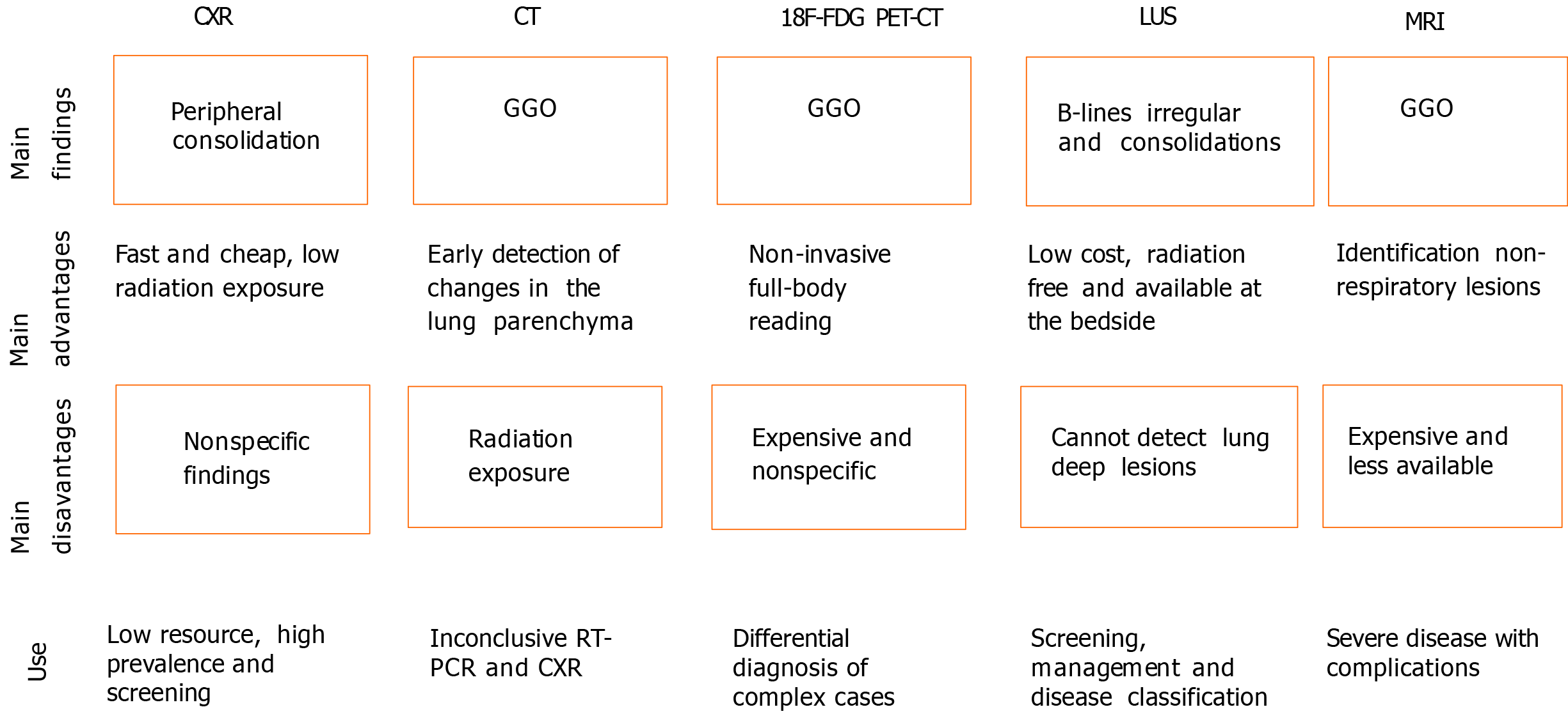
Figure 1 Highlights of imaging modalities in coronavirus disease 2019.
CXR: Chest X-ray; CT: Computed tomography; 18F-FDG PET/CT: 18F-fluorodeoxyglucose positron emission tomography/CT; LUS: Lung ultrasound; MRI: Magnetic resonance imaging; GGO: Ground-glass opacities; RT-PCR: Real-time reverse transcription polymerase chain reaction.
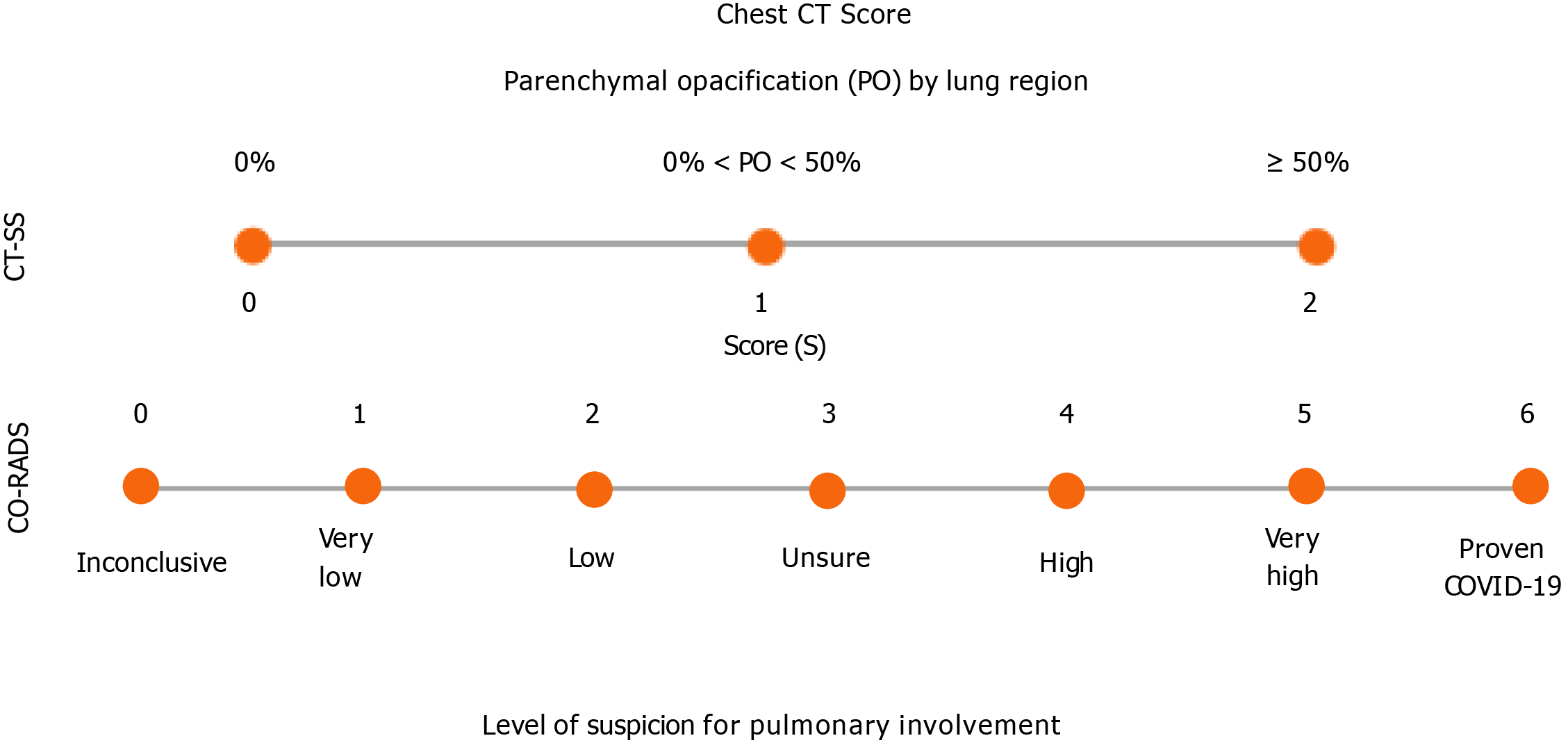
Figure 2 Summary of chest computed tomography scores to assess coronavirus disease 2019.
CT-SS: Computed tomography severity score; COVID-19: Coronavirus disease 2019; CO-RADS: COVID-19 Reporting and Data System; PO: Parenchymal opacification.
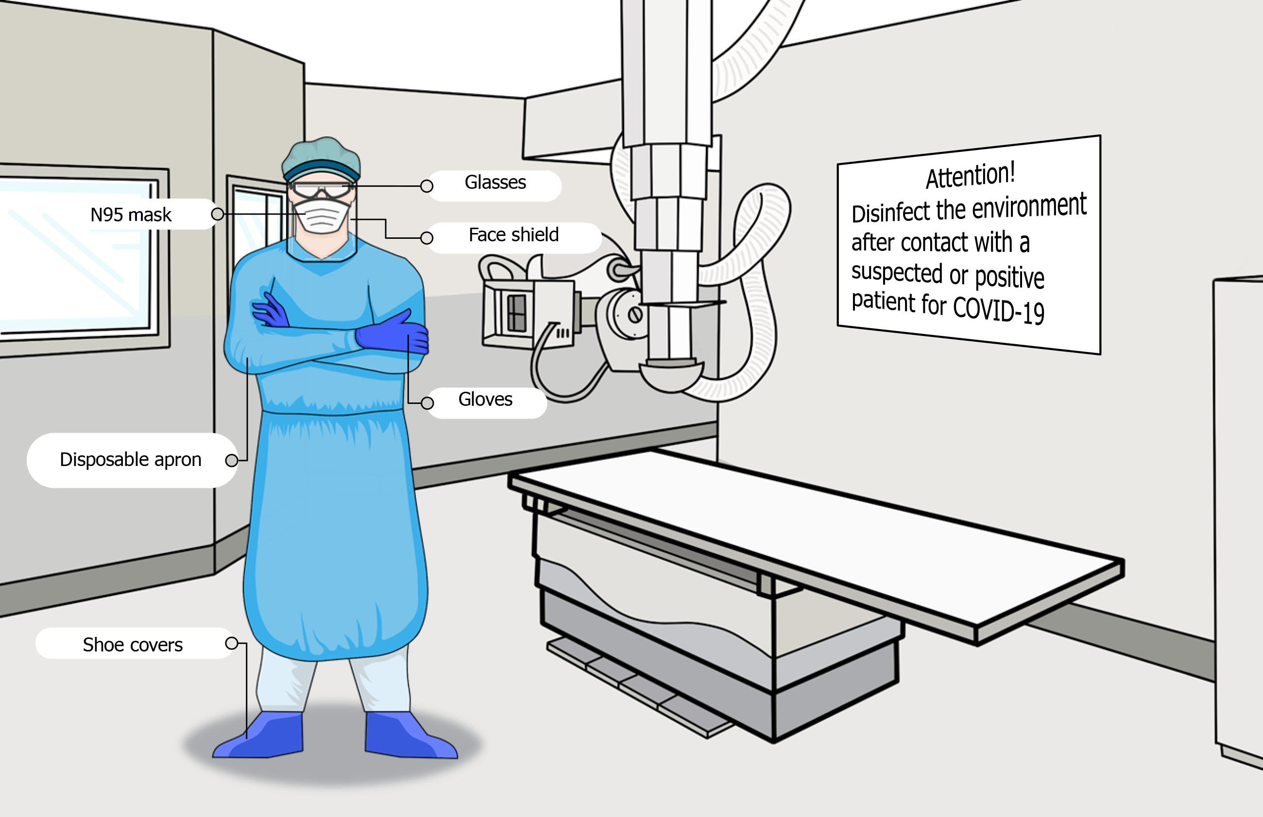
Figure 3 Safety measures to prevent infection in the radiology department.
COVID-19: Coronavirus disease 2019.
- Citation: de Carvalho LS, da Silva Júnior RT, Oliveira BVS, de Miranda YS, Rebouças NLF, Loureiro MS, Pinheiro SLR, da Silva RS, Correia PVSLM, Silva MJS, Ribeiro SN, da Silva FAF, de Brito BB, Santos MLC, Leal RAOS, Oliveira MV, de Melo FF. Highlighting COVID-19: What the imaging exams show about the disease. World J Radiol 2021; 13(5): 122-136
- URL: https://www.wjgnet.com/1949-8470/full/v13/i5/122.htm
- DOI: https://dx.doi.org/10.4329/wjr.v13.i5.122


