Copyright
©The Author(s) 2020.
World J Radiol. Aug 28, 2020; 12(8): 142-155
Published online Aug 28, 2020. doi: 10.4329/wjr.v12.i8.142
Published online Aug 28, 2020. doi: 10.4329/wjr.v12.i8.142
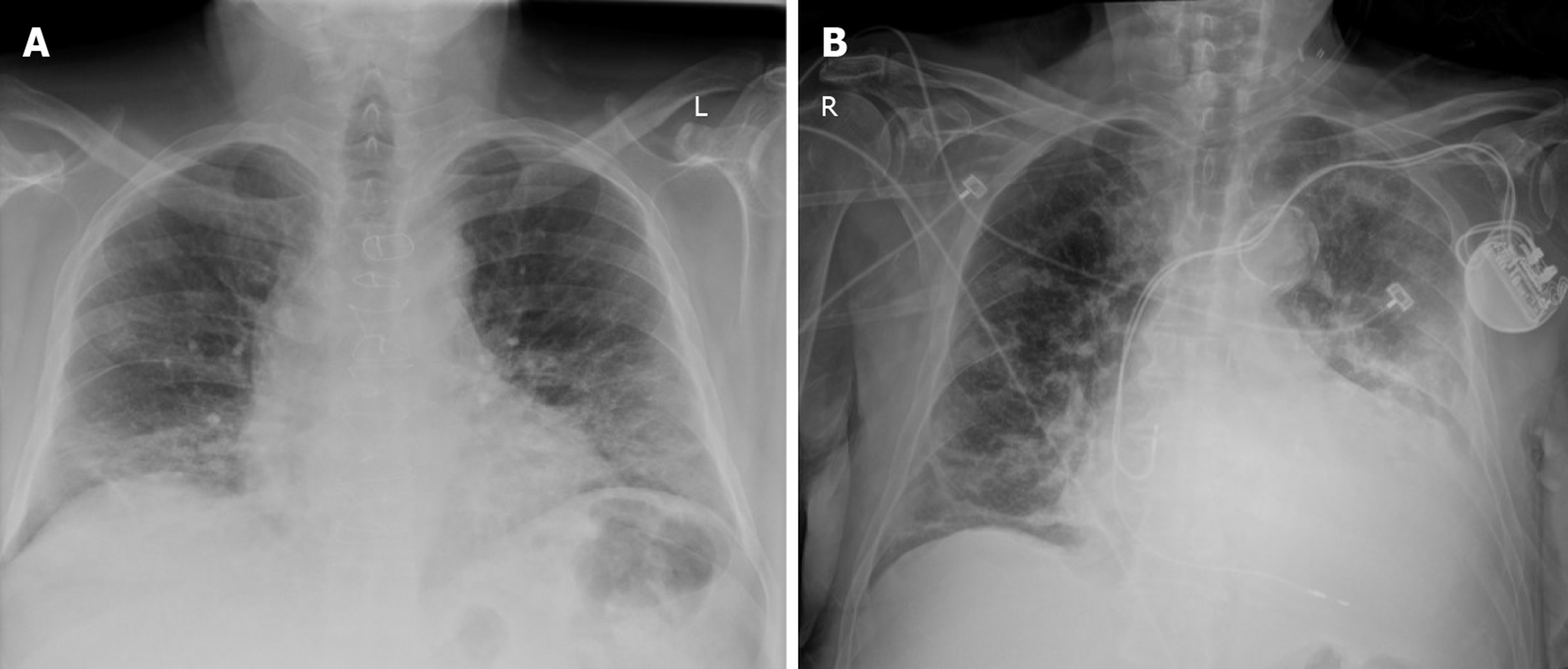
Figure 1 Anteroposterior chest radiographs in two patients with coronavirus disease 2019 pneumonia from our institution.
A: Interstitial infiltrates and ill-defined, patchy, rounded peripheral opacities in bilateral lung fields; B: Interstitial infiltrates with linear and patchy, rounded opacities in bilateral lung fields and heavily calcified aortic arch.
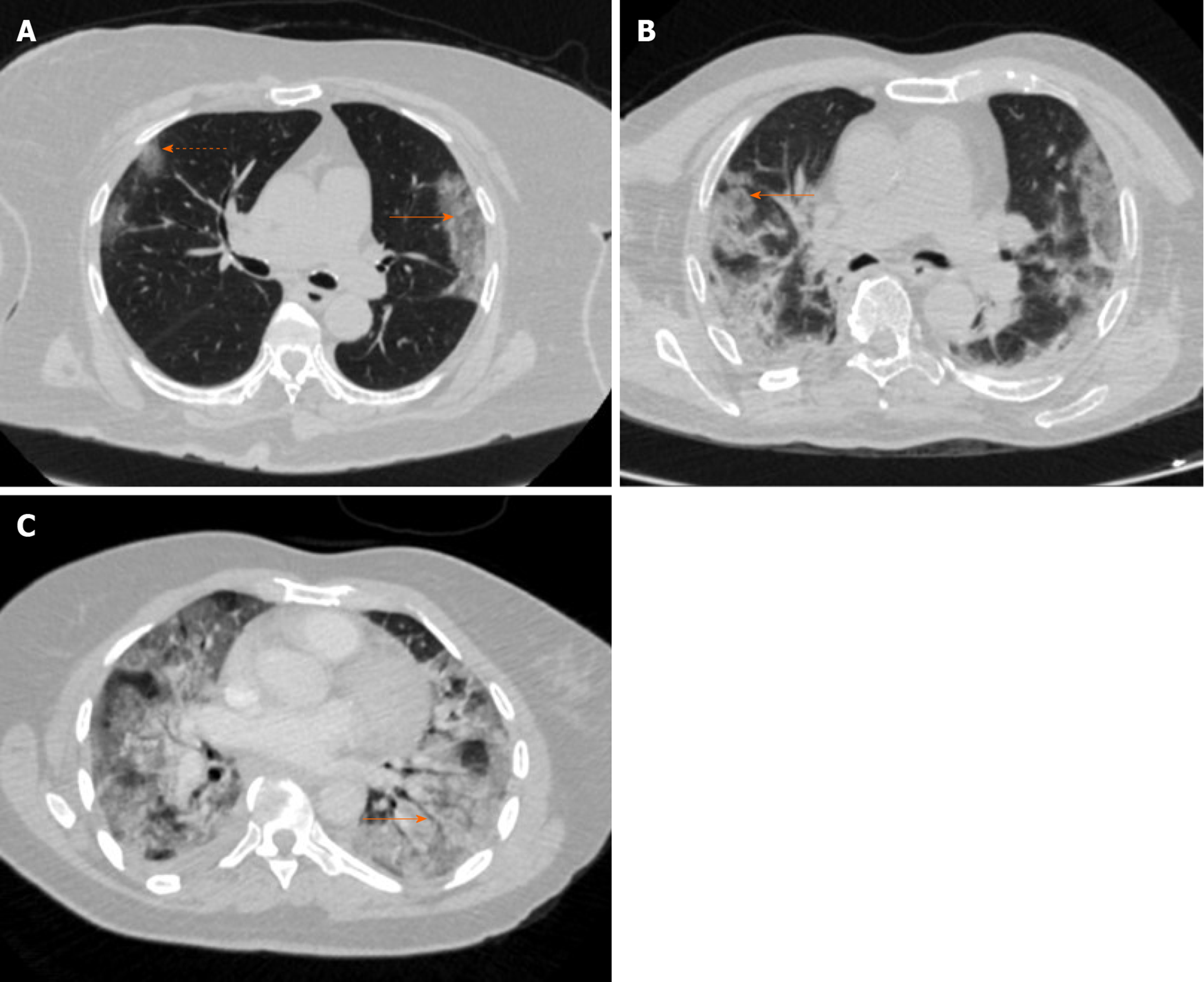
Figure 2 Axial thin-section chest computed tomography in three patients with coronavirus disease 2019 pneumonia from our institution.
A: Non-contrast exam with rounded, bilateral ground glass opacities (dashed arrow) and interlobular septal thickening giving appearance of crazy paving (solid arrow); B: Non-contrast exam with rounded, bilateral ground glass opacities and areas of surrounding consolidation giving appearance of reverse halo sign (solid arrow); and C: Contrast enhanced exam with ground glass and dense consolidation bilaterally and air bronchogram (solid arrow).
- Citation: Kaufman AE, Naidu S, Ramachandran S, Kaufman DS, Fayad ZA, Mani V. Review of radiographic findings in COVID-19. World J Radiol 2020; 12(8): 142-155
- URL: https://www.wjgnet.com/1949-8470/full/v12/i8/142.htm
- DOI: https://dx.doi.org/10.4329/wjr.v12.i8.142


