Copyright
©The Author(s) 2020.
World J Radiol. May 28, 2020; 12(5): 48-67
Published online May 28, 2020. doi: 10.4329/wjr.v12.i5.48
Published online May 28, 2020. doi: 10.4329/wjr.v12.i5.48
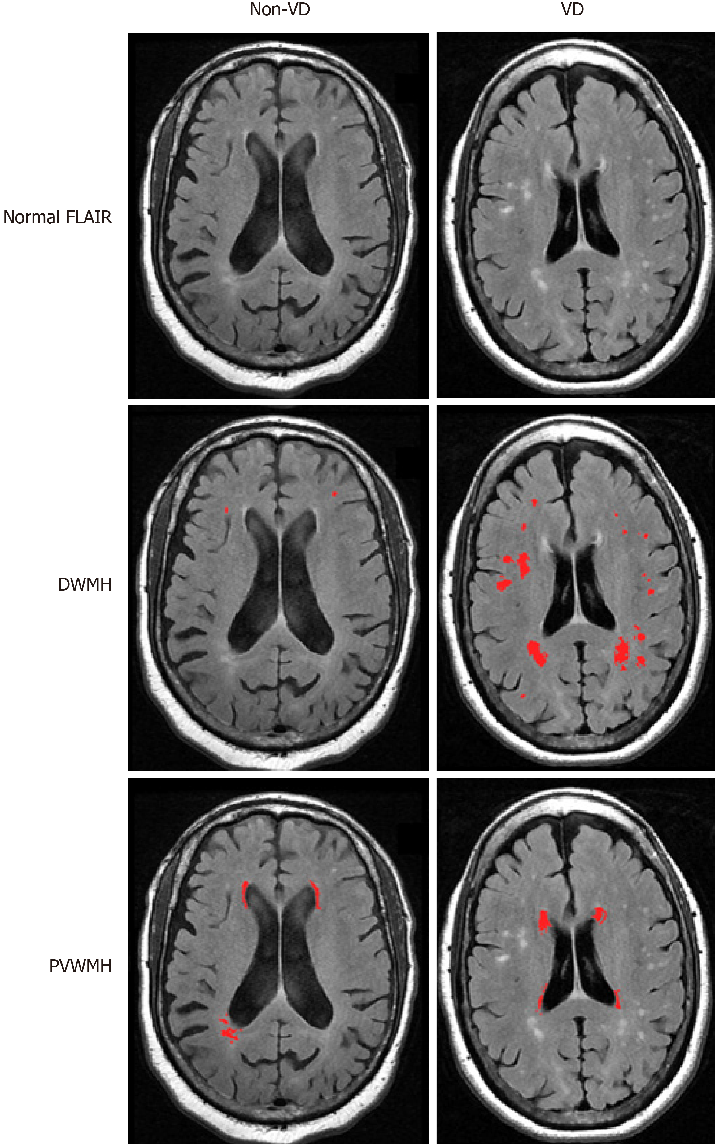
Figure 1 Discrimination of vascular depression and non-vascular depression white matter hyperintensities.
In non-vascular depression (non-VD) patients, deep white matter hyperintensities are occasionally present, though are few in number and do not begin to converge, as is common in VD patients. Periventricular white matter hyperintensities are more likely to be present in both VD and non-VD patients. VD: Vascular depression; DWMH: Deep white matter hyperintensities; PVWMH: Periventricular white matter hyperintensities.
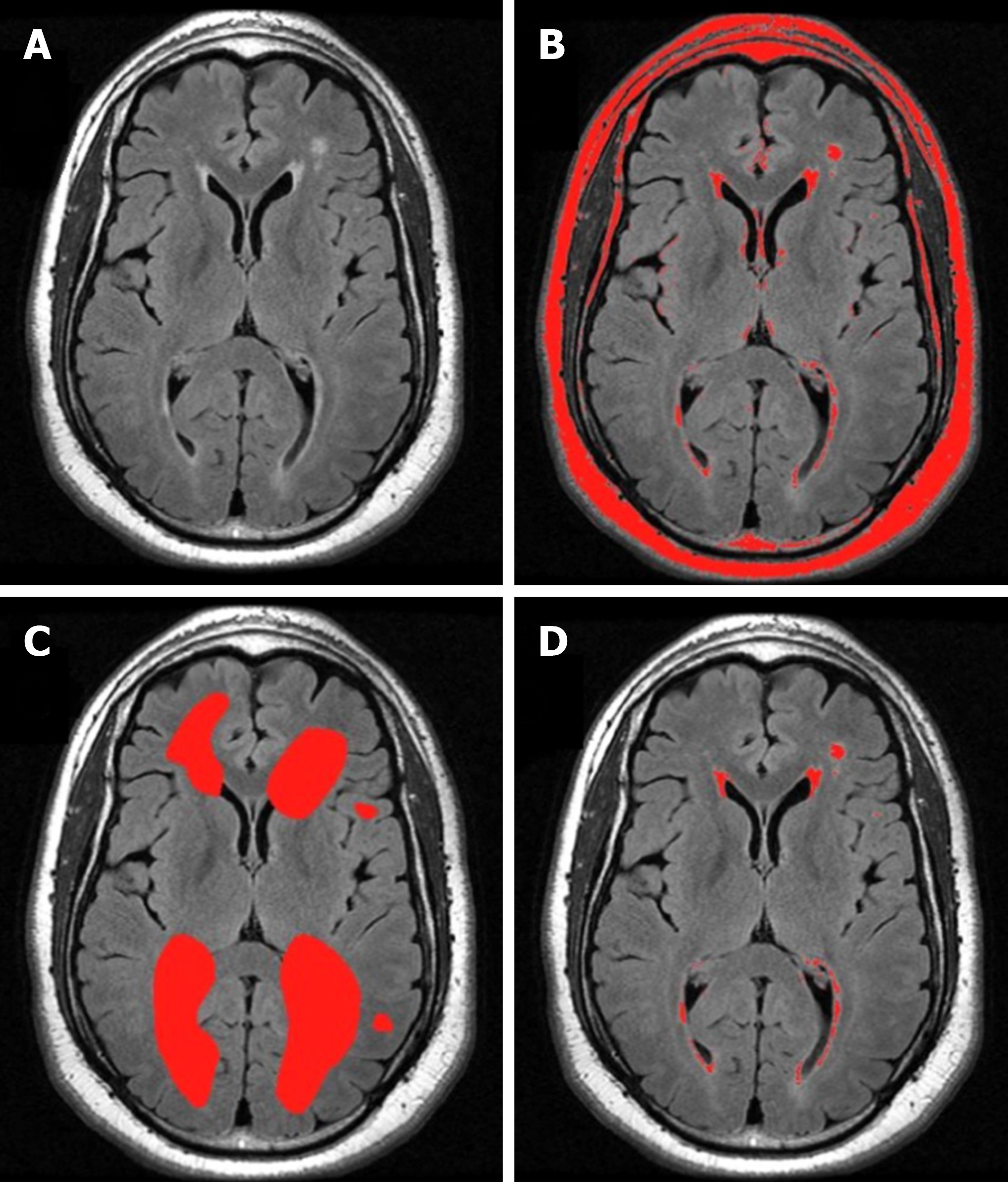
Figure 2 Semi-quantitative method.
Volumetric ratings made using the semi-automated software MRIcro. A: The original grayscale magnetic resonance imaging axial FLAIR image from a representative subject; B: The region of interest (ROI) map created by semiautomatic intensity thresholding; C: The ROI map created by gross manual outlining of hyperintensities; and D: The intersection of (B) and (C) yielding hyperintensities of interest.
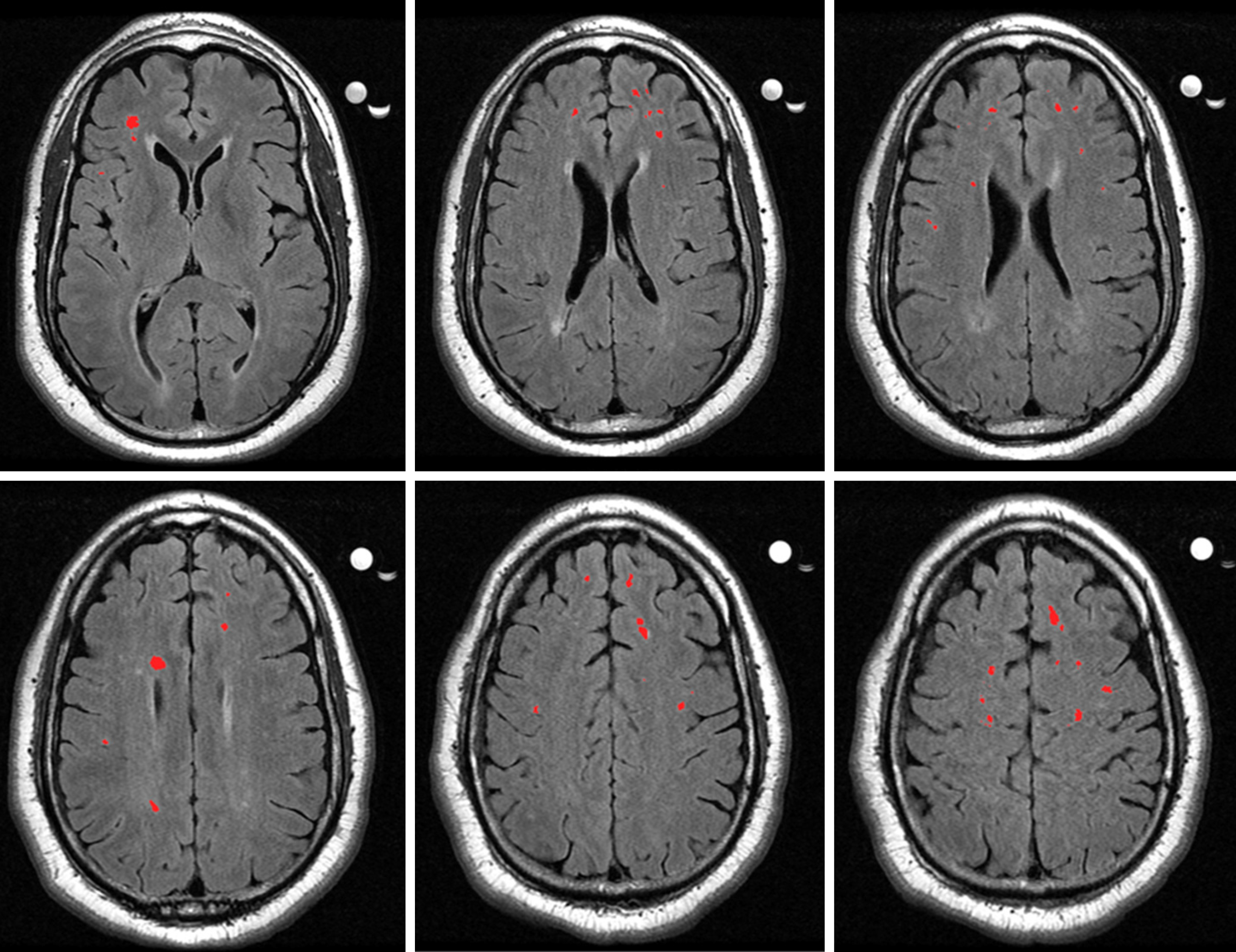
Figure 3 Axial slices from a magnetic resonance imaging scan of a vascular depression patient.
The highlighted sections are deep white matter hyperintensities, which have been graded as a severity of 2 on the Fazekas rating scale.
- Citation: Rushia SN, Shehab AAS, Motter JN, Egglefield DA, Schiff S, Sneed JR, Garcon E. Vascular depression for radiology: A review of the construct, methodology, and diagnosis. World J Radiol 2020; 12(5): 48-67
- URL: https://www.wjgnet.com/1949-8470/full/v12/i5/48.htm
- DOI: https://dx.doi.org/10.4329/wjr.v12.i5.48


