Copyright
©The Author(s) 2020.
World J Radiol. Nov 28, 2020; 12(11): 261-271
Published online Nov 28, 2020. doi: 10.4329/wjr.v12.i11.261
Published online Nov 28, 2020. doi: 10.4329/wjr.v12.i11.261
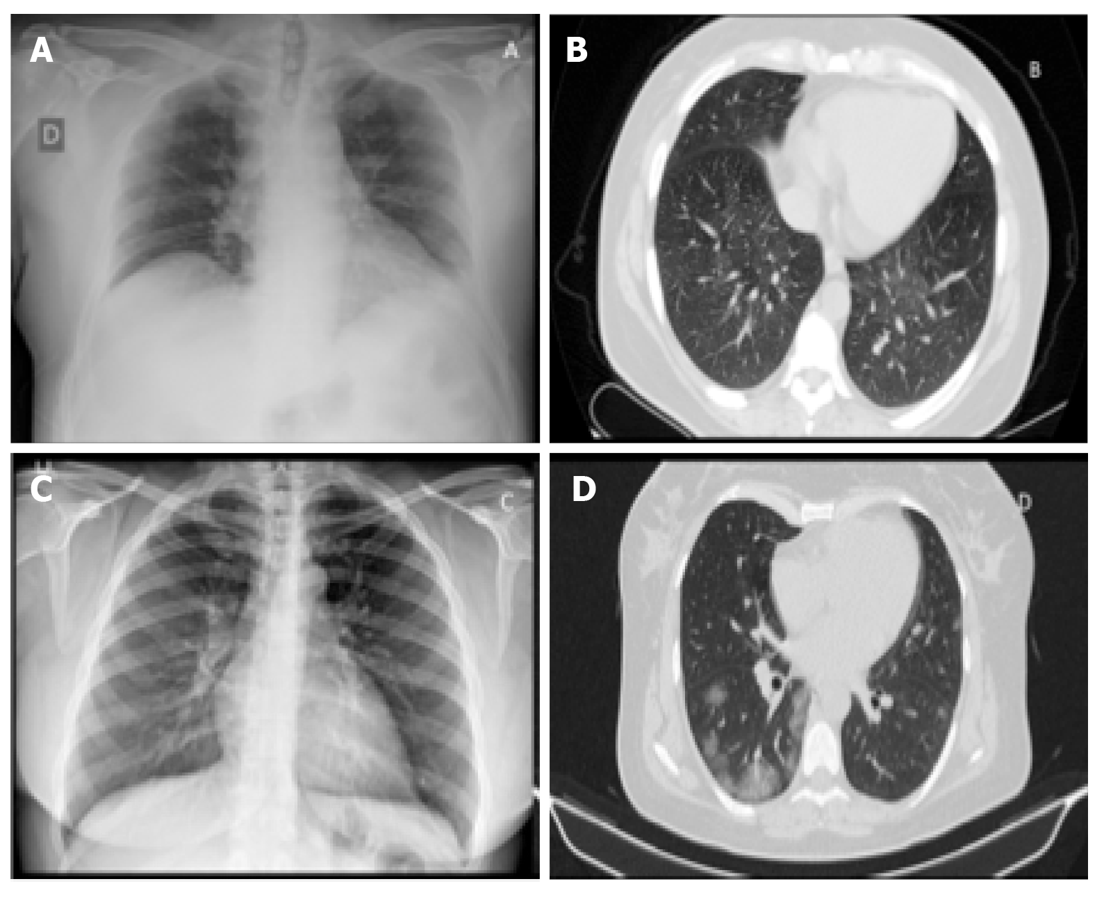
Figure 1 Standard chest X-ray and pulmonary computed tomography in a patient during early–stage and during the first week of coronavirus disease 2019 pneumonia.
A, B: Presymptomatic phase: Few areas of hyperdensity with ground glass on computed tomography, mainly in the lower and posterior fields; C, D: First week of symptoms: Bilateral ground glass opacities more confluent on computed tomography.
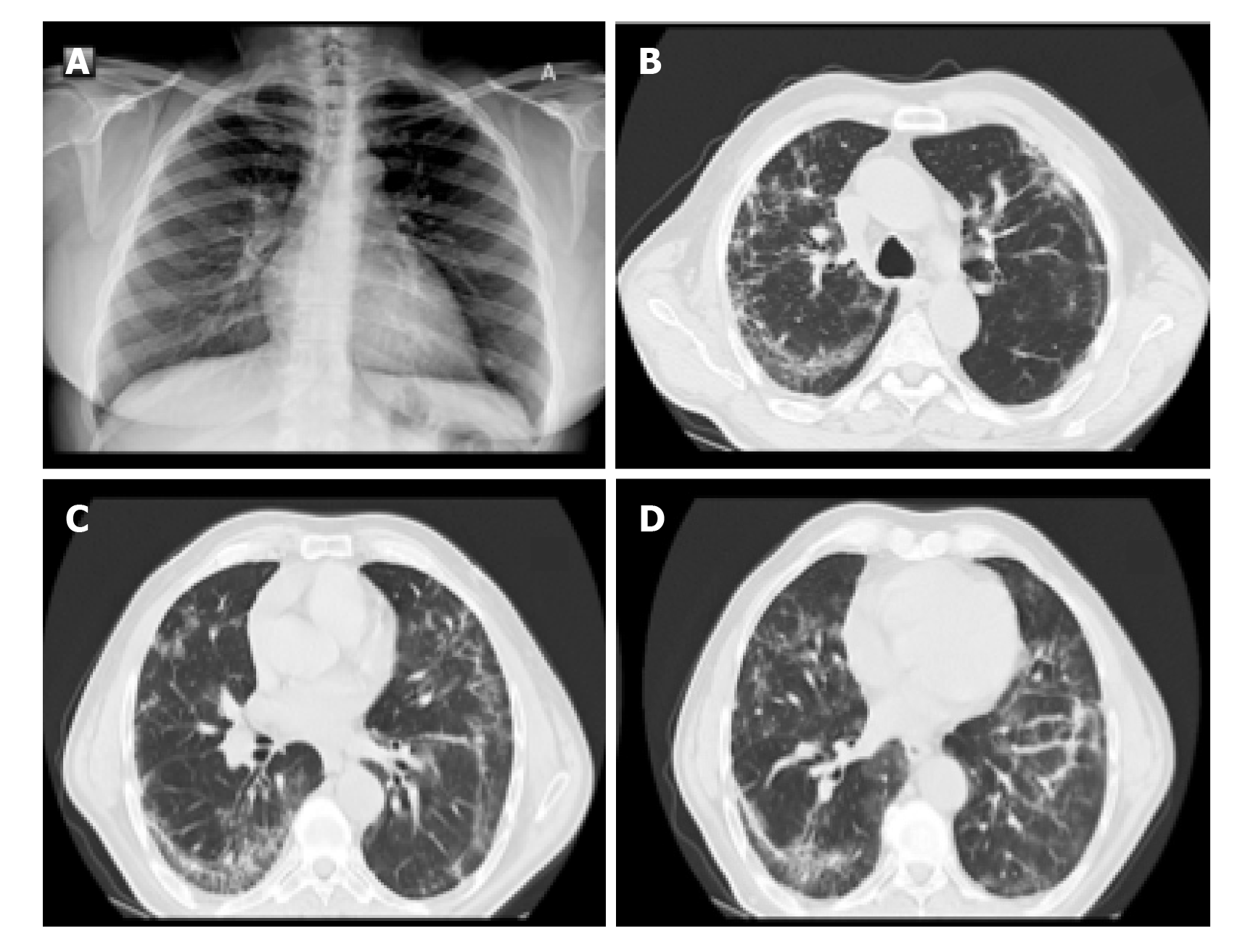
Figure 2 Standard chest X-ray and pulmonary computed tomography in a patient during the second week of severe coronavirus disease 2019 pneumonia.
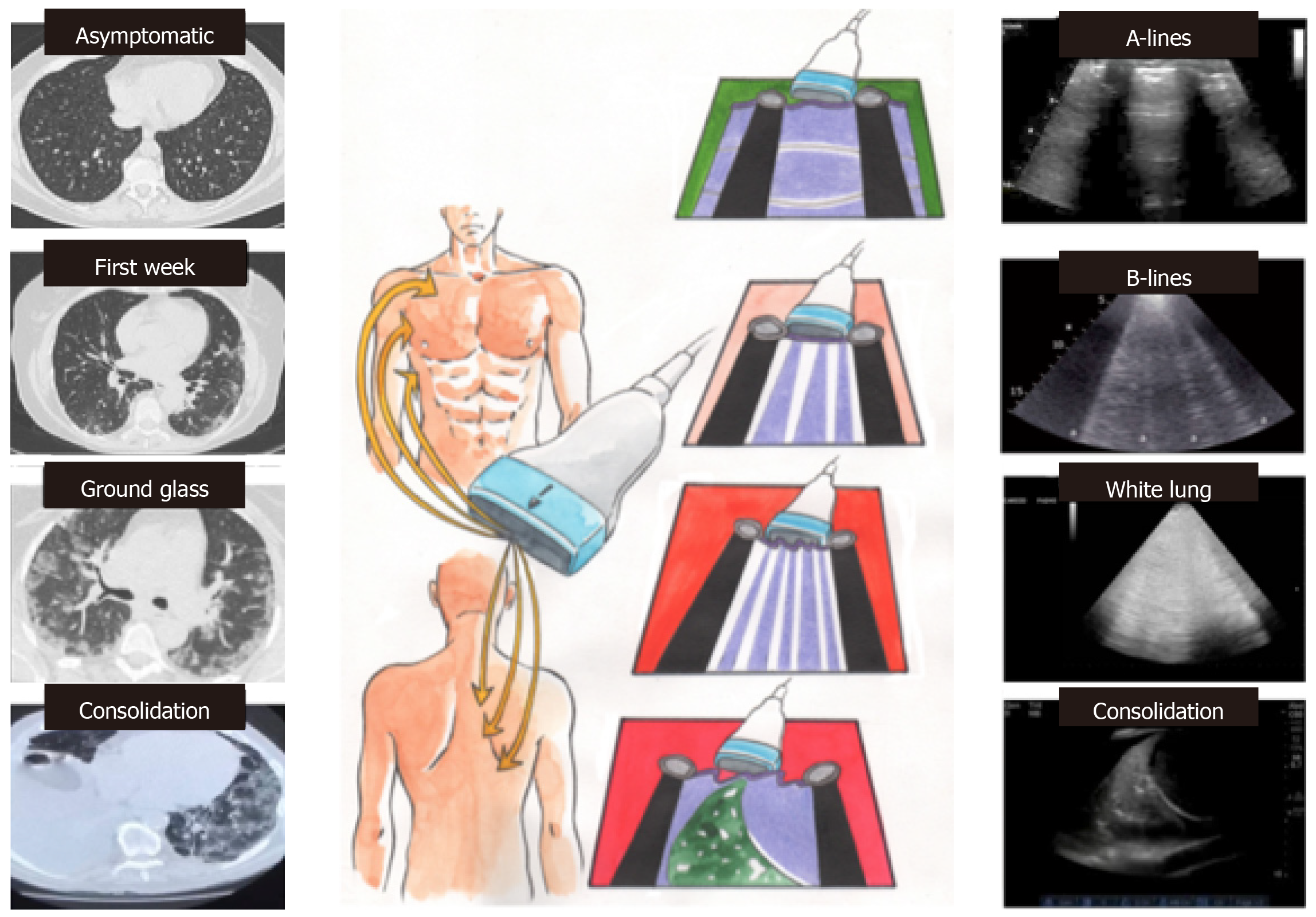
Figure 3 Correlations between bedside lung ultrasound and pulmonary computed tomography during the different phases of coronavirus disease 2019 pneumonia.
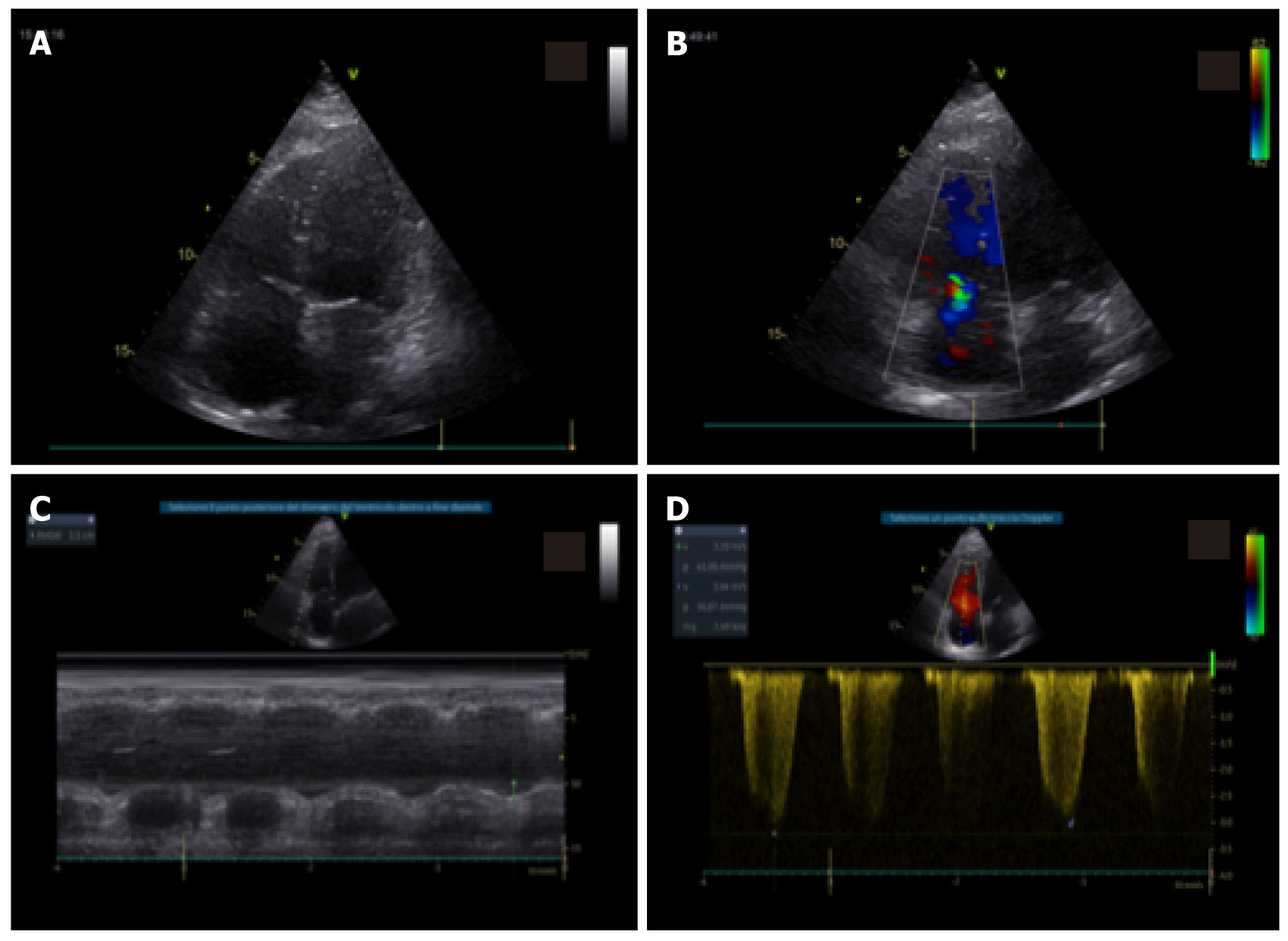
Figure 4 Standard echocardiography in a patient with coronavirus disease 2019 pneumonia and right ventricular impairment.
A: In apical four-chamber view, mild right ventricular dilatation; B: Mild tricuspid regurgitation; C: With moderate impairment of right ventricular function with reduced tricuspid annular plane systolic excursion; D: Increase in pulmonary pressures.
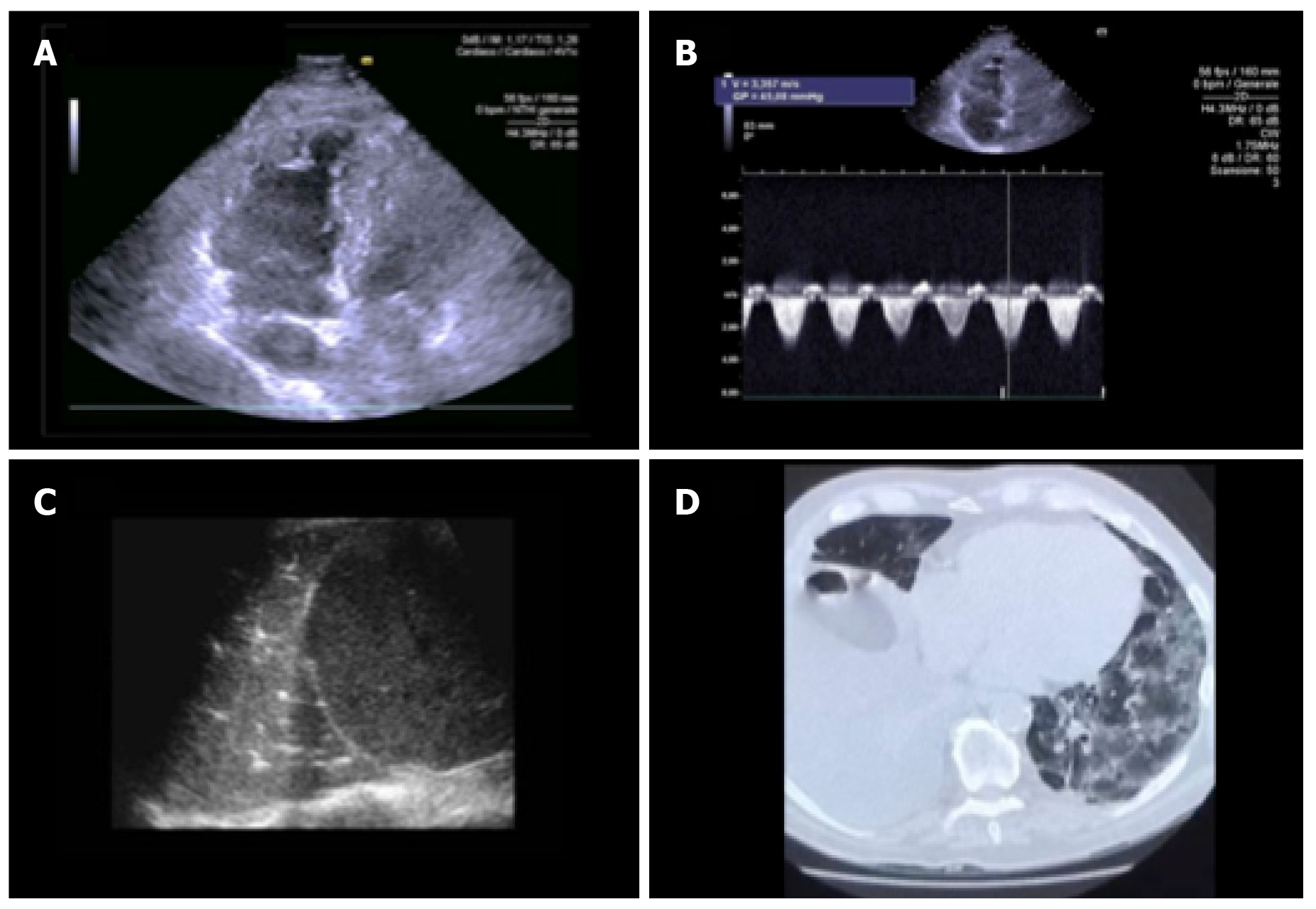
Figure 5 Standard echocardiography, lung ultrasound and pulmonary computed tomography in a patient with coronavirus disease 2019 pneumonia.
A: In apical four-chamber view, mild right ventricular dilatation; B: With increase in pulmonary pressures; C: By lung ultrasound; D: Pulmonary computed tomography ground glass with presence of consolidation areas.
- Citation: D'Andrea A, Radmilovic J, Carbone A, Forni A, Tagliamonte E, Riegler L, Liccardo B, Crescibene F, Sirignano C, Esposito G, Bossone E. Multimodality imaging in COVID-19 patients: A key role from diagnosis to prognosis. World J Radiol 2020; 12(11): 261-271
- URL: https://www.wjgnet.com/1949-8470/full/v12/i11/261.htm
- DOI: https://dx.doi.org/10.4329/wjr.v12.i11.261


