Copyright
©The Author(s) 2019.
World J Radiol. May 28, 2019; 11(5): 74-80
Published online May 28, 2019. doi: 10.4329/wjr.v11.i5.74
Published online May 28, 2019. doi: 10.4329/wjr.v11.i5.74
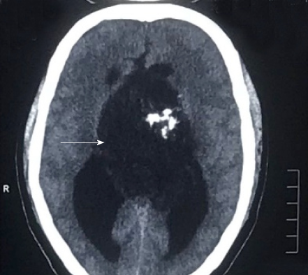
Figure 1 A non-contrast computed tomography scan of the head showing a mass arising from the third ventricle appearing hypodense and showing coarse nodular as well as curvilinear calcifications.
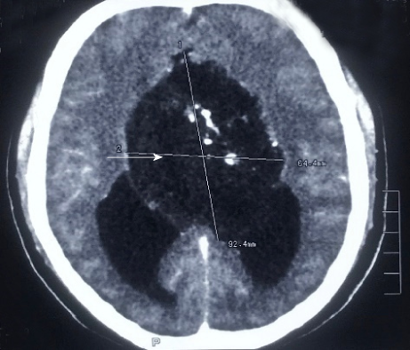
Figure 2 A contrast-enhanced computed tomography of the head demonstrates minimal perilesional enhancement.
The lesion obstructs the hydrocephalus of lateral ventricles (Figure 2). The maximum size of lesion measures 77 mm × 57 mm on computed tomography scan.
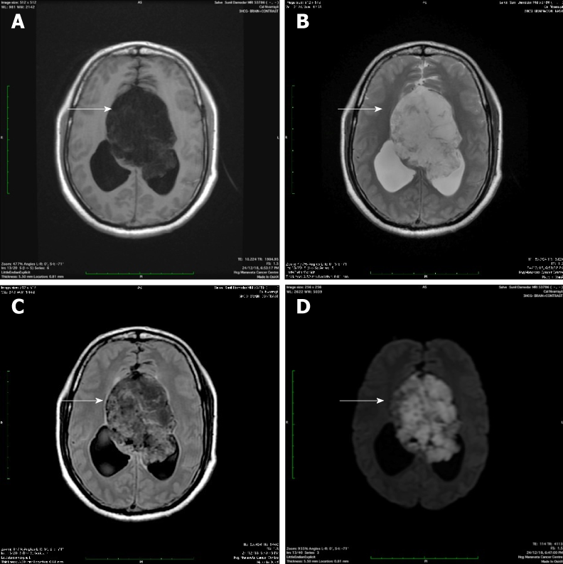
Figure 3 Magnetic resonance imaging.
Magnetic resonance imaging revealed a large, lobulated and septated lesion arising from the third ventricle that was: A: Hypointense on T1 weighted imaging; B: Hyperintense on T2 weighted imaging; C: Heterogeneously hyperintense on T2 axial FLAIR imaging; D: Appeared diffusely restricted on diffusion weighted imaging.
Figure 4 Minimal peripheral enhancement without any demonstrable solid enhancing component.
The maximum size of the lesion measures 73 mm × 65 mm × 64 mm in size.
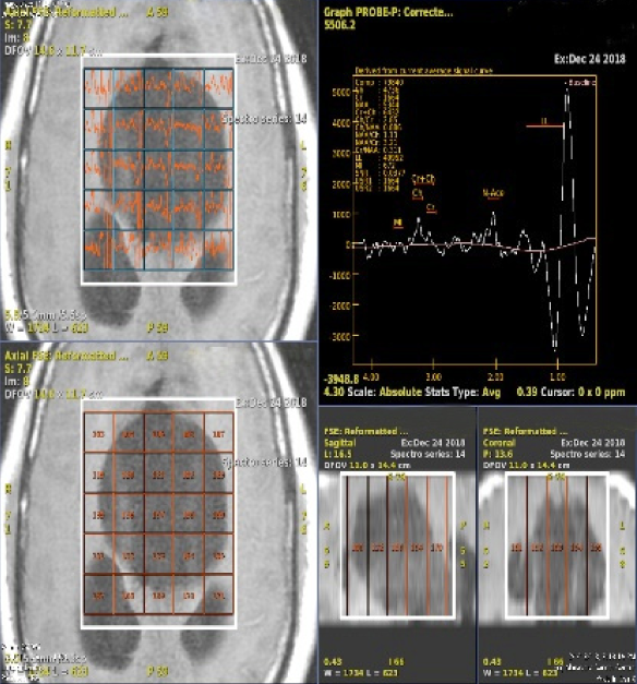
Figure 5 MR spectroscopy.
High lipid/lactate peak with comparatively lower N-acetylaspartate and choline levels. Maximum choline/creatine ratio was 2.85 within the lesion and choline/N-acetylaspartate ratio being 0.766.
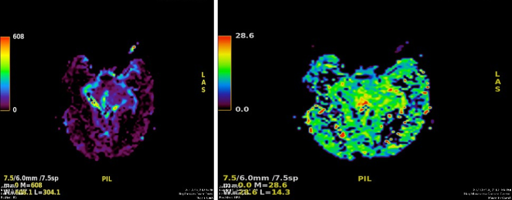
Figure 6 MR perfusion derived relative cerebral blood volume maps demonstrate heterogeneous perfusion abnormalities.
Mild higher normalized relative cerebral blood volume ratios in comparison with white matter are noted in the peripheral region of the cyst, which corresponds to malignant transformation.
- Citation: Pawar S, Borde C, Patil A, Nagarkar R. Malignant epidermoid arising from the third ventricle: A case report. World J Radiol 2019; 11(5): 74-80
- URL: https://www.wjgnet.com/1949-8470/full/v11/i5/74.htm
- DOI: https://dx.doi.org/10.4329/wjr.v11.i5.74


