Copyright
©The Author(s) 2018.
World J Radiol. Sep 28, 2018; 10(9): 108-115
Published online Sep 28, 2018. doi: 10.4329/wjr.v10.i9.108
Published online Sep 28, 2018. doi: 10.4329/wjr.v10.i9.108
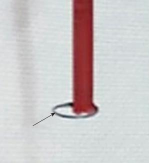
Figure 1 Distal loop of the indicator wire of the ExoSeal vascular closure device (black arrow).
A loop is automatically formed in the lumen of the blood vessel when the vascular closure device (VCD) is inserted into and attached to the vascular sheath. When the VCD/sheath assembly is pulled back, this loop contacts the inner surface of the vessel, causing the shape of the loop to change longitudinally.
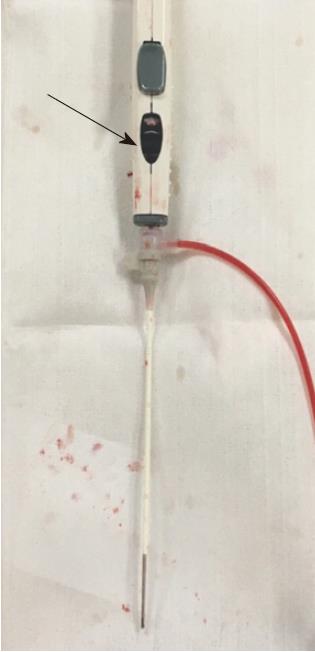
Figure 2 Appearance of the ExoSeal and vascular sheath complex when the polyglycolic acid plug has been normally deployed.
The color of the indicator window was black (black arrow), which means the plug is in the correct position, indicating that it is ready for the plug to be dropped.
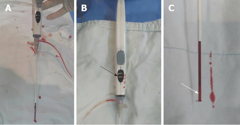
Figure 3 Representations of device appearances when failures occurred.
Normally, when the vascular closure devices (VCDs)/sheath assembly is pulled backward, the distal loop changes to a longitudinal shape and the plug is placed just above the arteriotomy. At this point, the indicator changes to black. When this indicator changes to black, pressing of the button deploys the plug. In this case, however, the indicator still did not turn black and the plug could not be dropped, even though the VCDs/sheath came out of the skin. A: Full view of VCDs/sheath in case of device failure; B: The color of the indicator window was white (black arrow); C: The polyglycolic acid plug (white arrow) remained in the delivery shaft of the ExoSeal.
- Citation: Han Y, Kwon JH, Park S. Korean single-center experience with femoral access closure using the ExoSeal device. World J Radiol 2018; 10(9): 108-115
- URL: https://www.wjgnet.com/1949-8470/full/v10/i9/108.htm
- DOI: https://dx.doi.org/10.4329/wjr.v10.i9.108


