Copyright
©The Author(s) 2018.
World J Radiol. Jul 28, 2018; 10(7): 65-77
Published online Jul 28, 2018. doi: 10.4329/wjr.v10.i7.65
Published online Jul 28, 2018. doi: 10.4329/wjr.v10.i7.65
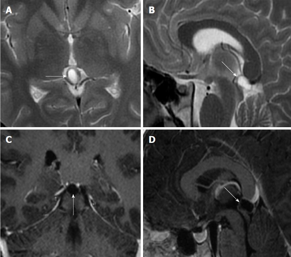
Figure 1 Eighteen years old male patient with typical pineal cyst (patient No.
43). A: Axial plane T2 weighted magnetic resonance imaging (MRI); B: Sagittal plane T2 weighted MRI; C: Axial plane T1 weighted contrast-enhanced MRI; D: Sagittal plane contrast-enhanced MRI. Typical pineal cyst is shown in the pineal region with unilocular homogeneous internal structure (arrows, A, B, C and D); Peripheral rim-like contrast-enhancement is shown (arrow, C and D).
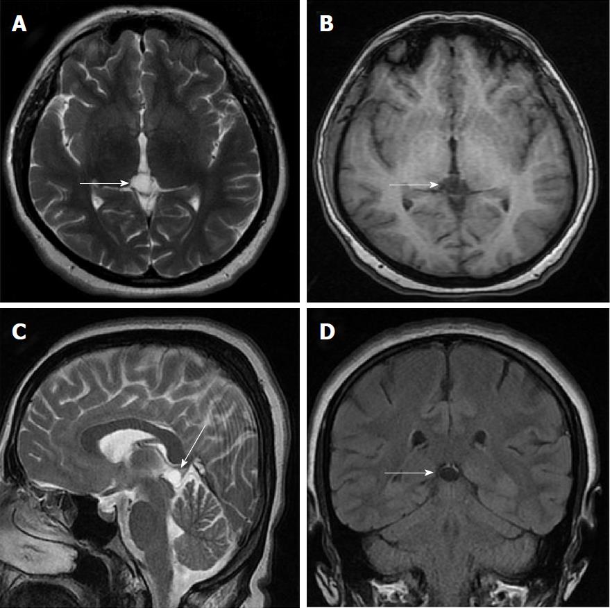
Figure 2 Thirty-three years old female patient with typical pineal cyst (patient No.
42). A: Axial plane T2 weighted magnetic resonance imaging (MRI); B: T1 weighted MRI; C: Sagittal plane T2 weighted MRI; D: Coronal plane FLAIR MRI. Typical pineal cysts are shown in the pineal region with unilocular homogeneous internal structure (white arrows, A, B, C and D); Isointense cyst with CSF (A, B and C); The slightly hyperintense pineal cyst compared to CSF is shown on FLAIR sequence (D). CSF: Cerebrospinal fluid; FLAIR: Fluid-attenuated inversion recovery.
Figure 3 Thirty-five years old female patient with typical pineal cyst (patient No.
34). A: Axial plane T2 weighted magnetic resonance imaging (MRI); B: T1 weighted MRI; C: Sagittal plane T2 weighted MRI; D: Coronal plane FLAIR MRI. Typical pineal cyst is shown in the pineal region with unilocular homogeneous internal structure (white arrows, A, B, C and D); The isointense pineal cyst compared to CSF is shown in the pineal region (A, B, C and D). CSF: Cerebrospinal fluid; FLAIR: Fluid-attenuated inversion recovery.
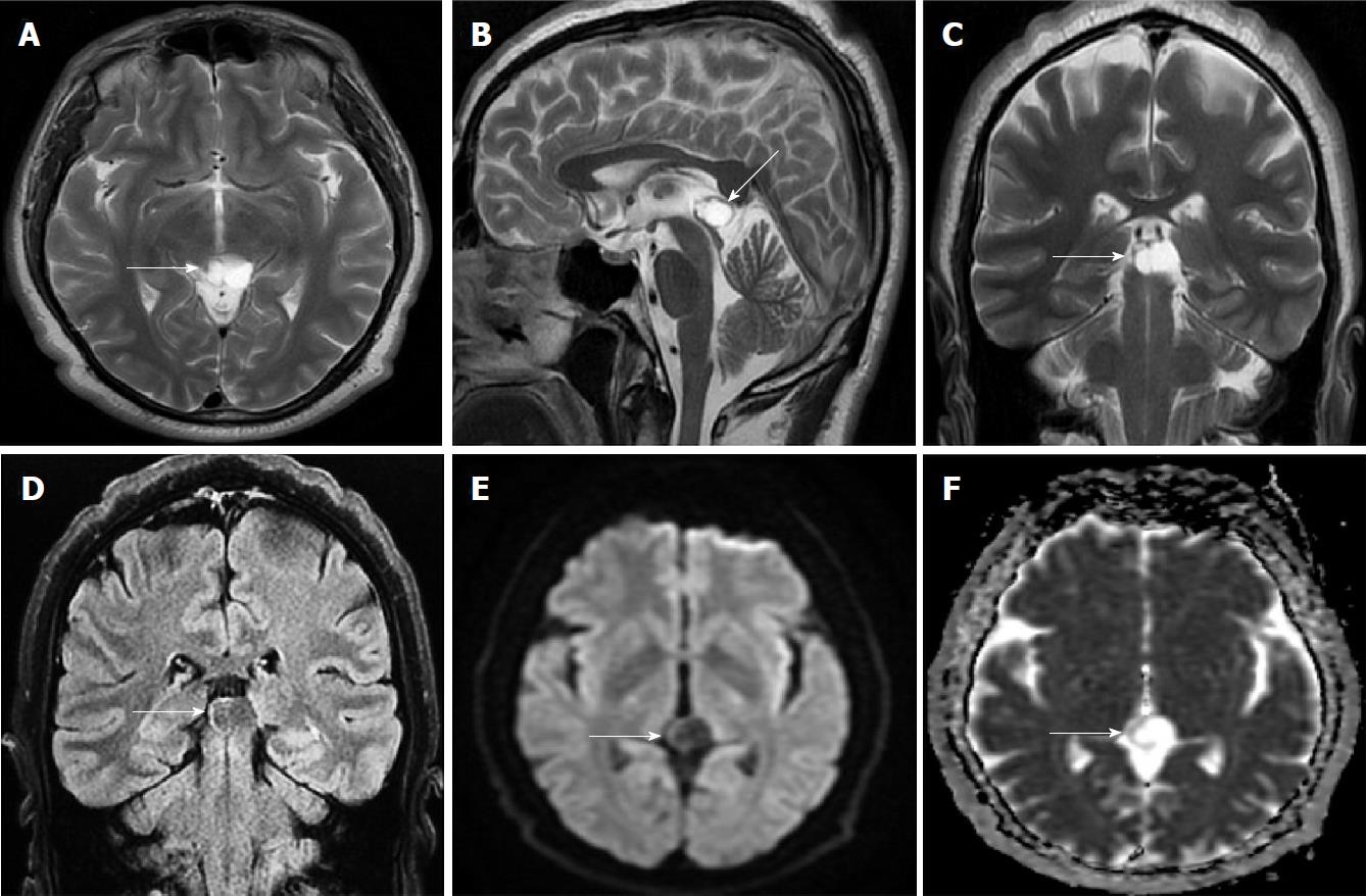
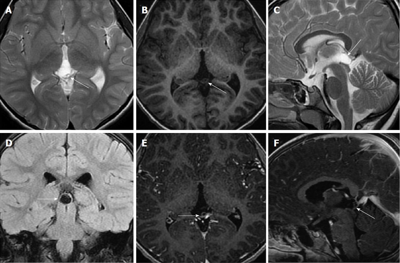
Figure 4 Fifty years old male patient with atypical pineal cyst (patient No.
40). Axial plane (A) sagittal plane magnetic resonance imaging (MRI) (B) coronal plane T2 weighted (C) and coronal plane FLAIR magnetic resonance imaging (D) axial plane diffusion weighted imaging (E) axial plane apparent diffusion coefficient map (F). A, B, C, D, E and F: Bilocular, lobule contoured atypical pineal cyst is shown in pineal area (white arrows); D: The slightly hyperintense pineal cyst compared to CSF is shown on FLAIR sequence; E and F: No diffusion restriction. CSF: Cerebrospinal fluid; FLAIR: Fluid-attenuated inversion recovery.
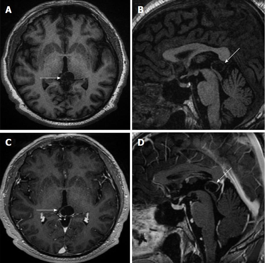
Figure 5 Fifty years old male patient with atypical pineal cyst (patient No.
40). Axial plane (A) sagittal plane T1 weighted (B) axial plane (C) sagittal plane contrast-enhanced magnetic resonance imaging (D). A, B, C and D: Bilocular, lobule contoured atypical pineal cyst is shown in pineal area (white arrows); C and D: Partial rim-like enhancement is shown (white arrows).
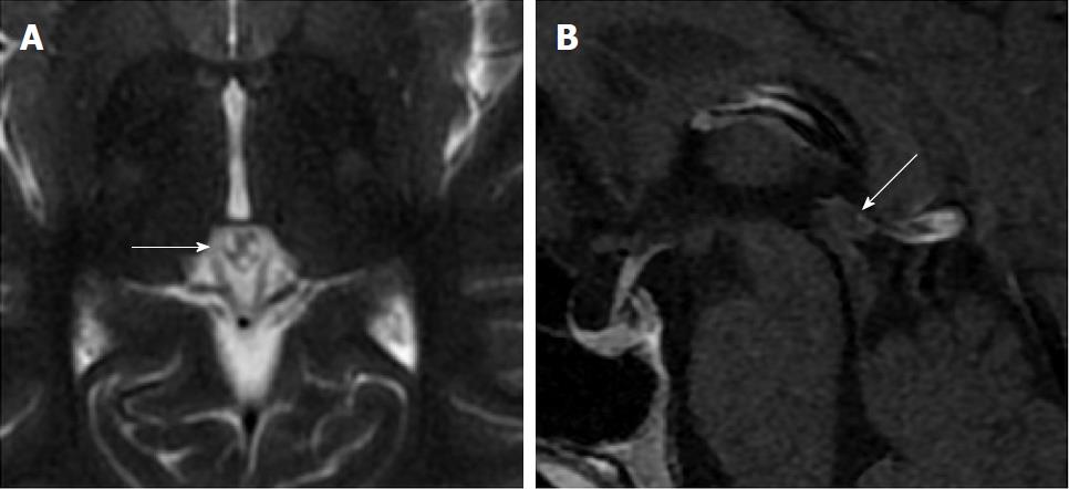
Figure 6 Thirty-six years old female patient with atypical pineal cyst (patient No.
2). Axial plane T2 weighted (A) sagittal plane (B) contrast-enhanced magnetic resonance imaging. A and B: Atypical pineal cysts are shown in the pineal region; A: Trilocular cyst (white arrow); B: Peripheral rim-like enhancement is shown (white arrow).
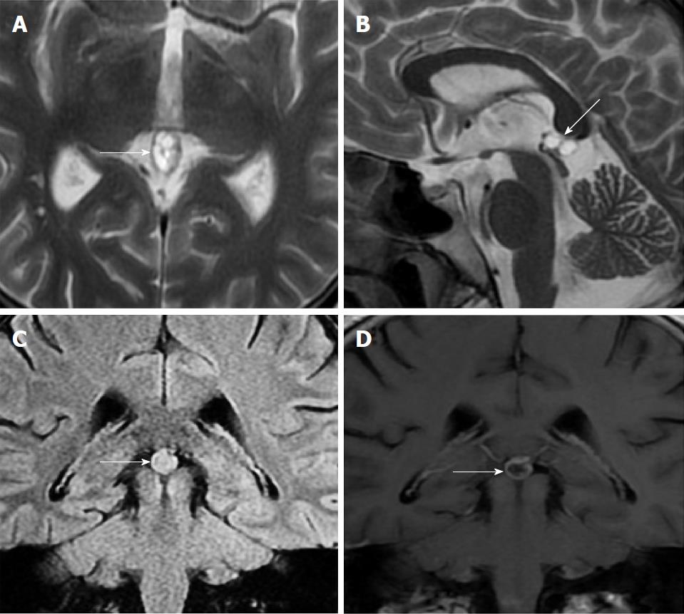
Figure 7 Thirty-three years old male patient with atypical pineal cyst (patient No.
41). Axial plane (A) sagittal plane T2 weighted (B) coronal plane FLAIR (C) coronal plane (D) contrast-enhanced magnetic resonance imaging. A and B: Atypical multilocular pineal cysts are shown in the pineal region (white arrows); C: The hyperintense pineal cyst compared to CSF is shown in the pineal region (white arrows); D: Peripheral rim-like and septal enhancement is shown (white arrows). CSF: Cerebrospinal fluid; FLAIR: Fluid-attenuated inversion recovery.
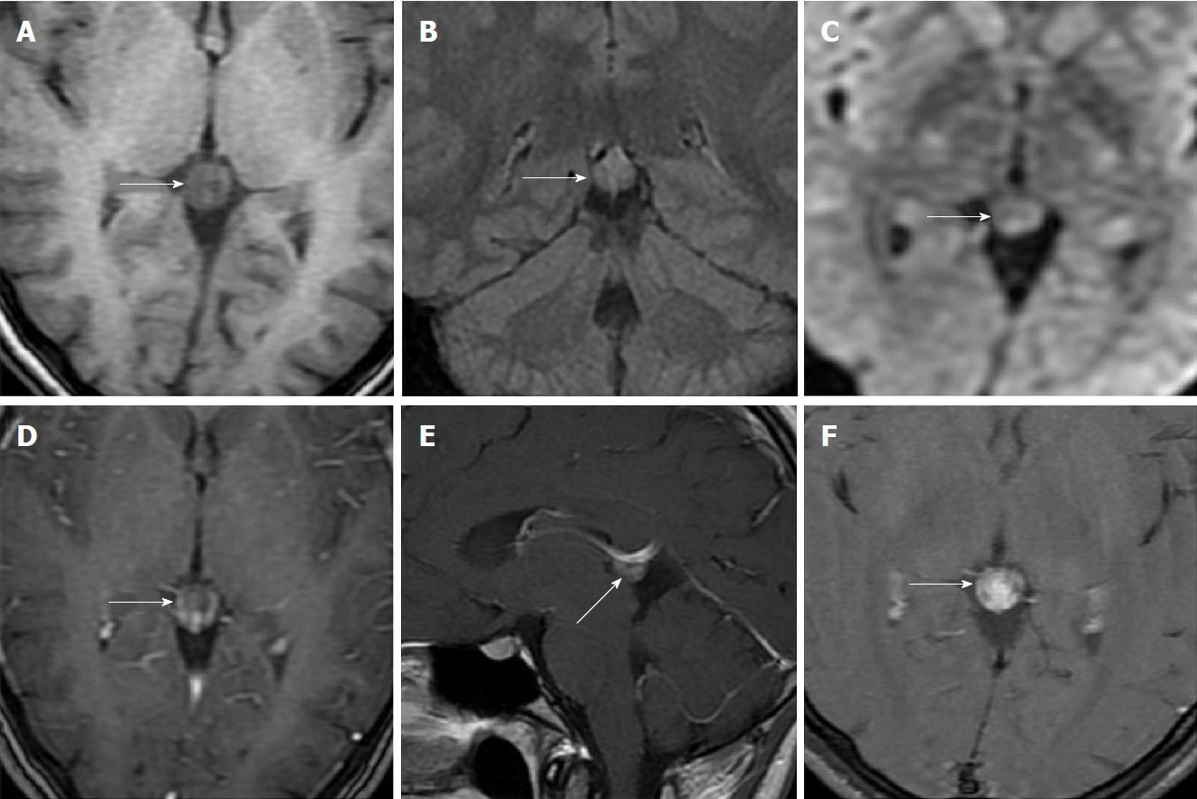
Figure 8 Twenty-two years old female patient with atypical pineal cyst (patient No.
50). Axial plane T1 weighted (A) coronal plane FLAIR (B) axial plane diffusion weighted (C) consecutive contrast-enhanced magnetic resonance imaging (D, E and F). A, B, C, D, E and F: Atypical pineal cyst or pineocytoma is not distinguished radiologically (white arrows); C: No diffusion restriction; D, E and F: Increasing gradually heterogeneous enhancement is shown in pineal region (white arrows). FLAIR: Fluid-attenuated inversion recovery.
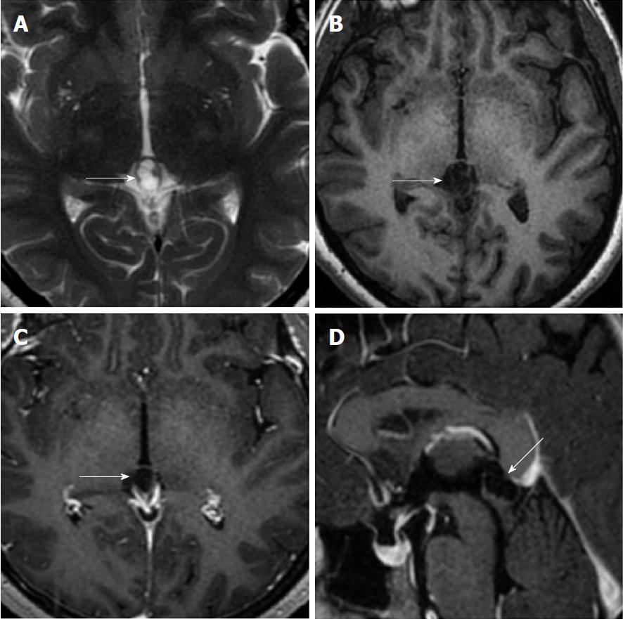
Figure 9 Thirty-three years old female patient with atypical pineal cyst (patient No.
53). Axial plane (A) T2 weighted (B) T1 weighted (C) contrast-enhanced (D) sagittal plane contrast-enhanced magnetic resonance imaging. A, B, C and D: Multilocular, lobule contoured, atypical pineal cyst is observed in pineal area (white arrows); C and D: Peripheral rim-like enhancement is shown (white arrows).
Figure 10 Five years old female patient with atypical pineal cyst (patient No.
44). Axial plane (A) T2 weighted (B) T1 weighted (C) sagittal plane T2 weighted (D) coronal plane FLAIR (E) axial plane (F) sagittal plane contrast-enhanced magnetic resonance imaging. A, B, C, D, E and F: Multilocular, lobule contoured, atypical pineal cyst is observed in pineal area (white arrows); D: The isointense pineal cyst compared to CSF is shown on FLAIR sequence; E and F: Peripheral rim-like and septal (white arrows) enhancement is shown. CSF: Cerebrospinal fluid; FLAIR: Fluid-attenuated inversion recovery.
- Citation: Gokce E, Beyhan M. Evaluation of pineal cysts with magnetic resonance imaging. World J Radiol 2018; 10(7): 65-77
- URL: https://www.wjgnet.com/1949-8470/full/v10/i7/65.htm
- DOI: https://dx.doi.org/10.4329/wjr.v10.i7.65


