Copyright
©The Author(s) 2018.
World J Radiol. Nov 28, 2018; 10(11): 150-161
Published online Nov 28, 2018. doi: 10.4329/wjr.v10.i11.150
Published online Nov 28, 2018. doi: 10.4329/wjr.v10.i11.150
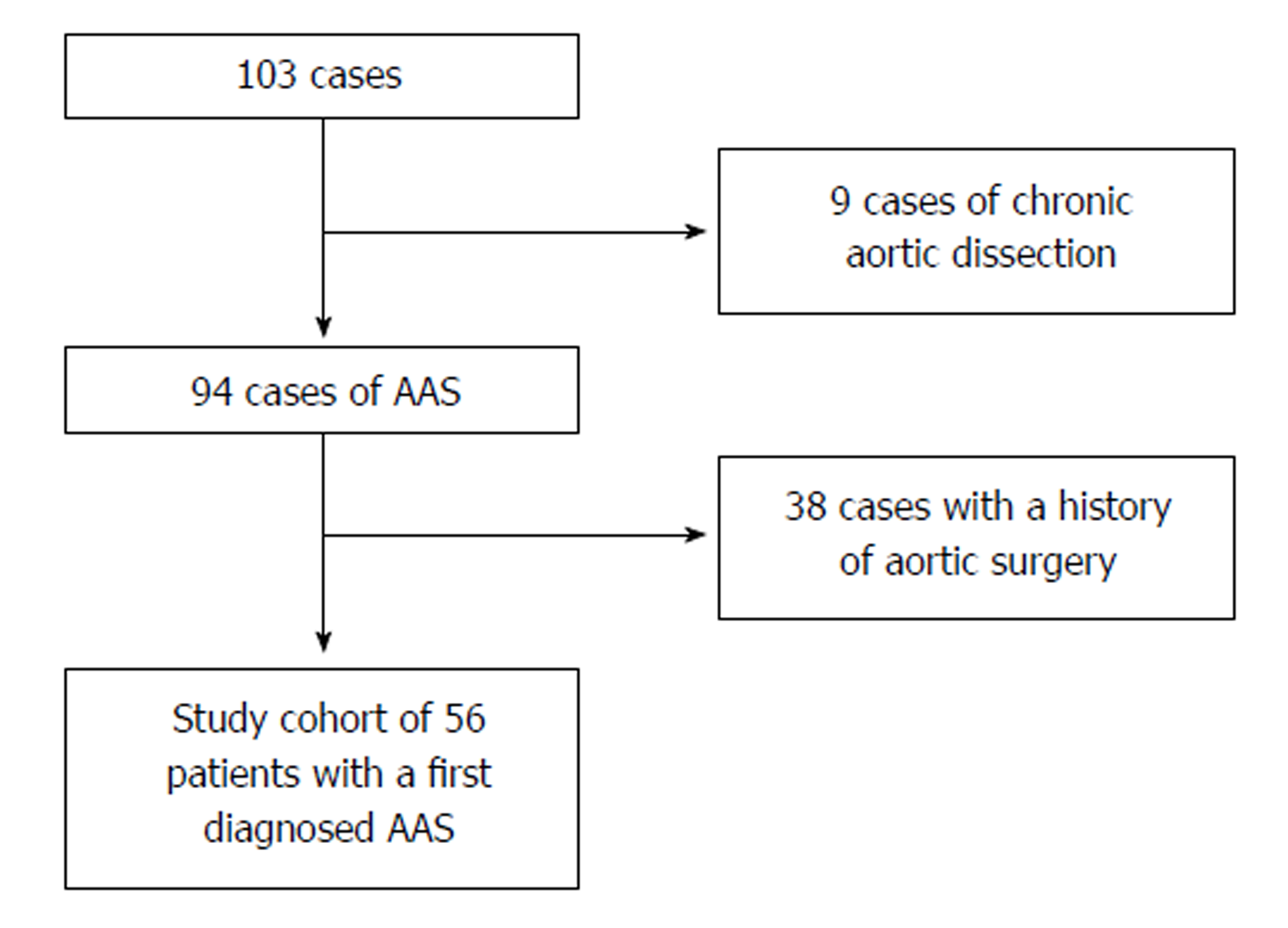
Figure 1 Selection process of the study cohort.
After the exclusion of chronic dissections and patients with a history of aortic surgery, 56 cases made up the study cohort of individuals with a newly diagnosed acute aortic syndrome. AAS: Acute aortic syndrome.
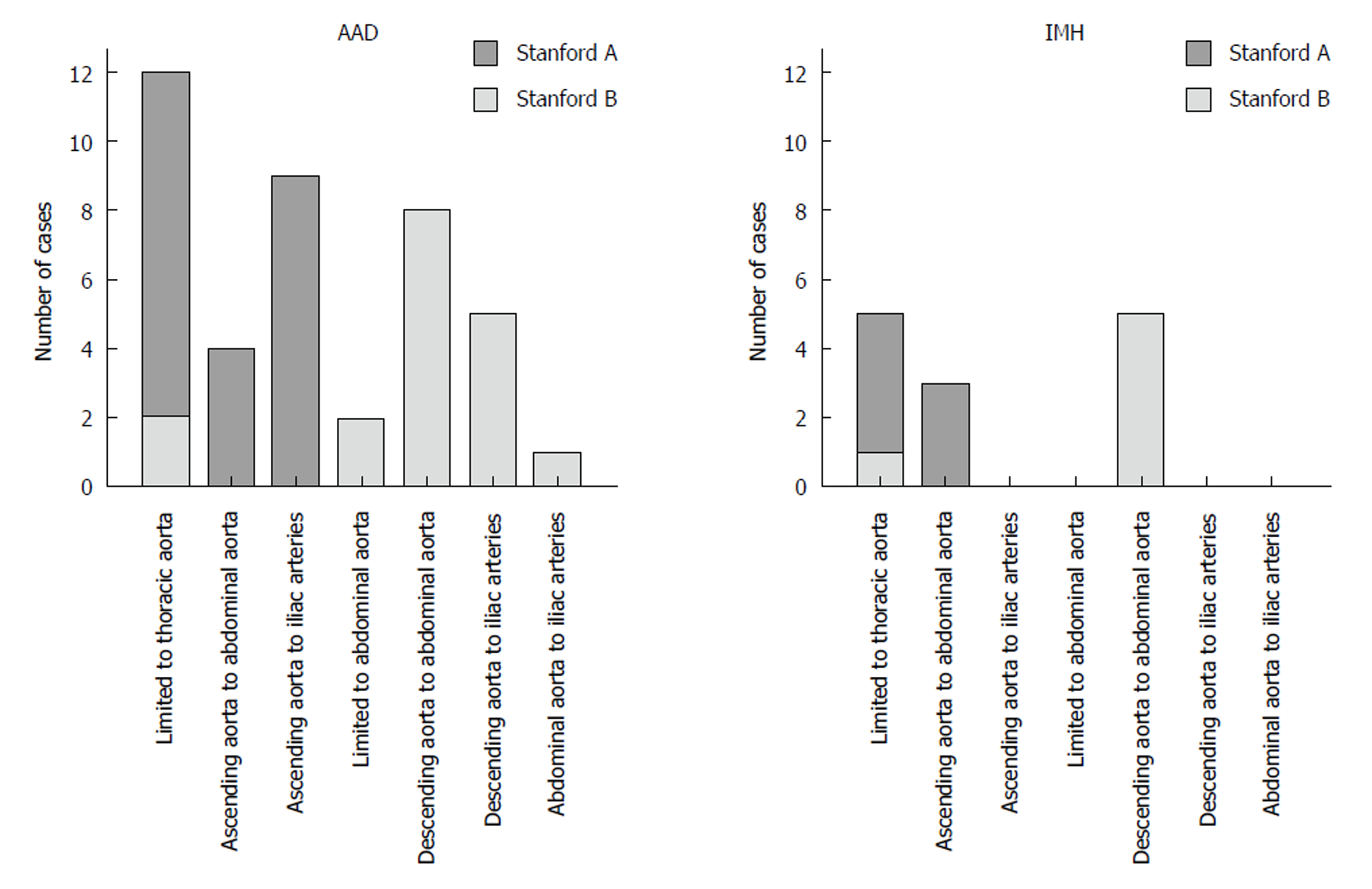
Figure 2 Extent of dissection and intramural hematoma.
Nearly one third of acute aortic syndrome was limited to the thoracic aorta. There was no case in which the intramural hematoma extended into the iliac arteries. AAD: Acute aortic dissection; IMH: Intramural hematoma.
Figure 3 Boundaries of acute aortic dissection and intramural hematoma.
Anatomic features detain a distal progression of acute aortic dissection and intramural hematoma. AAD: Acute aortic dissection; IMH: Intramural hematoma.
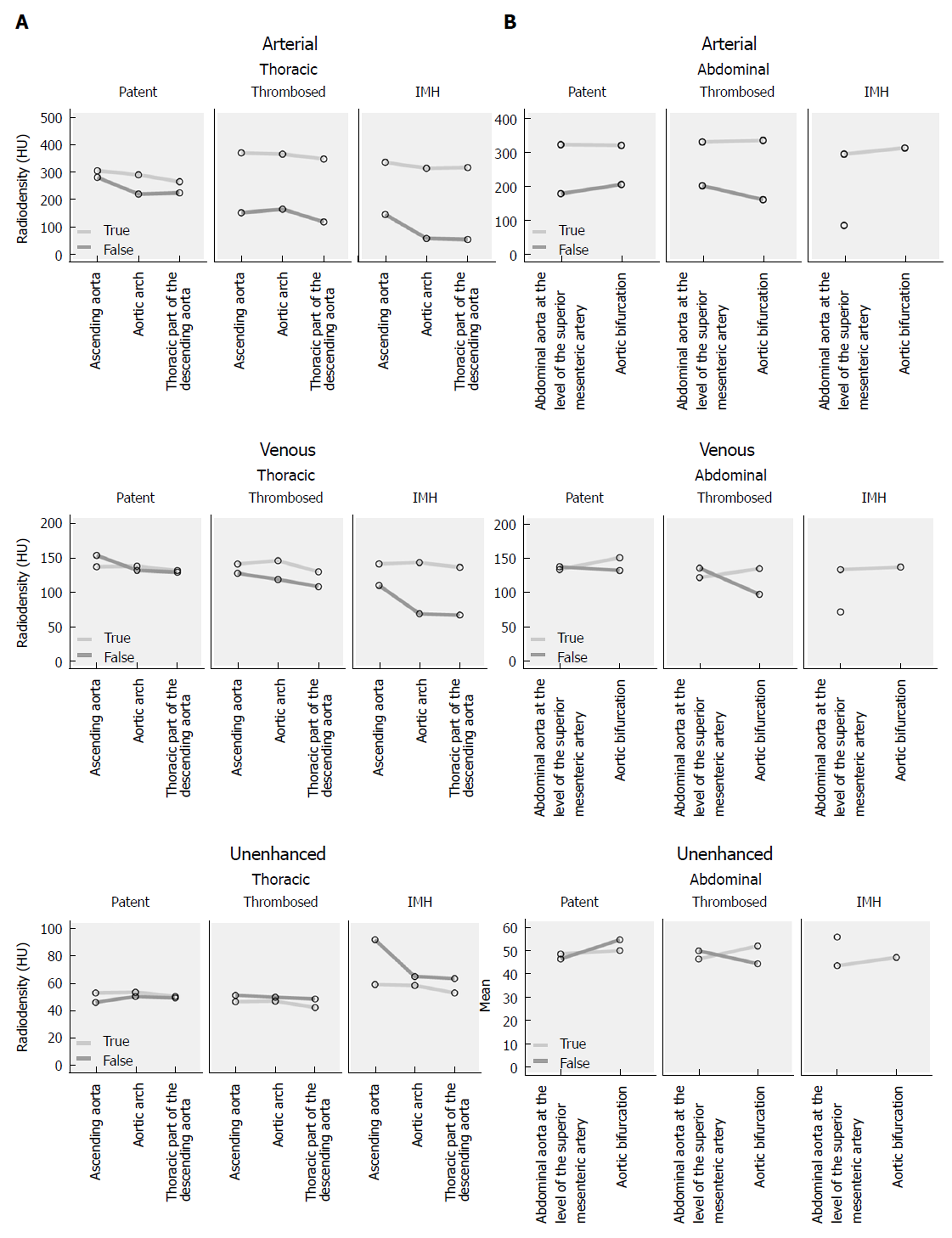
Figure 4 Density measurements, absolute values.
Absolute values of the density measurements in true and false lumen/intramural hematoma in arterial, venous and unenhanced phase of (A) thoracic and (B) abdominal aorta. IMH: Intramural hematoma.
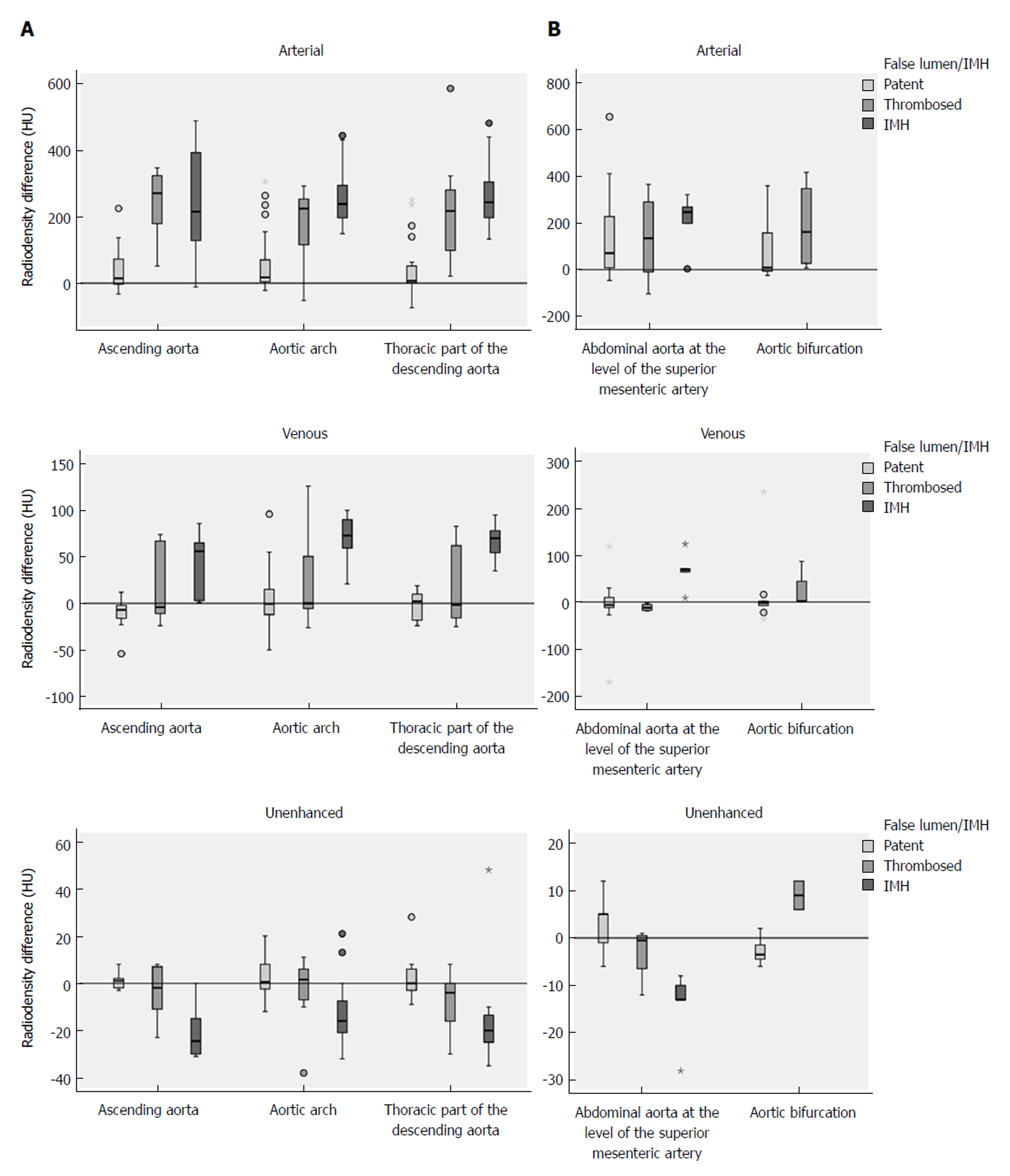
Figure 5 Density measurements, differences.
Density differences between true and false lumen/intramural hematoma (IMH) in arterial, venous and unenhanced phase of (A) thoracic and (B) abdominal aorta. Differences in radiodensity between true lumen and IMH as well as true lumen and thrombosed false lumen, respectively, were only significant in the unenhanced acquisition (P = 0.035). IMH: Intramural hematoma.
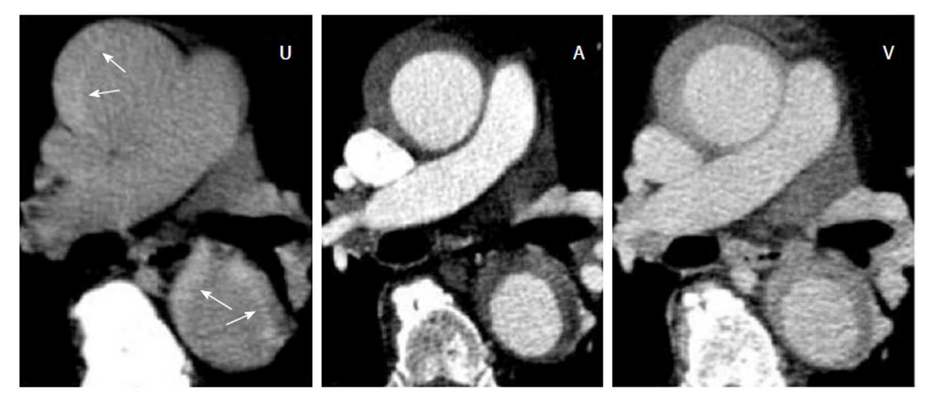
Figure 6 Acute intramural hematoma.
Triphasic computed tomography angiography with an acute intramural hematoma (IMH) type Stanford A in the ascending and descending aorta. The unenhanced scan (U) shows a hyperdense wall thickening compared to the lumen (arrows). In the arterial (A) and venous (V) phase of the enhanced scans, the IMH con not be differentiated from a thrombotic layer.
- Citation: Panagiotopoulos N, Drüschler F, Simon M, Vogt FM, Wolfrum S, Desch S, Richardt D, Barkhausen J, Hunold P. Significance of an additional unenhanced scan in computed tomography angiography of patients with suspected acute aortic syndrome. World J Radiol 2018; 10(11): 150-161
- URL: https://www.wjgnet.com/1949-8470/full/v10/i11/150.htm
- DOI: https://dx.doi.org/10.4329/wjr.v10.i11.150


