Published online Feb 26, 2017. doi: 10.4330/wjc.v9.i2.191
Peer-review started: October 17, 2016
First decision: November 14, 2016
Revised: November 28, 2016
Accepted: December 13, 2016
Article in press: December 15, 2016
Published online: February 26, 2017
Processing time: 133 Days and 9.7 Hours
Aberrant right subclavian artery (arteria lusoria) is the most common congenital root anomaly, remaining asymptomatic in most cases. Nevertheless, some of the 20%-40% of those affected present tracheo-esophageal symptoms. We report on a 6-year-old previously healthy girl presenting with progressive dysphagia over 4 wk. Diagnostics including barium swallow, echocardiography and magnetic resonance angiography (MRA) revealed a retro-esophageal compression by an aberrant right subclavian artery. Despite the successful, uneventful transposition of this arteria lusoria to the right common carotid via right-sided thoracotomy, the girl was suffering from persisting dysphagia. Another barium swallow showed the persistent compression of the esophagus on the level where the arteria lusoria had originated. As MRA showed no evidence of a significant re-obstruction by the transected vascular stump, we suspected a persisting ligamentum arteriosum. After a second surgical intervention via left-sided thoracotomy consisting of transecting the obviously persisting ligamentum and shortening the remaining arterial stump of the aberrant right subclavian artery, the patient recovered fully. In this case report we discuss the potential relevance of a persisting ligamentum arteriosum for patients with left aortic arch suffering from dysphagia lusoria and rational means of diagnosing, as well as the surgical options to prevent re-do surgery.
Core tip: We present a pediatric case of dysphagia caused by the common congenital root anomaly of an aberrant right subclavian artery. However, persisting symptoms after primary treatment via right-sided thoracotomy required redo-surgery via left-sided thoracotomy with transection of a persisting ligamentum arteriosum and shortening of the remaining lusorian arteries’ stump. Based on this experience, we want to emphasize the potential co-existence of a compressing ligamentum arteriosum even in patients with left aortic arch.
- Citation: Mayer J, van der Werf-Grohmann N, Kroll J, Spiekerkoetter U, Stiller B, Grohmann J. Dysphagia after arteria lusoria dextra surgery: Anatomical considerations before redo-surgery. World J Cardiol 2017; 9(2): 191-195
- URL: https://www.wjgnet.com/1949-8462/full/v9/i2/191.htm
- DOI: https://dx.doi.org/10.4330/wjc.v9.i2.191
Aberrant right subclavian artery is the most common congenital root anomaly in general population with a prevalence ranging from 0.5% to 1.8%[1]. The arteria lusoria was first described in 1794 by David Bayford[2]; it results from an atypical obliteration of the 4th aortic arch, whereby the right subclavian artery is formed by the persistent right dorsal aorta in connection with the 7th intersegmental artery. The resulting aberrant right subclavian artery has an atypical origin from the descending aorta and reveals a retro-esophageal course to supply the right arm with blood[1,3].
The aberrant right subclavian artery usually remains asymptomatic[4]. Nevertheless 20%-40% of patients have tracheo-esophageal symptoms, with dysphagia being the most frequent symptom in 90% of patients with clinical symptoms[1,5]. Respiratory symptoms like cough, dyspnea, stridor, increased respiratory infections or thoracic pain are more frequent in children than in adults[6].
Surgical treatment should be restricted to seriously symptomatic patients and is usually performed via right-sided thoracotomy. This operative intervention consists of mobilization and transection of the aberrant right subclavian artery from the descending aorta, and re-implantation into the right common carotid artery[7].
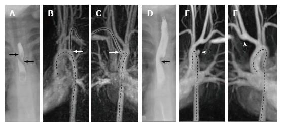
A 6-year-old girl presented with recurrent and progressive dysphagia over 4 wk. At first contact, she had lost 8 kg over 4 wk, showed severe difficulty swallowing solid foods and also suffered from recurrent thoracic pain. She underwent a barium swallow for differential diagnosis purposes which demonstrated a severe compression of the esophagus in its intermediate third, highly suspicious of vascular compression (Figure 1A). Subsequent echocardiography led to the presumptive diagnosis of an aberrant right subclavian artery with its origin in the descending aorta and a retro-esophageal course to the right side. The authors also diagnosed a truncus bicaroticus. Subsequent MRA confirmed the diagnosis of an isolated arteria lusoria (Figure 1B and C), which was considered as the proven cause for the girl’s dysphagia.
Because her dysphagia had worsened so rapidly (she could only swallow liquids), her discomfort and significant weight loss, the decision for an operative intervention was made. Via right-sided thoracotomy over the 4th intercostal space (ICS), the aberrant right subclavian artery was mobilized, transected behind the esophagus and transposed to the right common carotid by an end-to-side-anastomosis. There were no perioperative complications. Upon her discharge on day 12 after surgery, the patient was free of symptoms such as dysphagia or thoracic pain.
During follow-up a few weeks later, she returned suffering again from dysphagia, hypersalivation, dry cough and sore throat. Analgesics had not relieved her symptoms.
As the girl kept presenting with recurrent thoracic pain and mild symptoms of dysphagia accompanied by intermittent symptom-free periods and the lack of significant findings in clinical and diagnostic examinations, a somatoformic disorder was suspected as the origin of her symptoms.
Sixteen months after corrective surgery and following a symptom-free 7-mo interval, the patient presented again with severe dysphagia (no solid food) but no thoracic pain as described before. Another barium swallow was performed (Figure 1D) which showed persistent compression of the esophagus on the level where the arteria lusoria had originated. Subsequent MRA displayed the vascular stump with a maximum length of 10 mm and diameter of 4-5 mm (Figure 1E). There were no further changes compared to the images taken 12 mo earlier. The region of the vascular anastomosis showed no abnormalities (Figure 1F). Finally, the suspicion of two factors causing the ongoing compression of the esophagus and the recurrent symptoms arose: First, compression by the persisting stump of the aberrant right subclavian artery and second, an incomplete vascular ring due to a persisting ligamentum arteriosum, which was not transected.
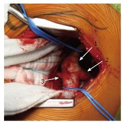
Therefore, redo-surgery was performed, this time over the 3th ICS via left-sided thoracotomy (Figure 2) and consisted of shortening the remaining lusorians’ stump and transecting the ligamentum arteriosum. The patient experienced an uneventful and complete recovery.
The barium swallow she underwent 6 wk after re-operation demonstrated no more signs of esophageal compression. At all subsequent clinical follow-ups over 6 mo after surgery, she had no more dysphagia and had regained weight.
We report on a 6-year-old girl presenting with dysphagia attributed to an aberrant right subclavian artery that unexpectedly caused persisting symptoms after corrective surgery via right-sided thoracotomy.
As the aberrant right subclavian artery often remains asymptomatic[3], surgical repair is restricted to highly-symptomatic patients, and it is usually very successful. Nevertheless, a small percentage of these patients return complaining of recurrent respiratory or swallowing problems[8].
There is a potential anatomical explanation for ongoing postoperative symptoms like dysphagia: Compared to a left-sided aortic arch, the right-sided aortic arch combined with an aberrant left subclavian artery maintains a persisting ligamentum arteriosum. In contrast, in those with a left-sided aortic arch and an aberrant right subclavian artery, a left-sided ligamentum arteriosum is much rarer but it remains a potential anatomical finding. Such a ligamentum arteriosum - a fibrous relict of the ductus arteriosus - leads from the proximal descending aorta to the left pulmonal artery. In the presence of a coexisting aberrant right subclavian artery, an incomplete vascular ring can form that compresses the esophagus[9].
We thus maintain that, to ensure optimum recovery after a surgical intervention for arteria lusoria, it is essential to be fully aware of the patient’s cardiothoracic anatomy beforehand, especially the existence of a persisting ligamentum arteriosum. In selecting the diagnostic tools, we suggest an age-dependent approach. In fetuses, newborns and infants presenting the incidental finding of an arteria lusoria, echocardiography has great potential to validate the cardiovascular system in detail, especially a vascular ring with a fibrous ductus arteriosus. Echocardiography remains highly informative even in symptomatic infants and children. Barium swallow and MRA are additional key diagnostic tools in this age group. In case of older children and adolescents, the first-line modality should be MRA. At that age, echocardiography becomes secondary because it precludes a thorough evaluation of the patient’s anatomy.
Another key factor for surgical success is the choice of surgical access. Regarding the preferred approach to the aberrant right subclavian artery in children, van Son et al[10] found that this vessel originates from the posteromedial side of the distal aortic arch. Therefore, a strong argument for currently-mandated right-sided thoracotomy in children is the vessel’s optimal mobilization, transection and reconnection.
However, assessing a ligamentum arteriosum is limited in this access path, thus in case of a left aortic arch with left-sided ligamentum arteriosum, the recommended surgical access to reach this usually fibrous strand is via left-sided thoracotomy.
In summary, we suggest considering a median thoracotomy to address both contrary structural conditions and to effectively treat a right arteria lusoria in combination with a left ligamentum arteriosum at the same time. Via this median access, the course of the aberrant right subclavian artery can be corrected, and the surgeon is able to explore and transect a persisting ligamentum arteriosum.
In conclusion, we suggest that our patient continued to suffer dysphagia after initial surgery of the aberrant right subclavian artery due to the persisting pathology of a ligamentum arteriosum causing further esophageal compression. Since this experience, our recommendation for other patients with a left-sided aortic arch and right arteria lusoria is first to focus on obtaining a detailed preoperative visualization of the individual’s anatomy by means of diagnostic imaging, especially to watch out for a ligamentum arteriosum. In case of a potential ligamentum arteriosum, we favor a median thoracotomy because of its optimal provision of intraoperative anatomical overview and accessibility to both the aberrant artery and ligamentum arteriosum.
We are grateful to Carole Cürten for language editing.
This is a rare pediatric case about a 6-year-old girl with an aberrant right subclavian artery unexpectedly presenting with persisting severe dysphagia after initial corrective surgery via right-sided thoracotomy.
Apart from mild symptoms such as hypersalivation and a dry cough, there were no other significant anomalies in clinical examination.
Ingested foreign bodies, esophageal infection or neoplasia, disorders in esophageal innervations or secondary to cardiovascular compression or thyroid disease.
The authors’ laboratory tests revealed no pathology.
The initial diagnosis of an aberrant right subclavian artery was confirmed via barium swallow, subsequent echocardiography and magnetic resonance angiography (MRA), whereas during the persistent dysphagia after her first intervention, only the barium swallow demonstrated a dorsal esophageal compression that led us to suspect an incomplete vascular ring due to a persisting ligamentum arteriosum.
Persisting esophageal compression after arteria lusoria dextra surgery caused by an incomplete vascular ring due to a persisting ligamentum arteriosum.
Redo-surgery via left-sided thoracotomy entailing the transection of a persisting ligamentum arteriosum and shortening of the remaining lusorian arteries’ stump.
Although arteria lusoria is the most common embryologic abnormality of the aortic arch and its potential esophageal compression can result in dysphagia, a case of persisting symptoms after corrective surgery because of a co-existing ligamentum arteriosum in patients with left aortic arch has never been reported in the literature so far.
Magnetic resonance angiography (MRA) is a type of magnetic resonance imaging scan to evaluate blood vessels and helps to identify vascular abnormalities by using a powerful magnetic field and pulses of radio wave energy.
Based on this experience, the authors wish to emphasize the potential co-existence of a compressing ligamentum arteriosum even in patients with left aortic arch; furthermore, the authors would like to inspire discussion about an age-dependent approach regarding which diagnostic tools are employed for pre-operative planning, as well as what constitutes the optimal surgical approach.
This is a rare case report about a pediatric case of dysphagia attributed to an aberrant right subclavian artery that unexpectedly caused persisting symptoms after corrective surgery via right-sided thoracotomy. The authors suggest considering a median thoracotomy to address both contrary structural conditions and to effectively treat a right arteria lusoria in combination with a left ligamentum arteriosum at the same time. This manuscript is nicely structured and well written.
Manuscript source: Invited manuscript
Specialty type: Cardiac and cardiovascular systems
Country of origin: Germany
Peer-review report classification
Grade A (Excellent): A
Grade B (Very good): B, B
Grade C (Good): C
Grade D (Fair): 0
Grade E (Poor): 0
P- Reviewer: Lin SL, Said SAM, Tan XR, Ueda H S- Editor: Ji FF L- Editor: A E- Editor: Lu YJ
| 1. | Levitt B, Richter JE. Dysphagia lusoria: a comprehensive review. Dis Esophagus. 2007;20:455-460. [RCA] [PubMed] [DOI] [Full Text] [Cited by in Crossref: 94] [Cited by in RCA: 103] [Article Influence: 5.7] [Reference Citation Analysis (0)] |
| 2. | Asherson N. David Bayford. His syndrome and sign of dysphagia lusoria. Ann R Coll Surg Engl. 1979;61:63-67. [PubMed] |
| 3. | Taylor M, Harris KA, Casson AG, DeRose G, Jamieson WG. Dysphagia lusoria: extrathoracic surgical management. Can J Surg. 1996;39:48-52. [PubMed] |
| 4. | Molz G, Burri B. Aberrant subclavian artery (arteria lusoria): sex differences in the prevalence of various forms of the malformation. Evaluation of 1378 observations. Virchows Arch A Pathol Anat Histol. 1978;380:303-315. [RCA] [PubMed] [DOI] [Full Text] [Cited by in Crossref: 82] [Cited by in RCA: 88] [Article Influence: 1.9] [Reference Citation Analysis (0)] |
| 5. | Janssen M, Baggen MG, Veen HF, Smout AJ, Bekkers JA, Jonkman JG, Ouwendijk RJ. Dysphagia lusoria: clinical aspects, manometric findings, diagnosis, and therapy. Am J Gastroenterol. 2000;95:1411-1416. [RCA] [PubMed] [DOI] [Full Text] [Cited by in RCA: 1] [Reference Citation Analysis (0)] |
| 6. | Ruzmetov M, Vijay P, Rodefeld MD, Turrentine MW, Brown JW. Follow-up of surgical correction of aortic arch anomalies causing tracheoesophageal compression: a 38-year single institution experience. J Pediatr Surg. 2009;44:1328-1332. [RCA] [PubMed] [DOI] [Full Text] [Cited by in Crossref: 79] [Cited by in RCA: 70] [Article Influence: 4.4] [Reference Citation Analysis (0)] |
| 7. | Atay Y, Engin C, Posacioglu H, Ozyurek R, Ozcan C, Yagdi T, Ayik F, Alayunt EA. Surgical approaches to the aberrant right subclavian artery. Tex Heart Inst J. 2006;33:477-481. [PubMed] |
| 8. | Backer CL, Mongé MC, Russell HM, Popescu AR, Rastatter JC, Costello JM. Reoperation after vascular ring repair. Semin Thorac Cardiovasc Surg Pediatr Card Surg Annu. 2014;17:48-55. [RCA] [PubMed] [DOI] [Full Text] [Cited by in Crossref: 35] [Cited by in RCA: 43] [Article Influence: 3.9] [Reference Citation Analysis (0)] |
| 9. | Allen HD, Driscoll DJ, Shaddy RE, Feltes TF. Moss and Adams’ Heart Disease in Infants, Children, and Adolescents: Including the Fetus and Young Adult. Philadelphia, USA: Lippincott Williams & Wilkins 2013; . |
| 10. | Van Son JA, Vincent JG, ten Cate LN, Lacquet LK. Anatomic support of surgical approach of anomalous right subclavian artery through a right thoracotomy. J Thorac Cardiovasc Surg. 1990;99:1115-1116. [PubMed] |


