Published online Aug 26, 2015. doi: 10.4330/wjc.v7.i8.499
Peer-review started: December 10, 2014
First decision: January 8, 2015
Revised: May 26, 2015
Accepted: June 9, 2015
Article in press: June 11, 2015
Published online: August 26, 2015
Processing time: 260 Days and 14.5 Hours
A 69-year-old man was admitted to our center to undergo catheter ablation of paroxysmal atrial fibrillation refractory to antiarrhythmic drug therapy. This procedure required access to the left atrium through the interatrial septum. During hospitalization, the patient performed routinely pre-procedure transthoracic echocardiography and gadolinium-enhanced cardiac magnetic resonance showing a normal anatomy of both the fossa ovalis and the interatrial septum. Access to the left atrium proved difficult and several unsuccessful attempts to perform the trans-septal puncture were made under both fluoroscopy and intracardiac echocardiography guidance, even with radiofrequency energy delivery. Finally, trans-septal puncture was successfully carried out using a novel nitinol J-shaped “SafeSept” trans-septal guidewire, designed to cross the interatrial septum through the trans-septal needle thanks to a special sharp tip. Moreover, thanks to its rounded J shape that reduces the risk of atrial perforation, the “SafeSept” guidewire, when advanced into the left atrium, becomes atraumatic.
Core tip: In recent years, the number of percutaneous therapeutic techniques requiring trans-septal catheterization has increased. We present the case of a 69-year-old man with a ten-year history of paroxysmal atrial fibrillation. Access to the left atrium proved difficult and several unsuccessful attempts to perform the trans-septal puncture were made under both fluoroscopy and intracardiac echocardiography guidance, even with radiofrequency energy delivery. Finally, trans-septal puncture was successfully performed using a novel nitinol “SafeSept” trans-septal guidewire. If the interatrial septum is thickened, scarred, fibrous, too mobile and/or aneurismal, the use of the “SafeSept” guidewire may be a safe and effective option.
- Citation: Zucchetti M, Casella M, Russo AD, Fassini G, Carbucicchio C, Russo E, Marino V, Catto V, Tondo C. Difficult case of a trans-septal puncture: Use of a “SafeSept” guidewire. World J Cardiol 2015; 7(8): 499-503
- URL: https://www.wjgnet.com/1949-8462/full/v7/i8/499.htm
- DOI: https://dx.doi.org/10.4330/wjc.v7.i8.499
Atrial fibrillation (AF) catheter ablation is a common therapeutic approach. The access to the left atrium, required to perform the procedure, is usually achieved through the interatrial septum. Trans-septal catheterization during catheter ablation results in high success rates with low complication incidence. Failure of the trans-septal approach is often related to unfavorable anatomical features of both the interatrial septum and the fossa ovalis[1,2]. In particular cases, trans-septal puncture can prove difficult even with the support of transesophageal or intracardiac echocardiographic imaging[3,4], or using radiofrequency energy to facilitate trans-septal puncturing[5,6]. In these cases, the use of the “SafeSept” trans-septal guidewire can be a valid alternative for achieving catheterization across the interatrial septum[7].
We present the case of a 69-year-old man with a ten-year history of paroxysmal AF. One year earlier, he had undergone catheter cryoablation of a common typical atrial flutter. After the procedure, several recurrences of paroxysmal AF refractory to antiarrhythmic drug therapy were recorded. The patient was then referred to our hospital for pulmonary vein disconnection by radiofrequency ablation. The patient had never undergone a previous procedure requiring trans-septal approach or heart surgery and did not have congenital heart defects.
During hospitalization, the patient underwent a baseline electrocardiogram that showed normal sinus rhythm and an echocardiogram that demonstrated a large left atrium (Ø 54 mm). The membrane of fossa ovalis was confirmed to be intact by gadolinium-enhanced cardiac magnetic resonance imaging, performed before ablation to assess left atrium and pulmonary vein anatomy and merge morphological and electroanatomic information during AF ablation.
At first a percutaneous trans-septal puncture was attempted. A decapolar catheter was inserted via femoral venous approach guided by fluoroscopy into the coronary sinus. A trans-septal needle (BRK, St Jude Medical Inc.) was advanced through a long sheath (SLO, 8 F, St. Jude Medical Inc., St. Paul, MN, United States) against the septum. By a percutaneous contralateral femoral venous approach, an ultrasound catheter (AcuNav, Siemens Healthcare, Mountain View, CA, United States) was advanced for intracardiac echocardiography monitoring. This technique was used to guide the needle to the correct position against the fossa ovalis. Normal interatrial septum anatomy was observed during the positioning.
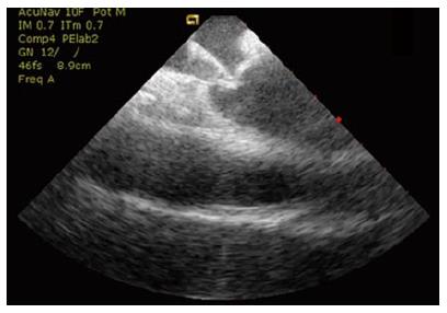
Several unsuccessful attempts to obtain a trans-septal puncture were performed by two expert electrophysiologists despite changing site of puncture, needle orientation, needle types and different curved sheaths. During needle puncture, a strong resistance against the septum was encountered; both fluoroscopy and intracardiac echocardiography showed tenting of the fossa ovalis, but puncturing was not achieved despite the correct location of the needle (Figure 1).
An electrosurgical cautery generator was used to facilitate trans-septal catheterization. A standard cautery pen was placed upon the proximal portion of the trans-septal needle, then a radiofrequency pulse was delivered for few seconds at 45 W. Two unsuccessful attempts with this technique were performed.
The previous techniques were unsuccessful because of the anatomical features of the interatrial septum (i.e., unusual thickness of the fossa ovalis).
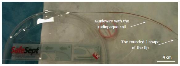
In order to get a successful fossa ovalis puncture, a special trans-septal guidewire (“SafeSept” Pressure Products, Inc., United States) was considered as an option (Figure 2).
The “SafeSept” is a nitinol trans-septal guidewire designed to easily cross the interatrial septum through the trans-septal needle thanks to a special sharp tip that allows it to penetrate the fossa ovalis without the use of a particular hard contact. Moreover, the “SafeSept” is non-traumatic when advanced into the left atrium thanks to its rounded J shape, thus reducing the perforation risk of the atrial wall. The trans-septal guidewire’s distal end can be easily visualized thanks to a radiopaque coil.
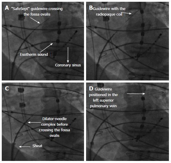
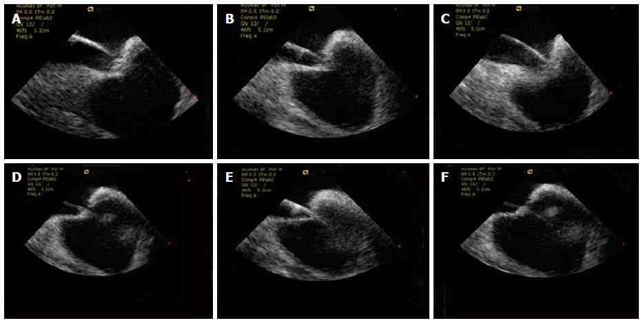
The trans-septal assembly (needle, dilator and sheath) was advanced over the guidewire. In particular, under fluoroscopic and intracardiac echo guidance, the “SafeSept” was advanced through the trans-septal needle; while tenting and maintaining constant force on the septum, the guidewire was advanced and easily crossed the interatrial septum (Figures 3 and 4). The wire (clearly visible thanks to its radiopaque coil) was further positioned in the left superior pulmonary vein.
The trans-septal needle was then advanced through the dilator and long sheath over the “SafeSept” across the fossa ovalis into the left atrium. Then the long sheath was placed in the left atrium and both trans-septal guidewire and dilator were pulled out.
Afterwards, unfractionated heparin was administered (100 U/kg) and pulmonary vein disconnection was successfully performed by radiofrequency ablation supported by the EnSite NavX electroanatomic mapping system.
In recent years the number of percutaneous therapeutic techniques requiring trans-septal catheterization has significantly increased[1,2]. The risks of complications related to perforation of the posterior atrial wall or aortic bulb remain limitations of this technique in the presence of anatomical alterations (distorted and/or thickened atrial septal tissue)[8].
Usually the trans-septal puncture is performed through fossa ovalis because it is the region offering the least resistance. This site can be located fluoroscopically and through the use of standard electrode catheters as anatomical landmarks (His region or, bulb of the aorta with a “pig-tail” catheter).
Intracardiac echocardiography was helpful to visualize the fossa ovalis in order to guide trans-septal puncture thus avoiding perforation of structures adjacent to the atrial septum or pericardial tamponade[3,4]. In the presence of unfavorable anatomy (i.e., a thickened atrial septum, extremely elastic or aneurysmal fossa ovalis, presence of fibrosis due to a previous catheterization), trans-septal puncture may be challenging even with the use of these methods.
A brief application of radiofrequency to the septum, either through a dedicated radiofrequency catheter system or the use of an electrosurgical cautery pen, could be helpful in facilitating both fluoroscopy and imaging guidance[5,6].
In extremely difficult puncturing, “SafeSept” could be a valid option to cross the interatrial septum[7,9,10]. After crossing the septum, the guidewire immediately bends into a J shape, so as to be atraumatic when advanced into the left atrium. Furthermore, a radiopaque coil is positioned on the distal end of the wire to provide a fluoroscopic visualization during every step of the procedure.
In conclusion, trans-septal catheterization may be challenging if the interatrial septum is thickened, scarred, fibrous, too mobile and/or aneurysmal. The use of fluoroscopy, intracardiac ultrasound and RF energy are helpful, but may sometimes not be enough to achieve trans-septal catheterization. In these cases, the use of the “SafeSept” trans-septal guidewire may be a safe and effective option.
The authors thank Dr. Viviana Biagioli for editorial assistance.
A 69-year-old man affected by palpitations with diagnosis of paroxysmal atrial fibrillation.
The patient had several recurrences of paroxysmal atrial fibrillation after the cryoablation of a common typical atrial flutter.
Difficult trans-septal puncture using fluoroscopy and intracardiac echocardiography, radiofrequency energy delivery and “SafeSept” trans-septal guidewire.
The patient had no alterations of hematological values.
Transthoracic echocardiography and gadolinium-enhanced cardiac magnetic resonance imaging showed a normal anatomy of the fossa ovalis and interatrial septum.
Access to the left atrium proved difficult and several unsuccessful attempts to perform the trans-septal puncture were made under both fluoroscopy and intracardiac echocardiography guidance, even with radiofrequency energy delivery.
Trans-septal puncture was successfully carried out using a nitinol J-shaped “SafeSept” trans-septal guidewire.
Very few cases of unsuccessful trans-septal puncture that require the use of nitinol J-shaped “SafeSept” trans-septal guidewire have been reported in the literature.
The novel nitinol J-shaped “SafeSept” trans-septal guidewire is designed to cross the interatrial septum through the trans-septal needle thanks to a special sharp tip but simultaneously is a non-traumatic device due to its rounded J-shape that reduces the risk of atrial wall perforation.
Trans-septal catheterization may be challenging if the interatrial septum is thickened, scarred, fibrous, too mobile and/or aneurysmal. The use of fluoroscopy, intracardiac ultrasound and radiofrequency energy are helpful, but may sometimes not be enough to achieve trans-septal catheterization. In these cases, the use of the “SafeSept” trans-septal guidewire may be a safe and effective aid.
This manuscript showed a case of paroxysmal atrial fibrillation in whom SafeSept was effective for trans-septal puncture. The case is peculiar and interesting.
P- Reviewer: De Ponti R, Santulli G, Satoh H S- Editor: Gong XM L- Editor: A E- Editor: Wu HL
| 1. | De Ponti R, Cappato R, Curnis A, Della Bella P, Padeletti L, Raviele A, Santini M, Salerno-Uriarte JA. Trans-septal catheterization in the electrophysiology laboratory: data from a multicenter survey spanning 12 years. J Am Coll Cardiol. 2006;47:1037-1042. [RCA] [PubMed] [DOI] [Full Text] [Cited by in Crossref: 136] [Cited by in RCA: 161] [Article Influence: 8.5] [Reference Citation Analysis (0)] |
| 2. | Fagundes RL, Mantica M, De Luca L, Forleo G, Pappalardo A, Avella A, Fraticelli A, Dello Russo A, Casella M, Pelargonio G. Safety of single transseptal puncture for ablation of atrial fibrillation: retrospective study from a large cohort of patients. J Cardiovasc Electrophysiol. 2007;18:1277-1281. [RCA] [PubMed] [DOI] [Full Text] [Cited by in Crossref: 54] [Cited by in RCA: 58] [Article Influence: 3.2] [Reference Citation Analysis (0)] |
| 3. | Daoud EG, Kalbfleisch SJ, Hummel JD. Intracardiac echocardiography to guide transseptal left heart catheterization for radiofrequency catheter ablation. J Cardiovasc Electrophysiol. 1999;10:358-363. [RCA] [PubMed] [DOI] [Full Text] [Cited by in Crossref: 114] [Cited by in RCA: 104] [Article Influence: 4.0] [Reference Citation Analysis (0)] |
| 4. | Dello Russo A, Casella M, Pelargonio G, Bonelli F, Santangeli P, Fassini G, Riva S, Carbucicchio C, Giraldi F, De Iuliis P. Intracardiac echocardiography in electrophysiology. Minerva Cardioangiol. 2010;58:333-342. [PubMed] |
| 5. | Casella M, Dello Russo A, Pelargonio G, Martino A, De Paulis S, Zecchi P, Bellocci F, Tondo C. Fossa ovalis radiofrequency perforation in a difficult case of conventional transseptal puncture for atrial fibrillation ablation. J Interv Card Electrophysiol. 2008;21:249-253. [RCA] [PubMed] [DOI] [Full Text] [Cited by in Crossref: 10] [Cited by in RCA: 11] [Article Influence: 0.6] [Reference Citation Analysis (0)] |
| 6. | McWilliams MJ, Tchou P. The use of a standard radiofrequency energy delivery system to facilitate transseptal puncture. J Cardiovasc Electrophysiol. 2009;20:238-240. [RCA] [PubMed] [DOI] [Full Text] [Cited by in Crossref: 30] [Cited by in RCA: 31] [Article Influence: 1.8] [Reference Citation Analysis (0)] |
| 7. | de Asmundis C, Chierchia GB, Sarkozy A, Paparella G, Roos M, Capulzini L, Burri SA, Yazaki Y, Brugada P. Novel trans-septal approach using a Safe Sept J-shaped guidewire in difficult left atrial access during atrial fibrillation ablation. Europace. 2009;11:657-659. [RCA] [PubMed] [DOI] [Full Text] [Cited by in Crossref: 13] [Cited by in RCA: 15] [Article Influence: 0.9] [Reference Citation Analysis (0)] |
| 8. | Cappato R, Calkins H, Chen SA, Davies W, Iesaka Y, Kalman J, Kim YH, Klein G, Natale A, Packer D. Prevalence and causes of fatal outcome in catheter ablation of atrial fibrillation. J Am Coll Cardiol. 2009;53:1798-1803. [RCA] [PubMed] [DOI] [Full Text] [Cited by in Crossref: 413] [Cited by in RCA: 443] [Article Influence: 27.7] [Reference Citation Analysis (0)] |
| 9. | De Ponti R, Marazzi R, Picciolo G, Salerno-Uriarte JA. Use of a novel sharp-tip, J-shaped guidewire to facilitate transseptal catheterization. Europace. 2010;12:668-673. [RCA] [PubMed] [DOI] [Full Text] [Cited by in Crossref: 21] [Cited by in RCA: 23] [Article Influence: 1.5] [Reference Citation Analysis (0)] |
| 10. | Wadehra V, Buxton AE, Antoniadis AP, McCready JW, Redpath CJ, Segal OR, Rowland E, Lowe MD, Lambiase PD, Chow AW. The use of a novel nitinol guidewire to facilitate transseptal puncture and left atrial catheterization for catheter ablation procedures. Europace. 2011;13:1401-1405. [RCA] [PubMed] [DOI] [Full Text] [Cited by in Crossref: 21] [Cited by in RCA: 25] [Article Influence: 1.8] [Reference Citation Analysis (0)] |


