Published online Jun 26, 2015. doi: 10.4330/wjc.v7.i6.357
Peer-review started: November 16, 2014
First decision: February 7, 2015
Revised: February 25, 2015
Accepted: April 1, 2015
Article in press: April 7, 2015
Published online: June 26, 2015
Processing time: 222 Days and 23.2 Hours
We are reporting a long-time magnetic resonance imaging (MRI) follow-up in a rare case of cardiac left lateral wall hypertrophy. Hypertrophic cardiomyopathy (HCM) is the most common genetic cardiovascular disorder and a significant cause of sudden cardiac death. Cardiac magnetic resonance (CMR) imaging can be a valuable tool for assessment of detailed information on size, localization, and tissue characteristics of hypertrophied myocardium. However, there is still little knowledge of long-term evolution of HCM as visualized by magnetic resonance imaging. Recently, our group reported a case of left lateral wall HCM as a rare variant of the more common forms, such as septal HCM, or apical HCM. As we now retrieved an old cardiac MRI acquired in this patient more than 20 years ago, we are able to provide the thrilling experience of an ultra-long MRI follow-up presentation in this rare case of left lateral wall hypertrophy. Furthermore, this case outlines the tremendous improvements in imaging quality within the last two decades of CMR imaging.
Core tip: Cardiac magnetic resonance imaging (MRI) can be a valuable tool for assessment of detailed information on size, localization, and tissue characteristics in cases of hypertrophic cardiomyopathy. We report the thrilling experience of an ultra-long magnetic resonance imaging follow-up presentation in a rare case of left lateral wall hypertrophy with an initial cardiac MRI of patient acquired more than 20 years ago. This case outlines the tremendous improvements in imaging quality within the last two decades of cardiac MR imaging.
- Citation: Gassenmaier T, Petritsch B, Kunz AS, Gkaniatsas S, Gaudron PD, Weidemann F, Nordbeck P, Beer M. Long term evolution of magnetic resonance imaging characteristics in a case of atypical left lateral wall hypertrophic cardiomyopathy. World J Cardiol 2015; 7(6): 357-360
- URL: https://www.wjgnet.com/1949-8462/full/v7/i6/357.htm
- DOI: https://dx.doi.org/10.4330/wjc.v7.i6.357
Hypertrophic cardiomyopathy (HCM) is the most common genetic cardiovascular disorder and a significant cause of sudden cardiac death. Cardiac magnetic resonance (CMR) imaging can be a valuable tool for assessment of detailed information on size, localization, and tissue characteristics of hypertrophied myocardium. However, there is still little knowledge of long-term evolution of HCM as visualized by magnetic resonance imaging (MRI). Recently, our group reported a case of left lateral wall HCM[1] as a rare variant of the more common forms, such as septal HCM, or apical HCM[2]. As this patient underwent initial cardiac MRI more than 20 years ago, we are able to provide the thrilling experience of an ultra-long MRI follow-up presentation in this rare case of left lateral wall hypertrophy, including the tremendous improvements in imaging quality within the last two decades of CMR imaging.
A 52-year-old male presented himself to the emergency department of our institution suffering an episode of nocturnal chest pain. As the electrocardiogram showed ventricular tachycardia, synchronized electrical cardioversion was performed, successfully terminating the arrhythmia. Afterwards, the patient was subjected to sequential clinical investigations, excluding ischemia as cause for the serious symptoms, and CMR imaging, revealing an atypical form of HCM.
CMR was performed five days after the initial arrhythmic event on a MAGNETOM® Trio 3.0 Tesla scanner (Siemens AG Sector Healthcare, Erlangen, Germany). In addition to these current investigations, we were now able to retrieve MR images from the year 1989 (1.5 Tesla Philips Gyroscan, Philips Medical System, Best, the Netherlands) which had been acquired in the pre-PACS era more than 23 years in advance of the current event. This enabled us to present a long term MRI follow-up of this rare manifestation of HCM and to point out the massive improvements in imaging quality of cardiac MRI, due to enormous technical development within the last two decades.
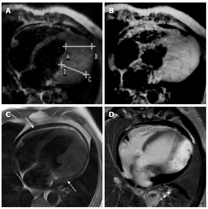
Past images from the year 1989 deliver some limited diagnostic information. Nevertheless, left ventricular wall thickening up to 45 mm was clearly seen in this patient even 23-years ago (Figure 1A). In addition, Gadolinum enhancement (GE) of the noticeable area of interest was depicted after iv contrast application (Figure 1B). In the late 1980s, late Gadolinium enhancement (LGE) had not yet been described as a non-invasive method for myocardial tissue characterization, including analysis for myocardial fibrosis.
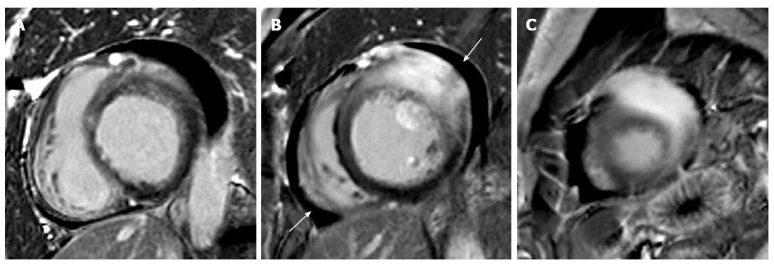
Current high-quality T1 weighted turbo-spin-echo images with dark blood technique confirmed an extensive, confined thickening of the left ventricular lateral wall up to 45 mm in the 4-chamber view (Figure 1C). Cine-SSFP sequences demonstrated a prolonged longitudinal relaxation of the lateral wall (not shown). After injection of 0.2 mmol/kg intravenous contrast agent (Gadovist®, Bayer HealthCare, Leverkusen, Germany) the myocardial mass showed homogenous contrast enhancement. LGE imaging was acquired 12 min after iv contrast administration. PSIR-SSFP images in the 4-chamber (Figure 1D) and short axis views (Figure 2A-C) revealed a homogenous enhancement, corresponding to the left ventricular lateral wall thickening, as it can be typically observed in other, more common forms of HCM.
In addition, a small accompanying circular pericardial effusion (indicated with arrows) was depicted in the present CMR images (Figure 1C and 2B).
Comparison of the studies revealed no significant change over time regarding the extent of wall thickening. It is difficult to judge whether significant change in myocardial fibrosis can be detected by CMR, as LGE techniques had not been described prior to 2001 and the baseline CMR was performed prior to that in 1989[3] (Figure 1, A/B vs C/D). In 1989, the contrast enhanced images were acquired about 3 min after Gd administration, a “late-enhancement stop” and the so-called “nulling” technique were not available/invented. However, as far as one can compare the images from 1989 to 2012, no significant change in fibrotic myocardium occurred during this time span. The diagnosis of myocardial fibrosis was already proven in 1989 by myocardial biopsy, showing massive hypertrophy but no signs of malignancy.
Therefore, this intensive myocardial thickening described above has to be considered a highly uncommon manifestation of HCM limited solely to the left ventricular lateral wall.
Finally, the patient could be discharged in good general condition after the implantation of an ICD for prevention of further episodes of ventricular tachycardia.
In a patient with atypical left wall cardiac hypertrophy, CMR was able to provide detailed information on size, localization, and tissue characteristics of the myocardial mass and allowed non-invasive long time assessment of these parameters. Even though limited to the left ventricular lateral wall, our variant of HCM showed some typical characteristics. For instance, LGE is frequently observed in HCM patients and reflects the presence of fibrosis within the myocardium[4,5]. However, the clinical significance of change of LGE over time in HCM is still under debate[6]. Nonetheless, the presence of LGE is an independent risk factor for adverse outcome in HCM and associated with an increased frequency of ventricular tachyarrhythmia, as was the case in our patient[7,8].
However, initial inspection of the LGE images with the atypical site of LGE and the considerable degree of myocardial hypertrophy raised concerns whether the patient might suffer from a malignant tumor of the myocardium. Pericardial effusion fortified this impression. However, only a short period after the current CMR, reports from the old, previous CMR from 1989 were retrieved. Finally, comparison of the images from 1989 to those of 2012 confirmed that the patient suffered from HCM and excluded a malignant tumor as a potential differential diagnosis.
For future studies, T1 maps might be helpful to differentiate between a cardiac entity (HCM) and non-cardiac entity (tumor). However, this was not performed in the current case, as the utilized MR scanner was not equipped with software applicable for T1 mapping.
A 52-year-old male with an episode of nightly chest pain.
Electrocardiogram showed ventricular tachycardia.
Ischemia, cardiac tumor, atypical hypertrophic cardiomyopathy (HCM).
Tests for exclusion of cardiac ischemia were within normal limits.
Cardiac magnetic resonance imaging showed an extensive, confined thickening of the left ventricular lateral wall up to 45 mm. Comparison with a previous study from 1989 revealed that this left ventricular wall thickening was clearly seen in this patient already 23-years ago.
Myocardial biopsy had shown massive hypertrophy but no signs of malignancy in 1989.
The patient underwent implantation of an ICD for prevention of further episodes of ventricular tachycardia.
Isolated left lateral wall hypertrophic cardiomyopathy is a rare variant of the more common forms, such as septal HCM, or apical HCM.
Late gadolinium enhancement is frequently observed in HCM patients and reflects the presence of fibrosis within the myocardium.
This case report outlines the tremendous improvements in imaging quality within the last two decades of cardiac MR imaging.
This article reported a long-time MRI follow-up in a rare case of cardiac left lateral wall hypertrophy. Cardiac magnetic resonance imaging is a valuable tool for assessment characteristics of hypertrophic cardiomyopathy. And this report provided detailed information on location, size and imaging characteristics of hypertrophic cardiomyopathy.
P- Reviewer: Kobza R, Yuan Z S- Editor: Ji FF L- Editor: A E- Editor: Zhang DN
| 1. | Gkaniatsas S, Gaudron PD, Gassenmaier T, Beer M, Weidemann F, Nordbeck P. Atypical hypertrophic cardiomyopathy of the left lateral wall leading to ventricular tachycardia. Eur Heart J. 2014;35:548. |
| 2. | Fattori R, Biagini E, Lorenzini M, Buttazzi K, Lovato L, Rapezzi C. Significance of magnetic resonance imaging in apical hypertrophic cardiomyopathy. Am J Cardiol. 2010;105:1592-1596. |
| 3. | Simonetti OP, Kim RJ, Fieno DS, Hillenbrand HB, Wu E, Bundy JM, Finn JP, Judd RM. An improved MR imaging technique for the visualization of myocardial infarction. Radiology. 2001;218:215-223. |
| 4. | Ho CY, López B, Coelho-Filho OR, Lakdawala NK, Cirino AL, Jarolim P, Kwong R, González A, Colan SD, Seidman JG. Myocardial fibrosis as an early manifestation of hypertrophic cardiomyopathy. N Engl J Med. 2010;363:552-563. |
| 5. | Noureldin RA, Liu S, Nacif MS, Judge DP, Halushka MK, Abraham TP, Ho C, Bluemke DA. The diagnosis of hypertrophic cardiomyopathy by cardiovascular magnetic resonance. J Cardiovasc Magn Reson. 2012;14:17. |
| 6. | Todiere G, Aquaro GD, Piaggi P, Formisano F, Barison A, Masci PG, Strata E, Bacigalupo L, Marzilli M, Pingitore A. Progression of myocardial fibrosis assessed with cardiac magnetic resonance in hypertrophic cardiomyopathy. J Am Coll Cardiol. 2012;60:922-929. |
| 7. | Adabag AS, Maron BJ, Appelbaum E, Harrigan CJ, Buros JL, Gibson CM, Lesser JR, Hanna CA, Udelson JE, Manning WJ. Occurrence and frequency of arrhythmias in hypertrophic cardiomyopathy in relation to delayed enhancement on cardiovascular magnetic resonance. J Am Coll Cardiol. 2008;51:1369-1374. |
| 8. | O’Hanlon R, Grasso A, Roughton M, Moon JC, Clark S, Wage R, Webb J, Kulkarni M, Dawson D, Sulaibeekh L. Prognostic significance of myocardial fibrosis in hypertrophic cardiomyopathy. J Am Coll Cardiol. 2010;56:867-874. |


