Copyright
©The Author(s) 2015.
World J Cardiol. Oct 26, 2015; 7(10): 703-706
Published online Oct 26, 2015. doi: 10.4330/wjc.v7.i10.703
Published online Oct 26, 2015. doi: 10.4330/wjc.v7.i10.703
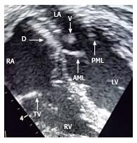
Figure 1 Two dimensional echocardiography image showing a large mobile vegetation (V) attached to the lower surface of an Amplatzer device (D) near the mitral valve.
AML: Anterior mitral leaflet; PML: Posterior mitral leaflet; LV: Left ventricle; RV: Right ventricle; LA: Left atrium; TV: Tricuspid valve; RA: Right atrium.
- Citation: Jha NK, Kiraly L, Murala JS, Tamas C, Talo H, Badaoui HE, Tofeig M, Mendonca M, Sajwani S, Thomas MA, Doory SAA, Khan MD. Late endocarditis of Amplatzer atrial septal occluder device in a child. World J Cardiol 2015; 7(10): 703-706
- URL: https://www.wjgnet.com/1949-8462/full/v7/i10/703.htm
- DOI: https://dx.doi.org/10.4330/wjc.v7.i10.703


