Published online Oct 26, 2015. doi: 10.4330/wjc.v7.i10.700
Peer-review started: February 22, 2015
First decision: April 10, 2015
Revised: June 7, 2015
Accepted: July 16, 2015
Article in press: July 17, 2015
Published online: October 26, 2015
Processing time: 255 Days and 14.5 Hours
Intra-hisian atrioventricular (AV) block is not a common phenomenon, but it is important for the development of advanced or complete AV block. We observed a 77-year-old female patient with the 2:1 AV block due to an intra-hisian block. In this case we tried to detect the block site, but an alternating pattern of the AH conduction was noted on the His-electrogram in the electrophysiological study (EPS). The cause of the confusing finding might have been the instability of the catheter to record a His potential. We could detect a splitting of the His-electrogram with an intra-hisian block after minimal manipulation of the catheter. The authors’ observations suggest that catheter stability is important for a precise recording in the EPS and radiofrequency catheter ablation procedure.
Core tip: Intra-hisian atrioventricular (AV) block associated with 2:1 AV block is an uncommon phenomenon, but it is important for the development of complete AV block. We observed a 77-year-old female with 2:1 AV block due to an intra-hisian block. An alternating pattern of the AH conduction was noted on the His-electrogram. The cause of that confusing finding might have been the instability of the catheter for recording the His potential. We could detect a splitting of the His-electrogram with intra-hisian block after minimal manipulation of the catheter. The authors’ observations suggest that catheter stability is important for a precise recording.
- Citation: Hong SP, Park YW, Lee YS. Intra-His bundle block in 2:1 atrioventricular block. World J Cardiol 2015; 7(10): 700-702
- URL: https://www.wjgnet.com/1949-8462/full/v7/i10/700.htm
- DOI: https://dx.doi.org/10.4330/wjc.v7.i10.700
Catheter stability is very important for achieving a precise electrophysiological study (EPS); 2:1 atrioventricular (AV) block is usually a disease of various levels such as supra-, intra- and infra-hisian[1,2]. We report a case with some confusion caused by the instability of the catheter used for the His-electrogram recording during 2:1 AV block due to an intra-hisian block.
A 77-year-old female complained of dizziness. Her past history had hypertension, and had been taking dihydrophyridine calcium channel blockers for 10 years. The chest radiograph showed cardiomegaly. The echocardiography revealed a normal function and wall motion. A resting electrocardiogram (ECG) revealed 2:1 AV block and right bundle branch block. After an intravenous injection of atropine and an exercise treadmill test, the ECG exhibited persistent 2:1 AV block.
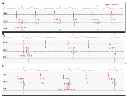
The patient underwent an EPS to determine the position of the conduction block. EPS catheters located in right ventricle, His bundle and right atrium. Firstly, we found an infra-hisian block with a normal A-H interval (70 ms) and long H-V interval (70 ms) on the intracardiac ECG (Figure 1A), and then a supra-hisian block with longer A-H interval (100 ms) and shorter H-V interval (40 ms) (Figure 1B). After a while, another intracardiac ECG revealed a splitting of the His potential (H-H’) in the A-V conduction and only the proximal activation of the His potential during A-V block, which meant an intra-His bundle block (Figure 1C).
The patient was performed the implantation of permanent pacemaker as a result of the intra-His bundle block. The patient has no other symptoms until now.
In our case, we tried to detect the block site but an alternating pattern of the AH conduction was noted in the His-electrogram during the EPS. The cause of this confusing finding might have been due to the instability of the catheter to record a His potential. However, the His potential was distinct in the first recoding of the His bundle (Figure 1A). In addition, the His potential spontaneously moved and the AH interval prolonged during the AV conduction, which concluded that the catheter was not stable.
A consistent intracardiac electrogram reflects a stable catheter position. Among the several catheters used during the EPS, the catheter used for the His-electrogram recoding is the most unstable in a beating heart. Catheter stability is important for obtaining a precise recording during the EPS and radiofrequency catheter ablation procedure. Instability of catheters can contribute to a misdiagnosis and procedural complications especially with AV nodal reentrant tachycardia. Recently, a remote magnetic navigation system is able to provide better catheter stability during the radiofrequency catheter ablation procedure[3]. However, that system cannot be used during diagnostic procedures such as in our study because of the high cost. Fortunately, we could detect the splitting of the His-electrogram with an intra-hisian block after a minimal manipulation of the catheter.
Intra-hisian AV block during 2:1 AV block is not a common phenomenon, but it tends to develop into advanced or complete AV block[4]. We should try to obtain a precise His electrogram and find the exact block site when conducting an EPS for 2nd degree high grade AV block.
A 77-year-old female complained of dizziness.
Intra-hisian atrioventricular (AV) block detected a splitting of the His-electrogram after a correction of the catheter instability during the electrophysiological study (EPS).
Second-degree Mobitz 1 block with 2:1 AV is easily misdiagnosed as general two to one conductive rate regardless of block site.
The laboratory test results were unremarkable.
The intracardiac electrocardiogram revealed splitting of the His potential of the AV conduction during the EPS.
A permanent pacemaker was implanted as a result of intra-hisian AV block.
To best of our knowledge, intra-hisian AV block is an uncommon phenomenon.
Intra-hisian AV block in His-electrogram exhibited a splitting of the His potential during the AV conduction and only the proximal activation potential of the His bundle during the AV block, which meant an intra-His bundle block.
The authors should try to achieve a precise recording of the His electrogram and find the exact block site when conducting an EPS for 2nd degree high grade AV block.
It is an interesting case report.
P- Reviewer: Falconi M, Ho KM, Lazzeri C S- Editor: Tian YL L- Editor: A E- Editor: Lu YJ
| 1. | Narula OS, Samet P. Wenckebach and Mobitz type II A-V block due to block within the His bundle and bundle branches. Circulation. 1970;41:947-965. [RCA] [PubMed] [DOI] [Full Text] [Cited by in Crossref: 145] [Cited by in RCA: 160] [Article Influence: 2.9] [Reference Citation Analysis (0)] |
| 2. | Lee YS, Kim SY, Kim KS, Kim YN. Intra-His bundle block in second-degree Mobitz I atrioventricular block with right bundle branch block. Europace. 2009;11:1251-1252. [RCA] [PubMed] [DOI] [Full Text] [Cited by in RCA: 1] [Reference Citation Analysis (0)] |
| 3. | Armacost MP, Adair J, Munger T, Viswanathan RR, Creighton FM, Curd DT, Sehra R. Accurate and reproducible target navigation with the stereotaxis niobe® magnetic navigation system. J Cardiovasc Electr. 2007;18:S26-S31. [RCA] [DOI] [Full Text] [Cited by in Crossref: 27] [Cited by in RCA: 28] [Article Influence: 1.5] [Reference Citation Analysis (0)] |
| 4. | Ishikawa T, Sumita S, Kikuchi M, Nakagawa T, Ogawa H, Hanada K, Kobayashi I, Kosuge M, Shigemasa T, Endo T. Long term follow-up in patients with intra-hisian atrioventricular block. J Artif Organs. 2000;3:149-153. [RCA] [DOI] [Full Text] [Cited by in RCA: 1] [Reference Citation Analysis (0)] |


