Published online Oct 26, 2014. doi: 10.4330/wjc.v6.i10.1127
Revised: April 23, 2014
Accepted: July 25, 2014
Published online: October 26, 2014
Processing time: 224 Days and 13.6 Hours
We report the case of a 63-year-old woman affected by a severe form of systemic scleroderma with pulmonary involvement (interstitial fibrosis diagnosed by biopsy and moderate pulmonary hypertension) and cardiac involvement (paroxysmal atrial fibrillation, right atrial flutter treated by catheter ablation, ventricular tachyarrhythmias, previous dual chamber implantable cardioverter defibrillator implant). Because of recurrent electrical storms refractory to iv antiarrhythmic drugs the patient was referred to our institution to undergo catheter ablation. During electrophysiological procedure a 3D shell of cardiac anatomy was created with intracardiac echocardiography pointing out a significant right ventricular dilatation with a complex aneurysmal lesion characterized by thin walls and irregular multiple trabeculae. A substrate-guided strategy of catheter ablation was accomplished leading to a complete electrical isolation of the aneurism and to the abolishment of all abnormal electrical activities. The use of advanced strategies of imaging together with electroanatomical mapping added important information to the complex arrhythmogenic substrate and improved efficacy and safety.
Core tip: We report the case of a 63-year-old woman affected by a severe form of systemic scleroderma with cardiac involvement. Because of recurrent electrical storms the patient underwent catheter ablation. Intracardiac echocardiography pointed out a significant right ventricular dilatation with a complex aneurysmal lesion characterized by thin walls and irregular multiple trabeculae. A substrate-guided strategy of catheter ablation was accomplished leading to a complete electrical isolation of the aneurism. The use of advanced strategies of imaging together with electroanatomical mapping added important information to the complex arrhythmogenic substrate and improved efficacy and safety.
- Citation: Casella M, Carbucicchio C, Russo E, Pizzamiglio F, Golia P, Conti S, Costa F, Russo AD, Tondo C. Electrical storm in systemic sclerosis: Inside the electroanatomic substrate. World J Cardiol 2014; 6(10): 1127-1130
- URL: https://www.wjgnet.com/1949-8462/full/v6/i10/1127.htm
- DOI: https://dx.doi.org/10.4330/wjc.v6.i10.1127
Systemic sclerosis (SS) is a rare systemic infiltrative disorder characterized by a widespread damage to small blood vessels and connective tissue fibrosis resulting in multi-organ involvement. Ventricular tachyarrhythmias (VTs) are frequent clinical manifestation of SS-associated cardiovascular damage and a possible cause of sudden death. Antiarrhythmic therapy is usually limited by concomitant therapy or side effects. In many cases the use of an implantable cardioverter defibrillator (ICD) is mandatory. In this setting catheter ablation (CA) has been proposed as an alternative option but no evidence exists about the characteristics of the arrhythmogenic substrate.
A 63-year-old woman affected by a severe form of SS was referred to our institution for management of her VTs. She presented both an advanced pulmonary and cardiac involvement. Since June 2006 she has been suffering from multiple episodes of sustained VT; pharmacological therapy by beta blockers, sotalol or amiodarone was ineffective and limited by side effects (Raynaud’s phenomenon and pulmonary interstitial fibrosis) thus the patient underwent a dual chamber ICD implant; on September 2013 the patient developed recurrent electrical storms refractory to i.v. antiarrhythmic drugs.
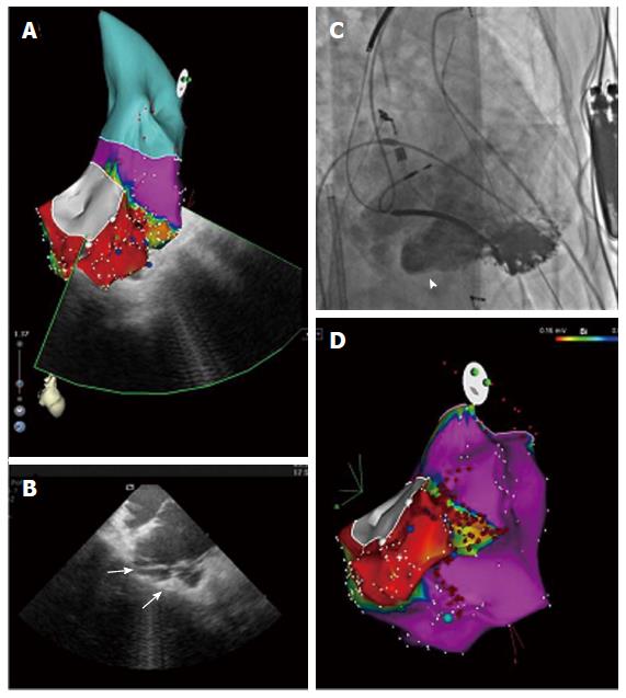
On the second day after admission, CA was performed. First, a 3D shell of cardiac anatomy was created on an electroanatomic mapping system integrated with intracardiac echocardiography (ICE) (Cartosound, Biosense-Webster, United States)[1]. ICE allowed to detect a significant right ventricular (RV) dilatation with a complex lesion characterized by thin walls and irregular multiple trabeculae, expanding from the inferior to the lateral RV wall in perivalvular basal segments (Figure 1A and B) this aneurysmal dilatation was easily identified angiographically, too (Figure 1C, Figure 2).
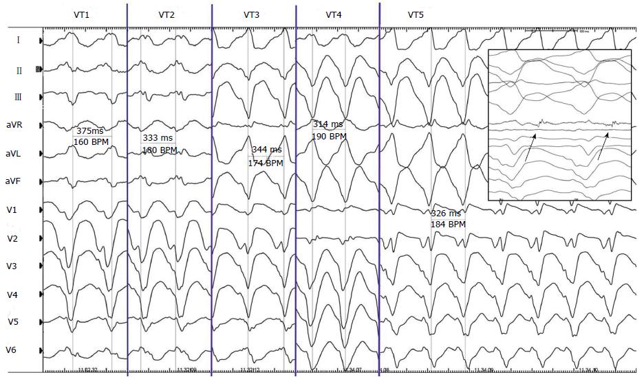
A complete electroanatomical map (EAM) of the aneurysm was obtained by ICE, showing an area of dense scar surrounded by near-scar tissue with abnormal electrical activities (AEAs) all along the borders of the aneurism. Five VT morphologies spontaneously occurred (Figure 3); only 2 of them were effectively mapped creating an activation map and identifying a mid-diastolic potential, both were terminated by radiofrequency (RF) delivery at the critical isthmus located at the border of the aneurysm. Then a substrate-guided CA was accomplished targeting all AEAs by sequential RF energy pulses (30 up to 40 Watts, SF Thermocool, Biosense-Webster, United States), to achieve the complete electrical isolation of the aneurysm. At the end of the procedure, a complete protocol of programmed electrical stimulation (drive 600 and 400 ms, up to three extrastimuli) from the RV apex was negative. No VT recurrence was observed in a 6 mo follow up period on amiodarone (1 g/wk) therapy.
Tachyarrhythmias appear as frequent clinical manifestations of SS-associated cardiovascular damage. Arrhythmias occurrence may be associated with poor outcome and represent 6% of the overall causes of death in the large European League Against Rheumatism Scleroderma Trials and up to 12% in Research (EUSTAR) database[2].
Since different classes of anti-arrhythmics are available and SS patients may have multiple organs involved and take concomitant drugs, the choice of treatment must be personalized to the patient. ICDs have been used effectively in selected patients to prevent sudden cardiac death. There is no specific recommendation in VT treatment in SS patients, and the use of CA has been reported anecdotally[3,4]. We present the first case of SS patient with multiple VT morphologies undergoing successful CA guided by an integrated approach aiming at the electro-anatomical characterization of RV cardiomyopathy. Based on our experience CA should be considered as an adjunctive treatment in patients with sustained, monomorphic VTs refractory to pharmacological therapy. Our experience adds new pieces on the knowledge of the arrhythmic substrate in SS. First of all, CA approach requires an accurate imaging of the RV acquired by both angiography and real time echo (ICE, as in our case, or transoesophageal) to identify the area of interest and to reduce potential risk as RF delivery in very thin tissue[5]. Secondary, a combined high density mapping of the whole scar and peri-scar area allows a substrate-guided abolition of all AEAs leading to a successful procedure.
Malignant VTs may be expression of an advanced form of RV disease in patients with SS and CA may be proposed as a therapeutic option for VT treatment. The combination of advanced strategies of imaging together with EAM should be preferred due to the complex arrythmogenic substrate to improve efficacy and safety.
The authors thank Dr. Viviana Biagioli for editorial assistance.
A 63-year-old woman affected by a severe form of systemic sclerosis (SS) with previous dual chamber implantable cardioverter defibrillator implant presented with drug-refractory multiple forms of ventricular tachycardias.
Identification of aneurismal lesion of the right ventricular (RV) with the critical isthmus from which the five morphologies of ventricular tachyarrhythmias (VTs) arise.
Arrhythmogenic right ventricular dysplasia, myocarditis.
Positive Antinuclear Antibodies; metabolic panel and liver function test were within normal limits.
Intracardiac echocardiography allowed to detect a significant RV dilatation with a complex lesion characterized by thin walls and irregular multiple trabeculae.
A complete electroanatomical map of the aneurysm showed an area of dense scar surrounded with abnormal electrical activities.
Five VTs morphologies were terminated by radiofrequency (RF) delivery at the critical isthmus located at the border of the aneurysm.
There is no specific recommendation in VT treatment in SS patients, and the use of CA has been reported anecdotally.
CARTO system is a non fluoroscopic mapping system allowing to accurately determine the location of arrhythmia origin, define cardiac chamber geometry in 3D, delineate areas of anatomic interest.
This is the first case of systemic sclerosis patient with multiple VT morphologies undergoing successful CA guided by an integrated approach aiming at the electro-anatomical characterization of RV cardiomyopathy.
This case report describes a case of SS complicated by multiple VTs. This very rare case nicely demonstrates image findings together with important electrophysiologic features in this entity.
P- Reviewer: Kettering K, Nam GB S- Editor: Ji FF L- Editor: A E- Editor: Wu HL
| 1. | Dello Russo A, Casella M, Pelargonio G, Bonelli F, Santangeli P, Fassini G, Riva S, Carbucicchio C, Giraldi F, De Iuliis P. Intracardiac echocardiography in electrophysiology. Minerva Cardioangiol. 2010;58:333-342. [PubMed] |
| 2. | Tyndall AJ, Bannert B, Vonk M, Airò P, Cozzi F, Carreira PE, Bancel DF, Allanore Y, Müller-Ladner U, Distler O. Causes and risk factors for death in systemic sclerosis: a study from the EULAR Scleroderma Trials and Research (EUSTAR) database. Ann Rheum Dis. 2010;69:1809-1815. [RCA] [DOI] [Full Text] [Cited by in Crossref: 1037] [Cited by in RCA: 968] [Article Influence: 64.5] [Reference Citation Analysis (0)] |
| 3. | Chung HH, Kim JB, Hong SH, Lee HJ, Joung B, Lee MH. Radiofrequency Catheter Ablation of Hemodynamically Unstable Ventricular Tachycardia Associated with Systemic Sclerosis. J Korean Med Sci. 2012;27:215-221. [RCA] [DOI] [Full Text] [Full Text (PDF)] [Cited by in Crossref: 9] [Cited by in RCA: 9] [Article Influence: 0.7] [Reference Citation Analysis (0)] |
| 4. | Lacroix D, Brigadeau F, Marquié C, Klug D. Electroanatomic mapping and ablation of ventricular tachycardia associated with systemic sclerosis. Europace. 2004;6:336-342. [PubMed] |
| 5. | Khouzam RN, D’Cruz IA, Arroyo M, Minderman D. Systemic scleroderma with moderate to severe mitral regurgitation: unusual three-dimensional echocardiographic features. Can J Cardiol. 2008;24:152. [PubMed] |


