Published online Apr 26, 2013. doi: 10.4330/wjc.v5.i4.109
Revised: March 3, 2013
Accepted: March 8, 2013
Published online: April 26, 2013
Processing time: 165 Days and 3.9 Hours
Persistent left superior vena cava (LSVC) can be incidentally detected during pacemaker implantation through left pectoral side. There is technical difficulty of optimal site pacing and lead stability for right ventricle lead in such situation. We hereby report a case of successful single-chamber implantable cardioverter defibrillator (ICD) implantation in a 50 years-old male with LSVC. The practical issues related with right ventricle lead implantation and pacing/defibrillation parameters for ICD device are discussed.
Core tip: Persistent left superior vena cava (LSVC) can be incidentally detected during pacemaker implantation through left pectoral side. we hereby report a case of persistent LSVC, who had successful single chamber implantable cardioverter defibrillator (ICD) implantation with dual coil active fixation lead. We achieved good functional parameters of the ICD and had uneventful 6 mo of follow-up.
- Citation: Vijayvergiya R, Shrivastava S, Kumar A, Otaal PS. Transvenous defibrillator implantation in a patient with persistent left superior vena cava. World J Cardiol 2013; 5(4): 109-111
- URL: https://www.wjgnet.com/1949-8462/full/v5/i4/109.htm
- DOI: https://dx.doi.org/10.4330/wjc.v5.i4.109
The presence of persistent left superior vena cava (LSVC) can be incidentally detected during pacemaker implantation from left pectoral side. There is technical difficulty for optimal site pacing and lead stability of right ventricle (RV) lead in such a situation. There is also a concern about optimal vector for defibrillation potential following implantable cardioverter defibrillator (ICD) implantation. We hereby report a case of single chamber ICD implantation in a 50 years-old male, who had persistent LSVC. The issues related to RV lead implantation and defibrillation threshold is discussed.
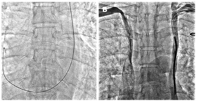
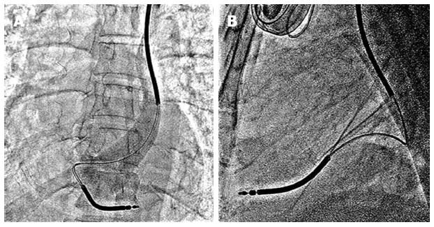
A 50-years-old hypertensive, chronic smoker male had anterior wall myocardial infarction in November 2007, for which he underwent coronary stenting of proximal left anterior descending and proximal left circumflex arteries. Later in January 2012, he had inferior wall myocardial infarction, for which he underwent coronary stenting of mid right coronary artery. One month later, he presented with hemodynamically unstable monomorphic ventricular tachycardia of rate 200 beats/min, which was reverted by electrical cardioversion. His left ventricle (LV) ejection fraction was 0.30. A repeat coronary angiography revealed patent stents in all three coronary arteries. He was taken up for ICD implantation for secondary prevention of sudden cardiac death. After conventional left subclavian vein puncture, the guide wire took the unusual course by descending across the left heart border to reach the right atrium, suggesting a persistent LSVC (Figure 1A). A contrast injection from left ante-cubital vein confirmed the presence of LSVC. There was no communicating vein between left and right superior vena cava (Figure 1B). An active fixation, dual coil RV lead (Medtronic Sprint Quattro Secure, Model No.6947, length 65 cm, 8.2 F) was passed through the LSVC-coronary sinus for implantation. A U shaped stylet was used to direct the lead from right atrium to RV. A wide loop of the lead was made in right atrium and the tip of the lead was directed towards tricuspid valve with the help of curved stylet. After few manipulations, the lead could be positioned at RV apex (Figure 2). The lead parameters were satisfactory-pacing threshold was 1.2 V at 0.5 milli-seconds pulse width, with a pacing impedance of 1098 Ω, R wave amplitude was 7.8 milli-Volts with slew rate of > 2.0 Volts/s. The RV and SVC defibrillation impedance was 41 and 46 ohms, respectively. The device (Medtronic Maximo II VR, Model D284VRC) was connected with the lead and implanted in left pectoral region. The total fluoroscopy time for the procedure was 12 min. An initial 15 J shock could revert the ventricular fibrillation to normal sinus rhythm during defibrillation testing (DFT), with SVC coil in on-mode. During 6 mo of follow-up, he did not have any shock or anti-tachycardia pacing for ventricular tachycardia.
The persistent LSVC is a remnant of embryologic venous system[1]. Its prevalence is 0.3%-0.5% in normal population[2,3]. There is considerable anatomic variation in venous drainage with persistent LSVC. A right side superior vena cava (SVC) may be absent, or both right and left SVC if present may or may not be connected with a bridging innominate vein[3-5]. It is technically challenging to direct the pacing lead at appropriate RV site in the presence of LSVC[3,6], as forward movement of the lead in right atrium is commonly towards SVC or IVC side and remain away from the tricuspid valve. As reported by others, we also made a manual U turn in the stylet to direct the active fixation lead from right atrium to right ventricle. After few manipulations, we could successfully screw the active fixation lead at RV apex and achieved satisfactory pacing parameters. The fluoroscopy time of 12 min was also comparable with others[4]. We have used active fixation RV lead to have adequate lead stability, as being used by other operators[3-6]. There is also a concern about optimum vector for defibrillation potential in these patients, as SVC coil is in coronary sinus and on the left side, instead of its usual right SVC position[4,7]. Few operators have used additional subcutaneous patch[8,9] or defibrillation coil[10] for the optimal defibrillation potential. Even, Tauras et al[11] had performed innominate vein angioplasty to put defibrillation lead via right SVC in a patient with LSVC. As there was absent bridging innominate vein in the index case (Figure 1B), we could not approach to right SVC from left side. Various authors have used a single coil lead in such a situation to avoid high defibrillation threshold[4,7], however we achieved effective 15 J defibrillation threshold (DFT) even with dual coil lead. A newer generation device like the one we implanted (Medtronic Maximo II, Model No. D284VRC), have an option to turn-off the SVC coil, thus make it functional as a single coil lead. This option can be tried in the index case, if tachycardia therapy is not effective at follow-up.
In conclusion, we hereby report a case of persistent LSVC, who had successful single chamber ICD implantation with dual coil active fixation lead. We achieved good functional parameters of the ICD and had uneventful 6 mo of follow-up.
P- Reviewer Providência R S- Editor Wen LL L- Editor A E- Editor Zhang DN
| 1. | Nsah EN, Moore GW, Hutchins GM. Pathogenesis of persistent left superior vena cava with a coronary sinus connection. Pediatr Pathol. 1991;11:261-269. [RCA] [PubMed] [DOI] [Full Text] [Cited by in Crossref: 95] [Cited by in RCA: 99] [Article Influence: 2.9] [Reference Citation Analysis (0)] |
| 2. | Albert M, Geissler W. [Persistent left superior vena cava and mitral stenosis]. Z Gesamte Inn Med. 1956;11:865-874. [PubMed] |
| 3. | Biffi M, Boriani G, Frabetti L, Bronzetti G, Branzi A. Left superior vena cava persistence in patients undergoing pacemaker or cardioverter-defibrillator implantation: a 10-year experience. Chest. 2001;120:139-144. [RCA] [PubMed] [DOI] [Full Text] [Cited by in Crossref: 155] [Cited by in RCA: 158] [Article Influence: 6.6] [Reference Citation Analysis (0)] |
| 4. | Biffi M, Bertini M, Ziacchi M, Martignani C, Valzania C, Diemberger I, Branzi A, Boriani G. Clinical implications of left superior vena cava persistence in candidates for pacemaker or cardioverter-defibrillator implantation. Heart Vessels. 2009;24:142-146. [RCA] [PubMed] [DOI] [Full Text] [Cited by in Crossref: 34] [Cited by in RCA: 35] [Article Influence: 2.2] [Reference Citation Analysis (0)] |
| 5. | Gasparini M, Mantica M, Galimberti P, Coltorti F, Simonini S, Ceriotti C, Gronda E. Biventricular pacing via a persistent left superior vena cava: report of four cases. Pacing Clin Electrophysiol. 2003;26:192-196. [RCA] [PubMed] [DOI] [Full Text] [Cited by in Crossref: 19] [Cited by in RCA: 22] [Article Influence: 1.0] [Reference Citation Analysis (0)] |
| 6. | Paulussen GM, van Gelder BM. Implantation of a biventricular pacing system in a patient with a persistent left superior vena cava. Pacing Clin Electrophysiol. 2004;27:1014-1016. [RCA] [PubMed] [DOI] [Full Text] [Cited by in Crossref: 13] [Cited by in RCA: 14] [Article Influence: 0.7] [Reference Citation Analysis (0)] |
| 7. | Arora V, Singh J, Kler TS. Implantable cardioverter defibrillatory implantation in a patient with persistent left superior vena cava and right superior vena cava atresia. Indian Heart J. 2005;57:717-719. [PubMed] |
| 8. | Mattke S, Markewitz A, Dorwarth U, Hoffmann E, Steinbeck G. Defibrillator implantation in a patient with a persistent left superior vena cava. Pacing Clin Electrophysiol. 1995;18:117-120. [RCA] [PubMed] [DOI] [Full Text] [Cited by in Crossref: 13] [Cited by in RCA: 15] [Article Influence: 0.5] [Reference Citation Analysis (0)] |
| 9. | Favale S, Bardy GH, Pitzalis MV, Dicandia CD, Traversa M, Rizzon P. Transvenous defibrillator implantation in patients with persistent left superior vena cava and right superior vena cava atresia. Eur Heart J. 1995;16:704-707. [PubMed] |
| 10. | Leng CT, Crosson JE, Calkins H, Berger RD. Lead configuration for defibrillator implantation in a patient with congenital heart disease and a mechanical prosthetic tricuspid valve. Pacing Clin Electrophysiol. 2001;24:1291-1292. [RCA] [PubMed] [DOI] [Full Text] [Cited by in Crossref: 14] [Cited by in RCA: 16] [Article Influence: 0.7] [Reference Citation Analysis (0)] |
| 11. | Tauras JM, Palma EC. Venoplasty of innominate bridge during implantation of single-chamber ICD in a patient with a persistent left-sided superior vena cava. Pacing Clin Electrophysiol. 2008;31:1077-1078. [RCA] [PubMed] [DOI] [Full Text] [Cited by in Crossref: 2] [Cited by in RCA: 4] [Article Influence: 0.2] [Reference Citation Analysis (0)] |


