Published online Jul 26, 2012. doi: 10.4330/wjc.v4.i7.240
Revised: March 10, 2012
Accepted: March 17, 2012
Published online: July 26, 2012
Ebstein’s anomaly (EA) is a rare cardiac congenital malformation with displacement of septal and posterior tricuspid leaflets, resulting in atrialization of the right ventricle. We report a case of EA in which the etiology of a malfunctioning prosthetic tricuspid valve is depicted on cardiac computed tomography to be as a result of thrombus lodged in the valve.
- Citation: O’Neill AC, Kelly RM, McCarthy CJ, Martos R, McCreery C, Dodd JD. Thrombosed prosthetic valve in Ebstein's anomaly: Evaluation with echocardiography and 64-slice cardiac computed tomography. World J Cardiol 2012; 4(7): 240-241
- URL: https://www.wjgnet.com/1949-8462/full/v4/i7/240.htm
- DOI: https://dx.doi.org/10.4330/wjc.v4.i7.240
Ebstein’s anomaly (EA) is a rare cardiac congenital malformation characterized by apical displacement of the septal and posterior tricuspid leaflets, resulting in atrialization of the right ventricle. Surgical replacement of the tricuspid valve is performed in cases of severe tricuspid regurgitation. Follow-up of tricuspid prosthetic valves is typically performed with transthoracic echocardiography. We describe the case of a malfunctioning thrombosed tricuspid valve in a patient with EA. Cardiac computed tomography allowed the detection, localization and extent of the thrombus to be depicted.
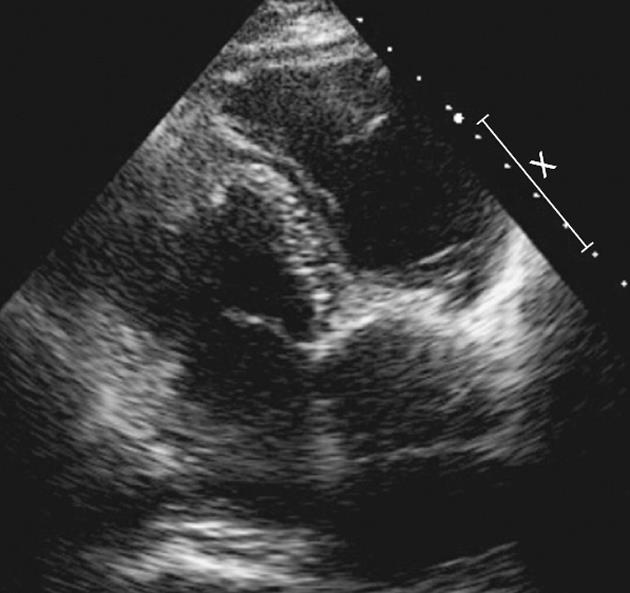
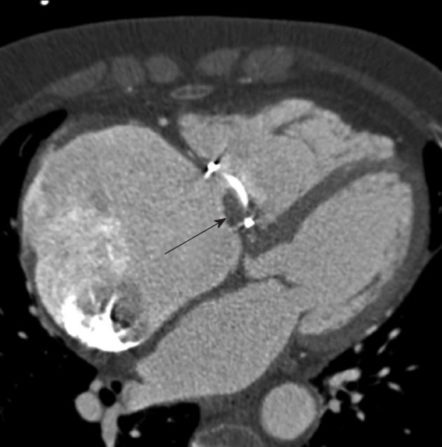
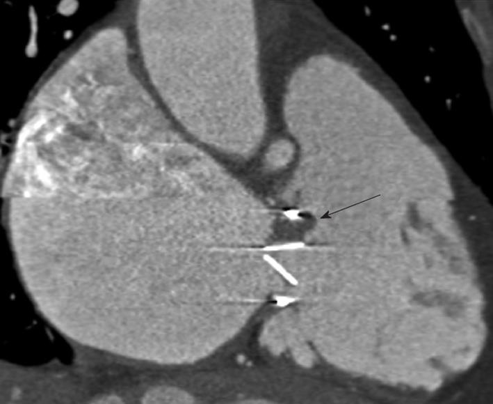
A 68-year-old woman with known EA underwent echocardiography for evaluation of dyspnea. She complained of a 3-mo history of progressive breathlessness, reduced exercise tolerance (New York Heart Association grade 2) and increasing leg swelling. She had been diagnosed with EA at the age of 50 years and had been treated successfully with an angiotensin-converting-enzyme inhibitor until 2001, when her symptoms deteriorated. An echocardiogram demonstrated severe tricuspid regurgitation, and she underwent tricuspid valve replacement. Subsequent echocardiography at 6 mo post-procedure demonstrated malfunction of the prosthesis leaflets with fixation throughout the cardiac cycle. On the current admission, a 64-slice cardiac computed tomography (CT) was undertaken to exclude hemodynamically significant coronary disease. It demonstrated no evidence of coronary atherosclerosis, and clearly depicted the characteristic features of EA including an enlarged right atrium and atrialization of the right ventricle (Figure 1). Thrombus was identified lodged between the medial valve flap and the valve ring (Figures 2 and 3). Multiphasic cine CT throughout the cardiac cycle demonstrated immobility of the tricuspid valve leaflets. The patient refused redo-valve replacement surgery. Her symptoms remain moderate.
EA is a rare congenital cardiac malformation characterized by apical displacement of the septal and posterior tricuspid leaflets, resulting in atrialization of the right ventricle[1,2]. Medical management includes treatment for heart failure and complex cardiac arrhythmias. Surgical tricuspid valve repair is undertaken in severe cases of tricuspid regurgitation[3]. Echocardiography is the investigation of choice for valve appraisal. Recent technical advances in cardiac CT, using multiphasic electrocardiography-gated image reconstruction, have allowed evaluation of both native and prosthetic cardiac valves. In the current report, echocardiography suggested the underlying abnormality, but the thrombus was more clearly depicted using cardiac CT. Cardiac CT also excluded obstructive coronary artery disease as a potential etiology for the patient’s symptoms. Our report highlights the increasing versatility of cardiac CT in detecting non-coronary, cardiac pathology.
Peer reviewers: Dr. Thomas Hellmut Schindler, PD, Department of Internal Medecine and Cardiology, Rue Gabrielle-Perret-Gentil, 4, 1211 Geneva, Switzerland; Dr. Ole Dyg Pedersen, Department of Cardiology, Bispebjerg Bakke 23, 2400 Copenhagen, Denmark
S- Editor Cheng JX L- Editor Kerr C E- Editor Li JY
| 1. | Correa-Villaseñor A, Ferencz C, Neill CA, Wilson PD, Boughman JA. Ebstein's malformation of the tricuspid valve: genetic and environmental factors. The Baltimore-Washington Infant Study Group. Teratology. 1994;50:137-147. [PubMed] |
| 2. | Frescura C, Angelini A, Daliento L, Thiene G. Morphological aspects of Ebstein's anomaly in adults. Thorac Cardiovasc Surg. 2000;48:203-208. [PubMed] |
| 3. | Attenhofer Jost CH, Connolly HM, Dearani JA, Edwards WD, Danielson GK. Ebstein's anomaly. Circulation. 2007;115:277-285. [PubMed] |


