Published online Nov 26, 2012. doi: 10.4330/wjc.v4.i11.309
Revised: November 16, 2012
Accepted: November 22, 2012
Published online: November 26, 2012
Left ventricle (LV) pseudoaneurysm is a late mechanical complication of myocardial infarction. A giant LV pseudoaneurysm is a rare presentation. We report a case of giant LV pseudoaneurysm in a post-MI patient who presented with gross congestive heart failure. The patient had a successful surgical repair of the aneurysm and had a favorable 3-mo outcome. The imaging modality and surgical treatment of the pseudoaneurysm are discussed.
- Citation: Vijayvergiya R, Kumar A, Rana SS, Singh H, Puri GD, Singhal M. Post-myocardial infarction giant left ventricular pseudoaneurysm presenting with severe heart failure. World J Cardiol 2012; 4(11): 309-311
- URL: https://www.wjgnet.com/1949-8462/full/v4/i11/309.htm
- DOI: https://dx.doi.org/10.4330/wjc.v4.i11.309
Left ventricle (LV) pseudoaneurysm is a contained cardiac rupture, which is sealed by layers of organized thrombus and hematoma. It is encircled by a thin layer of adherent pericardium without any myocardial layer, which makes it susceptible to rupture. The clinical presentation of pseudoaneurysm is variable. A timely diagnosis and early surgical treatment are key factors in the management. We hereby report an unusual case of giant LV pseudoaneurysm in a 42-year-old man following anterior wall myocardial infarction (MI). The role of various imaging modalities and the surgical treatment of pseudoaneurysm are discussed.
A 42-year-old male had an acute anterior wall MI in March 2012. During the inpatient admission at a local hospital, he had hemorrhagic pericardial effusion with tamponade, which was drained by pigtail catheter insertion. At 2 mo of follow-up, he was diagnosed as having a large LV pseudoaneurysm, for which he was referred to our center for further management.
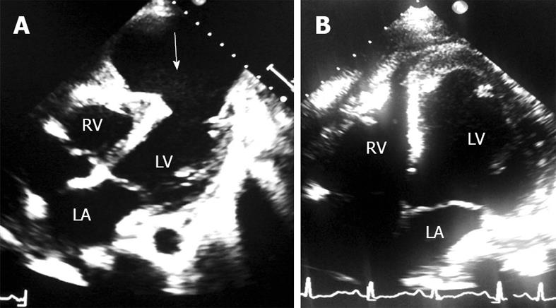
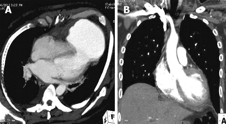
On admission at our institute, the patient was in gross congestive heart failure with New York Heart Association functional class IV. His blood pressure was 100/70 mmHg, pulse rate 100/min, and systemic oxygen saturation at room air was 95%. His cardiac examination revealed a LV 3rd heart sound, and chest examination revealed basal crepitations in both the lung fields. An electrocardiogram revealed normal sinus rhythm and QS pattern in V1-V4 chest leads. An echocardiogram revealed a large LV apical aneurysm of size 60 mm × 90 mm, with a neck of 30 mm (Figure 1A). The volume of the pseudoaneurysm sac was 244 mL, and that of the LV cavity was 89 mL in diastole, as calculated by modified Simpson’s method. There was no mitral regurgitation. LV ejection fraction was 0.35, and a moderate tricuspid regurgitation was present with estimated pulmonary artery systolic pressure of 45 mmHg. A coronary angiography revealed 90% stenosis of the distal left anterior descending artery, and the rest of the coronaries were normal. LV end diastolic pressure was 50 mmHg. A computed tomography (CT) scan of the chest showed a large contrast-filled LV pseudoaneurysm arising from the LV apex, measuring 76 mm × 98 mm with a 28 mm neck (Figure 2A). After adequate decongestive medical therapy, he underwent surgical repair of the pseudoaneurysm.
During surgery, the LV apex was mobilized under cardio-pulmonary bypass. It was densely adherent to the pericardium and adjacent lingular segment of the left lung. Under cardioplegic arrest, the pseudoaneurysm was opened, leaving a small rim of sac wall towards the lung. The pseudoaneurysm had a circular gap of about 30 mm diameter through which it was connected to the LV cavity (Figure 3A). The defect was closed using a polytetrafluoroethylene (PTFE) patch with interrupted 4-0 prolene suture (Figure 3B). The false sac was tailored to make a flap which firmly covered the PTFE patch. Biological glue was spread between the two layers of sac before tying the last suture. The patient was weaned from cardiopulmonary bypass and had an uneventful postoperative recovery.
A postoperative CT scan of the chest revealed normal LV outline without any leakage (Figure 2B). An echocardiogram revealed a well-delineated LV apical wall with exclusion of pseudoaneurysm (Figure 1B). He was discharged on the 14th postoperative day. He remained asymptomatic at 3 mo of follow-up.
LV pseudoaneurysm is seen in patients having MI, cardiac infection, and following cardiac interventions or trauma[1]. MI is the most frequently observed etiology in LV pseudoaneurysm cases. It is a late mechanical complication of MI presenting within a few months of infarction, as happened in the index case. The clinical presentation may vary depending upon congestive heart failure, mitral regurgitation, ventricular tachy-arrhythmia, systemic thrombo-embolism and cardiac rupture[1,2]. In general, patients do not have specific symptoms pertaining to pseudoaneurysm[1], hence the diagnosis may be delayed. The index case was symptomatic since beginning with congestive heart failure; hence a diagnosis of giant LV pseudoaneurysm could be established. Blażejewski et al[3] have also reported a case of giant LV pseudoaneurysm presenting with severe heart failure. The index case possibly had impending cardiac rupture initially, when he presented with hemorrhagic pericardial effusion and tamponade. The leaking site could have been sealed by a thin layer of pericardium and organized thrombus, and later presented with giant LV pseudoaneurysm. Giant LV pseudoaneurysm has been reported to cause mitral regurgitation and compression of adjacent vascular structures[4-6]; however, there was no such complications in the index case. Both echocardiography and CT angiogram are good noninvasive imaging modalities for the diagnosis of pseudoaneurysm[1,3,6-8]. A CT scan can delineate the extent of pseudoaneurysm and also the involvement of adjacent cardiac and non-cardiac structures[8,9]. Surgery was indicated in the index case because of the symptomatic status, giant aneurysm size and an impending rupture. A conservative approach can be considered in asymptomatic cases, those with small aneurysms of less than 3 cm dimension, and those with a stable dimension during regular follow-up[2,3]. Surgery itself carries a high mortality[1]; nevertheless, we performed a successful PTFE patch repair of the aneurysm and the patient had an uneventful 3 mo of follow-up. As reported earlier by us[9], the surgical technique of aneurysm repair by PTFE patch augmentation has previously been effective with a favorable short term outcome.
In conclusion, giant LV pseudoaneurysm following MI can present with congestive heart failure. Echocardiography is a good imaging modality for the early diagnosis of pseudoaneurysm. Surgery is the definitive treatment of giant pseudoaneurysm, without which the prognosis is very poor.
Peer reviewers: Paul Erne, MD, Professor, Head, Department of Cardiology, Luzerner Kantonsspital, CH-6000 Luzern 16, Switzerland; Tevfik Fikret Ilgenli, MD, Associate Professor of Cardiology, Department of Cardiology, Gölcük Military Hospital, 41650-Gölcük, Kocaeli, Turkey; Jacob Joseph, MBBS, MD, Associate Professor of Medicine, Boston University School of Medicine, VA Boston Healthcare, Cardiology Section, 1400 VFW Parkway, West Roxbury, MA 02132, United States
S- Editor Cheng JX L- Editor Logan S E- Editor Li JY
| 1. | Frances C, Romero A, Grady D. Left ventricular pseudoaneurysm. J. Am Coll Cardiol. 1998;32:557-561. [RCA] [DOI] [Full Text] [Cited by in Crossref: 396] [Cited by in RCA: 409] [Article Influence: 15.1] [Reference Citation Analysis (0)] |
| 2. | Moreno R, Gordillo E, Zamorano J, Almeria C, Garcia-Rubira JC, Fernandez-Ortiz A, Macaya C. Long term outcome of patients with postinfarction left ventricular pseudoaneurysm. Heart. 2003;89:1144-1146. [PubMed] |
| 3. | Blażejewski J, Sinkiewicz W, Bujak R, Banach J, Karasek D, Balak W. Giant post-infarction pseudoaneurysm of the left ventricle manifesting as severe heart failure. Kardiol Pol. 2012;70:85-87. [PubMed] |
| 4. | Kansiz E, Hatemi AC, Tongut A, Cohcen S, Yildiz A, Kilickesmez K, Celiker C. Surgical treatment of a giant postero-inferior left ventricular pseudoaneurysm causing severe mitral insufficiency and congestive heart failure. Ann Thorac Cardiovasc Surg. 2012;18:151-155. [PubMed] |
| 5. | Fox SA, Templeton C, Hancock-Friesen C, Chen R. Commotio cordis and ventricular pseudoaneurysm. Can J Cardiol. 2009;25:237-238. [RCA] [PubMed] [DOI] [Full Text] [Cited by in Crossref: 3] [Cited by in RCA: 7] [Article Influence: 0.4] [Reference Citation Analysis (0)] |
| 6. | Lee YH, Hou CJ, Hung CL, Tsai CH. Silent and huge left ventricular pseudoaneurysm with left atrial compression: dedicated spatial resolution and geometry by 3-dimensional echocardiography. J Am Soc Echocardiogr. 2007;20:772.e5-772.e9. [PubMed] |
| 7. | Alizade E, Guler A, Acar G, Karakoyun S, Esen AM. Multimodality cardiac imaging of a giant left ventricular pseudoaneurysm. Eur J Echocardiogr. 2011;12:550. [PubMed] |
| 8. | Vijayvergiya R, Chongtham DS, Thingnam SK, Grover A, Lal A. Left ventricular pseudoaneurysm with infective pericarditis: a rare cause of intractable hemoptysis. Angiology. 2008;59:507-509. [PubMed] |
| 9. | Vijayvergiya R, Pattam J, Rana SS, Singh JD, Puri GD, Singhal M. Giant left ventricular pseudoaneurysm presenting with hemoptysis. World J Cardiol. 2012;4:218-220. [PubMed] |


