Revised: January 21, 2011
Accepted: January 27, 2011
Published online: February 26, 2011
A 42-year-old-woman presented with de novo crescendo angina. Thallium-scintigraphy showed inferior ischemia. Coronary angiogram revealed a right coronary artery (RCA), originating from the left sinus of Valsalva with a severe proximal systolic compression. She underwent successful transradial percutaneous coronary intervention with stent implantation. Multislice-computed tomography (MSCT) is usually used to evaluate coronary artery anomalies and can effectively show the anomalous RCA and the inter-arterial trajectory between the aorta and pulmonary arteries. Anomalies of the origin of the coronary arteries are rare, but can produce specific clinicopathological entities that should be diagnosed with accuracy. This case report illustrates the role of MSCT in the detailed description of an abnormal coronary artery and the use of stenting for symptoms relief.
- Citation: Bagur R, Gleeton O, Bataille Y, Bilodeau S, Rodés-Cabau J, Bertrand OF. Right coronary artery from the left sinus of valsalva: Multislice CT and transradial PCI. World J Cardiol 2011; 3(2): 54-56
- URL: https://www.wjgnet.com/1949-8462/full/v3/i2/54.htm
- DOI: https://dx.doi.org/10.4330/wjc.v3.i2.54
Coronary artery anomalies occur in approximately 1% of the population, often without other congenital cardiac malformations[1]. The abnormal origin of the right coronary artery (RCA) from the left aortic sinus of Valsalva is a very rare congenital anomaly, frequently coursing between the aorta and the pulmonary artery[2].
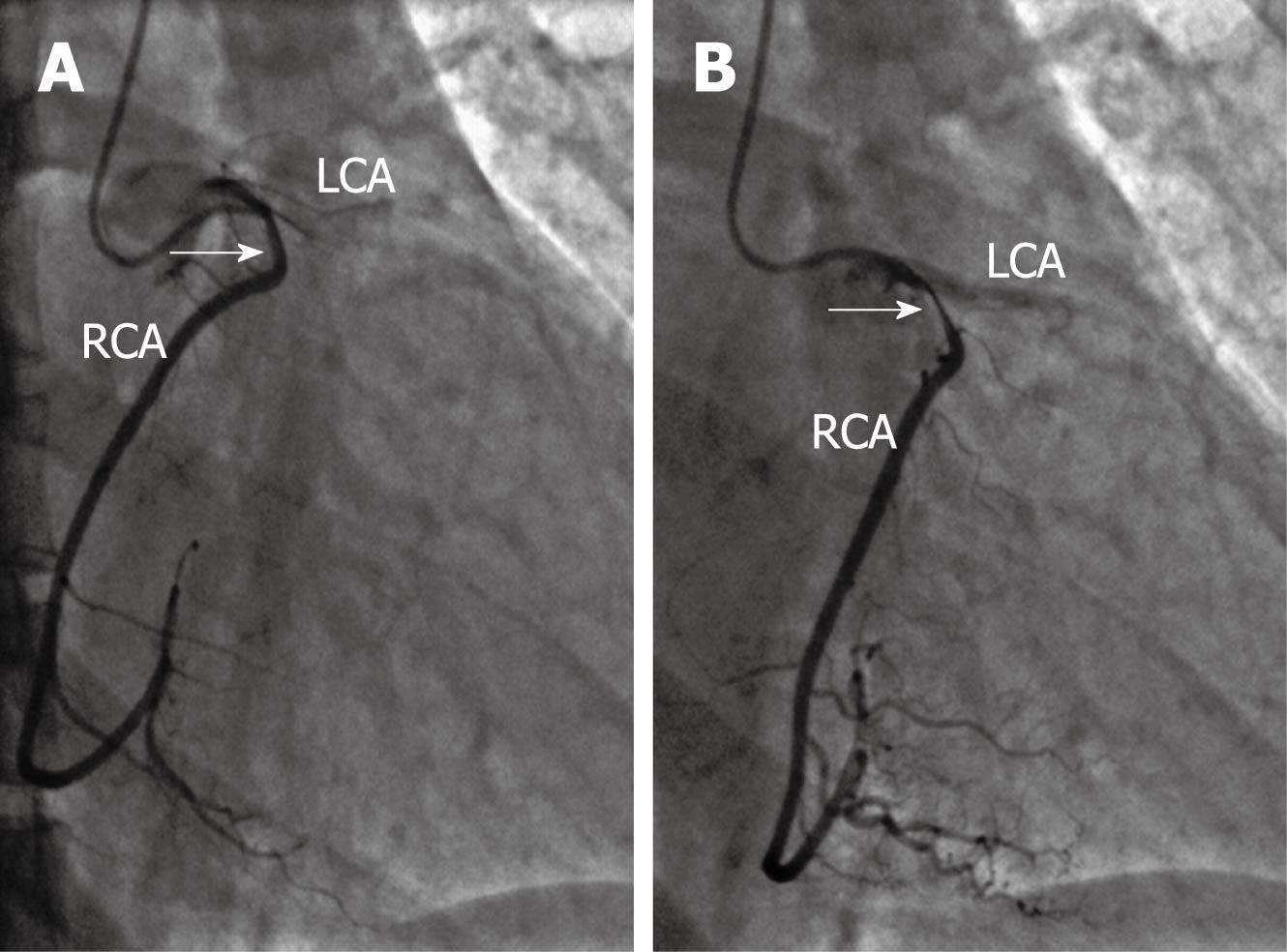
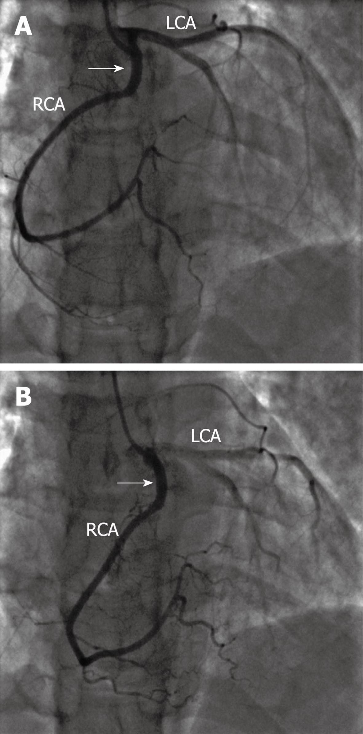
A 42-year-old woman presented with a history of de novo crescendo angina. The patient decided to enter the National Army Services. Following standard procedures, she began progressive and very stringent exercise training. During intense exercise sessions, she started to complain of typical angina. Despite no previous chest pain episodes, nor any cardiovascular risk factors, she underwent a complete workout to rule out atherosclerotic coronary artery disease. Thallium scintigraphy showed inferior ischemia. Therefore she was referred to our tertiary care center for diagnostic coronary angiography and possible revascularization therapy. The coronary angiogram showed a left coronary artery from the left sinus of Valsalva without significant lesion. The right coronary artery (RCA) originated from the left sinus and demonstrated a severe systolic compression (Figure 1A and B). Several injections/projections were performed after the injection of intracoronary nitroglycerine, and no changes nor improvement were observed. She underwent right radial percutaneous coronary angioplasty using a 6 Fr Amplatz Left 2 guiding catheter, and a 3.5 mm × 16 mm bare-metal stent (Liberté®, Boston Scientific Corporation, Natick, MA, US) was successfully implanted (Figure 2A and B).
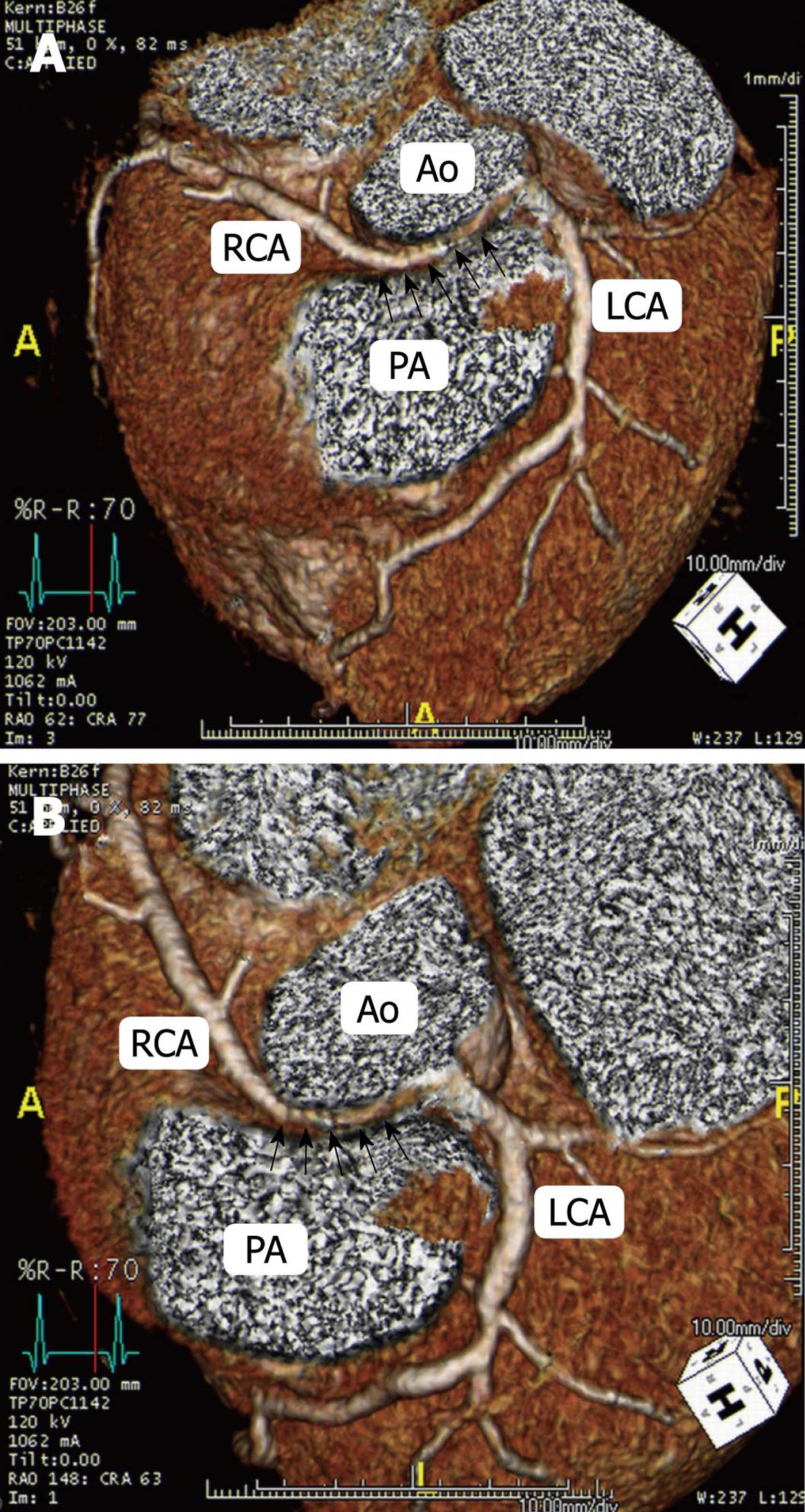
Multi-slice computed tomography (MSCT) is usually used and can effectively show the RCA arising from the left sinus of Valsalva and its inter-arterial trajectory
(Figure 3A and B, arrows) between the aorta and the pulmonary arteries. After 1 year follow-up, the patient has remained asymptomatic with a negative stress test.
In this case report, we describe the case of a young woman without cardiovascular risk factors who presented with typical effort angina and inferior ischemia. Angiography revealed dynamic compression of an abnormally arising right coronary artery and she was treated by transradial bare-metal stent implantation.
Anomalies of the origin of the coronary arteries are rare, but can provide specific clinicopathological entities that should be diagnosed with a high degree of accuracy. The origin of both coronary arteries from the left sinus of Valsalva is a very rare (0.28%) anomaly[3]. Manifestations vary from asymptomatic patients to those who present with angina pectoris, myocardial infarction, heart failure, syncope, arrhythmias, and also sudden death[1]. Myocardial ischemia in association with this anomaly is thought to be caused by an abnormal slit-like RCA ostium, acute angulations, and compression of the RCA between the aorta and pulmonary trunk during exercise[3]. Extrinsic compression of the left main coronary artery can occur in patients with severe pulmonary hypertension and enlarged pulmonary artery trunk[4,5]. It has usually been described in the setting of congenital defects such as atrial septal defect, ventricular septal defect, and, more rarely, isolated persistent ductus arteriosus[4,5]. MSCT allows 3-dimensional visualization of the coronary arteries with high spatial resolution, and may be the most promising imaging modality for diagnosing these anomalies[4-6].
Surgical correction or coronary artery bypass grafting can be carried out with minimal risk and good anatomic and functional results[7,8]. Although the risks of surgical intervention are low in young subjects, surgery requires opening the chest and may be complicated by aortic valve damage or neurological emboli.
This case-report illustrates the role of MSCT in the detailed description of an abnormal coronary artery and the use of stenting symptoms relief. In the presence of a symptomatic patient with an isolated RCA anomaly and no other atherosclerotic coronary disease, transradial percutaneous intervention could be an effective and safe option.
Peer reviewers: Masamichi Takano, MD, PhD, Cardiovascular Center, Chiba-Hokusoh Hospital, Nippon Medical School, 1715 Kamakari, Imba, Chiba, 270-1694, Japan; Mohammad-Reza Movahed, MD, PhD, FACC, FACP, FCCP, Associate Professor of Medicine, Director of Coronary Care Unit, University of Arizona Sarver Heart Center, 1501 North Campbell Ave., Tucson, AZ, 85724, United States; Pil-Ki Min, MD, PhD, Cardiology Division, Heart Center, Gangnam Severance Hospital, Yonsei University College of Medicine, 712 Eonjuro, Gangnam-gu, 135-720 Seoul, South Korea
S- Editor Cheng JX L- Editor Cant MR E- Editor Zheng XM
| 1. | Angelini P, Velasco JA, Flamm S. Coronary anomalies: incidence, pathophysiology, and clinical relevance. Circulation. 2002;105:2449-2454. |
| 2. | Ayalp R, Mavi A, Serçelik A, Batyraliev T, Gümüsburun E. Frequency in the anomalous origin of the right coronary artery with angiography in a Turkish population. Int J Cardiol. 2002;82:253-257. |
| 3. | Wilkins CE, Betancourt B, Mathur VS, Massumi A, De Castro CM, Garcia E, Hall RJ. Coronary artery anomalies: a review of more than 10,000 patients from the Clayton Cardiovascular Laboratories. Tex Heart Inst J. 1988;15:166-173. |
| 4. | Caldera AE, Cruz-Gonzalez I, Bezerra HG, Cury RC, Palacios IF, Cockrill BA, Inglessis-Azuaje I. Endovascular therapy for left main compression syndrome. Case report and literature review. Chest. 2009;135:1648-1650. |
| 5. | Safi M, Eslami V, Shabestari AA, Saadat H, Namazi MH, Vakili H, Movahed MR. Extrinsic compression of left main coronary artery by the pulmonary trunk secondary to pulmonary hypertension documented using 64-slice multidetector computed tomography coronary angiography. Clin Cardiol. 2009;32:426-428. |
| 6. | Ichikawa M, Sato Y, Komatsu S, Hirayama A, Kodama K, Saito S. Multislice computed tomographic findings of the anomalous origins of the right coronary artery: evaluation of possible causes of myocardial ischemia. Int J Cardiovasc Imaging. 2007;23:353-360. |
| 7. | Romp RL, Herlong JR, Landolfo CK, Sanders SP, Miller CE, Ungerleider RM, Jaggers J. Outcome of unroofing procedure for repair of anomalous aortic origin of left or right coronary artery. Ann Thorac Surg. 2003;76:589-595; discussion 595-596. |
| 8. | Krasuski RA, Magyar D, Hart S, Kalahasti V, Lorber R, Hobbs R, Pettersson G, Blackstone E. Long-term outcome and impact of surgery on adults with coronary arteries originating from the opposite coronary cusp. Circulation. 2011;123:154-162. |


