Copyright
©2010 Baishideng Publishing Group Co.
World J Cardiol. Jul 21, 2010; 2(7): 198-204
Published online Jul 21, 2010. doi: 10.4330/wjc.v2.i7.198
Published online Jul 21, 2010. doi: 10.4330/wjc.v2.i7.198
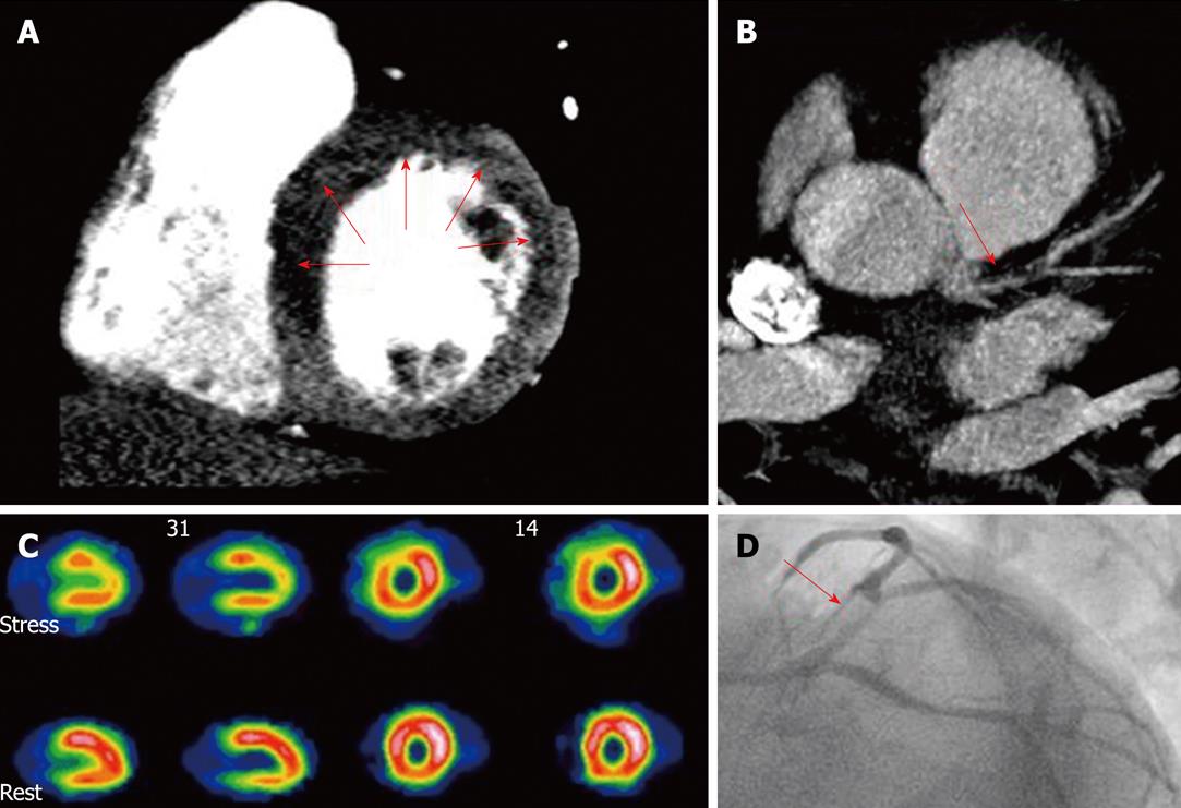
Figure 1 Combined assessment of myocardial perfusion at pharmacological (adenosine) stress and coronary angiography within a single-session cardiac computed tomography.
Cardiac computed tomography (CT) (panels A and B) showed diminished perfusion of the left ventricle anterior and septal wall (A, arrows) and a significant lesion at the proximal left anterior descending artery (B, arrow), findings confirmed by single photon emission computed tomography (SPECT) (C) and invasive coronary angiography (D, arrow). A: CT perfusion (stress); B: Coronary CT angiography; C: SPECT myocardial perfusion imaging; D: Invasive angiography. With permission of Blankstein et al[21], J Am Coll Cardiol 2009; 54: 1072-1084.
- Citation: Rodríguez-Granillo GA, Ingino CA, Lylyk P. Myocardial perfusion imaging and infarct characterization using multidetector cardiac computed tomography. World J Cardiol 2010; 2(7): 198-204
- URL: https://www.wjgnet.com/1949-8462/full/v2/i7/198.htm
- DOI: https://dx.doi.org/10.4330/wjc.v2.i7.198


