Published online Jun 26, 2010. doi: 10.4330/wjc.v2.i6.140
Revised: June 11, 2010
Accepted: June 18, 2010
Published online: June 26, 2010
Crosstalk between lipid peroxidation and inflammation is known to be a pathognomonic feature for the development of coronary heart disease (CHD). In this regard ligand activated liver X receptor (LXR)-α has emerged as a key molecular switch by its inherent ability to modulate an array of genes involved in these two fundamental cellular processes. In addition, LXR-α has also been found to play a role in hepatic lipogenesis and innate immunity. Although several lines of evidence in experimental model systems have established the atheroprotective nature of LXR-α, human subjects have been reported to possess a paradoxical situation in which increased blood cellular LXR-α gene expression is always accompanied by increased coronary occlusion. This apparent paradox was resolved recently by the finding that CHD patients possess a deregulated LXR-α transcriptome due to impaired ligand-receptor interaction. This blood cellular mutated LXR-α gene expression correlated specifically with the extent of coronary occlusion and hence need is felt to devise new synthetic ligands that could restore the function of this mutated LXR-α protein in order to modulate genes involved in reverse cholesterol transport and suppression of the inflammatory response leading to the effective treatment of CHD.
- Citation: Dave VP, Kaul D. Coronary heart disease: Significance of liver X receptor α genomics. World J Cardiol 2010; 2(6): 140-149
- URL: https://www.wjgnet.com/1949-8462/full/v2/i6/140.htm
- DOI: https://dx.doi.org/10.4330/wjc.v2.i6.140
In the last decade an accumulation of data from in vitro to in vivo model systems has established a pivotal role of liver X receptor (LXR) gene for its ability to regulate two fundamental cellular processes i.e. lipid metabolism and inflammation, which are the key components for the pathogenesis of coronary heart disease (CHD). There is a general recognition of the fact that co-operativity between lipid peroxidation and inflammation initiates a complex interaction between blood mononuclear cells, blood derived factors and the arterial wall leading to the development of CHD[1-3]. At the molecular level such a phenomenon arises due to interplay of genes involved in lipid metabolism and inflammation. In the light of this cross-talk, the LXR-α molecule has caught the imagination of researchers across the globe due to its inherent capacity to modulate an array of genes involved in various cellular metabolic processes[4]. In this review, an attempt will be made to evolve the interrelationship of LXR-α genomics with lipid peroxidation, inflammation and human atherosclerosis, a phenomenon vital for the design of an LXR-α gene based preventive/therapeutic strategy against CHD.
Much of the recent research on the origin of atherosclerosis has concentrated on the interplay between lipid metabolism, cytokines and cellular activity within arterial wall. Oxidative modification of low density lipoprotein (LDL) and ‘response to injury’ are two fundamental mechanisms proposed to be involved in the pathogenesis of atherosclerosis. Oxidized LDL has been shown to bypass the negative feedback mechanism in macrophages that inhibits the excessive uptake of native LDL[5]. Additionally, a vast number of oxidation products of lipids and proteins have been demonstrated in atherosclerotic lesions and in the plasma of patients with atherosclerosis[6-8]. On the other hand according to the ‘response to injury hypothesis[9], endothelial nudation is the initial step in atherosclerosis, however recent research has focused on endothelial dysfunction rather than endothelial nudation as being the first pathognomonic feature of atherosclerosis[10]. The risk factors influencing the development of atherosclerosis mainly involve hypercholesterolemia, altered LDL, hypertension, elevated levels of homocystine, genetic alteration, smoking, diabetes mellitus and infectious microorganisms such as Chlamydia pneumoniae[11].
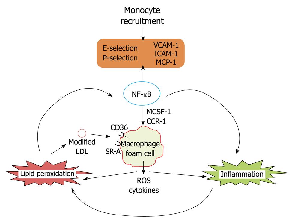
The first step in the pathogenesis of initial lesion development is lipid deposition followed by monocyte recruitment to the sub-endothelial space which is regulated by nuclear factor κB (NF-κB) induced gene expression[1]. At first LDL shuttling in and out of the vessel wall are trapped as a result of oxidative modifications by myeloperoxidase and 12/15 lipoxygenase enzymes[12,13]. Trapped LDL aggregates undergo further oxidation and oxidized LDL induces expression of adhesion factors in an NF-κB dependent manner[14]. Further monocyte attach to vascular endothelial cells which is mediated by selectins[15], vascular cell adhesion molecule-1 and intracellular cell adhesion molecule-1[16]. Monocyte transmigration into the sub-endothelial space is mediated by endothelium monocyte chemoattractant protein-1 (MCP-1) and its receptor CCR-2[17,18]. In the subendothelial space monocytes differentiate into macrophages under the influence of monocyte colony stimulating factor and express CD36, scavenger receptor-A which specifically recognizes oxidized LDL. Uptake of oxidized LDL by macrophages leads to foam cell formation, which is a crucial landmark for the pathogenesis of atherosclerosis. Lipid loaded macrophages in the subendothelial space release reactive oxygen species and cytokines which leads to further expression of adhesion factors and subsequent monocyte migration and exacerbation of vascular inflammation. Macrophages may also be an active player in both intracellular and extracellular lipid peroxidation. Foam cells also mediate the release of reactive oxygen species and inflammatory cytokines and oxidation of lipoprotein particles in the subendothelial space[19-23]. Thus co-operativity between lipid peroxidation and inflammation is a hallmark for the development of atherosclerosis (Figure 1).
In summary, endothelial dysfunction followed by cooperative action of lipid peroxidation and inflammation on vascular wall leads to the initial lesion development, mediated by the NF-κB dependent expression of various cell adhesion molecules and mediators which leads to the recruitment of monocytes and furthers their differentiation into macrophage foam cells is the hallmark feature for the development of CHD.
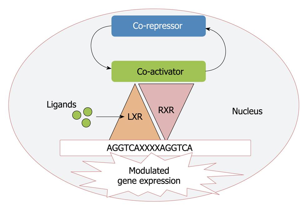
Based upon the sequence homology with other nuclear receptors LXRs (LXR-α, NR1H3 and LXR-β, NR1H2) were cloned a decade ago and considered originally as “orphan” nuclear receptors as their natural ligands were unknown[24,25]. LXR-α is highly expressed in liver, adipose tissue and macrophages whereas the β isoform is ubiquitously expressed in all tissues[26]. The LXRs are ligand activated transcription factors that form permissive heterodimers with retinoid X receptor (RXR). This heterodimer binds to LXR response element in the DNA consisting of direct repeats of the core sequence AGGTCA separated by four nucleotides[27]. In the absence of ligand, LXRs recruit complexes of co-repressors that are substituted by co-activators upon ligand binding[28] and thus regulate the gene expression (Figure 2).
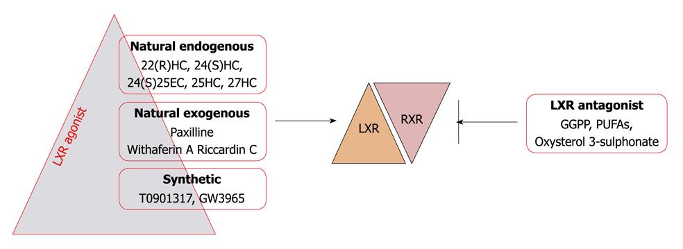
Mono-oxygenated derivatives of cholesterol or oxysterols are the major physiological LXR ligands. Intermediates between steroid hormone synthesis, 22(R) hydroxy cholesterol and 20(S) hydroxy cholesterol, have been shown to bind and stimulate LXR-α transcriptional activity in the physiological range. In the brain 24(S) hydroxy cholesterol and in the liver 24(S),25-epoxycholesterol is the abundant LXR agonist[29-32]. Apart from these, studies have demonstrated that D-glucose and D-glucose 6 phosphate are endogenous LXR agonists with equal efficacy to that of oxysterols[33], however recent findings have questioned this fact on the basis of inability of glucose and its metabolites to influence the interaction of cofactors with LXR and the lack of involvement of LXRs in the regulation of glucose sensitive genes in the liver[34]. Based upon molecular docking and in vitro studies, our recent finding have revealed withaferin A as a novel LXR-α agonist, which interacts in a similar fashion within the ligand binding domain, as its natural physiological ligands and these have potential to activate LXR-α[35]. An indol alkaloid, paxilline, produced by a fungus Penicillium paxilli was the first natural non oxysterol LXR agonist[36] but due to its toxicity in in vivo studies it is unsuitable. Riccardin C is also a natural nonsteroidal compound isolated from liverworts, which acts as an LXR-α agonist and LXR-β antagonist[37]. Synthetic LXR agonists T0901317 and GW3965 are commonly used in experimental studies. In addition to LXR agonists geranyl-geranyl pyrophosphate has been found to inhibit LXR activity by antagonizing their interaction with coactivators[38,39]. Further transcriptional activity of LXR has also been shown to be inhibited by oxidized cholesterol 3 sulphates (normally found in human plasma) and polyunsaturated fatty acids in various cell lines[40,41] (Figure 3).
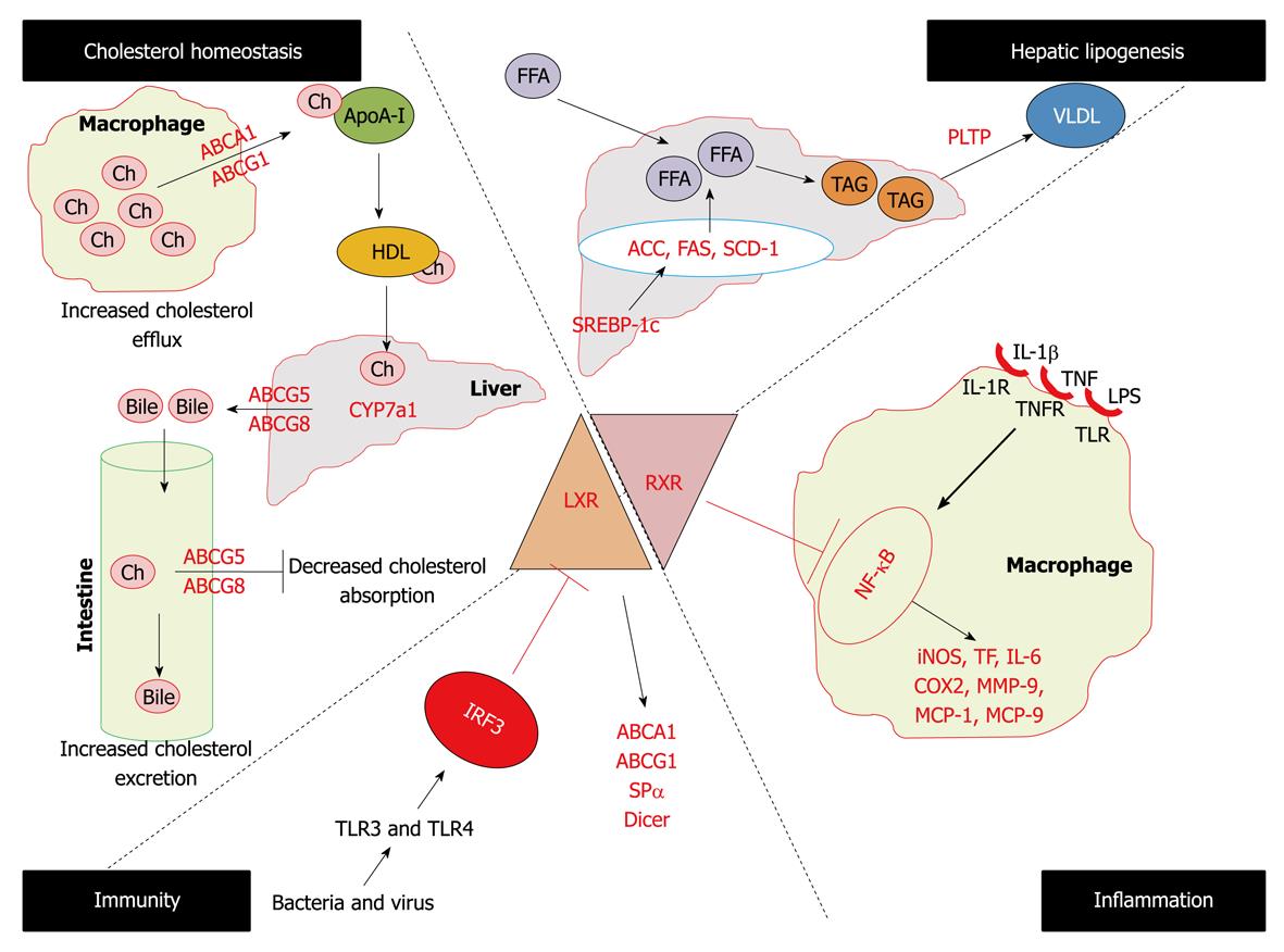
Studies on LXR-α-/- mice, but not LXR-β-/- mice, showed a marked cholesterol ester accumulation in the liver when fed with diets containing cholesterol[42]. This led to the identification of the first known LXR-α direct target CYP7A1 (the rate limiting enzyme in bile acid synthesis). The different phenotypes of the two knockout mice strains indicates that despite the considerable sequence homology between two LXR isoforms they have different distinct biological functions. With the identification of several LXR-α target genes, this molecule became a fascinating player for understanding its role in the mechanism of regulation of macrophage cholesterol metabolism, hepatic lipogenesis, and enterohepatic circulation by inhibiting cholesterol absorption. Apart from its role in atherosclerosis, recent data have uncovered the role of LXR-α in inflammation and immunity also leading to the integration of three fundamental cellular processes i.e. lipid metabolism, inflammation and immunity (Figure 4).
A primary function of LXR-α is to maintain cellular cholesterol homeostasis by participating in the process of reverse cholesterol transport[43]. In vivo activation of LXR-α with synthetic high affinity ligand increases HDL levels and net cholesterol secretion[44]. These activities are mediated by LXR-α by upregulating the expression of the ABC superfamily of membrane transporters including ABCA1, ABCG5, ABCG8 and ABCG1[45-51]. Mutations in the ABCA1 gene are the cause of Tangier disease, characterized by the complete absence of HDL in plasma of afflicted patients, resulting in the accumulation of cholesterol in tissue macrophages and an increased incidence of cardiovascular diseases[52]. In addition to ABC transporters, LXR-α driven reverse cholesterol is promoted by the induction of a subset of apolipoproteins that serves as cholesterol acceptors. It is now well recognized that in macrophages, which play a central role in the pathogenesis of atherosclerosis[53], ABCA1 facilitates the efflux of cholesterol and phospholipids to the lipid poor lipoproteins (apoA-I) and its induction may contribute to the increases in the plasma HDL level seen with LXR-α ligand treatment[54,55]. In addition LXR-α also induces the expression of apoE in macrophages and adipose tissues but not in liver[56], further LXR-α also induces apoC gene clusters in macrophages and apoD in adipose tissues[57,58]. The importance of the activation of apoC and apoD by LXR-α in lipoprotein metabolism are unknown but the protective role of apoE in atherogenesis has been uncovered. Loss of macrophage apoE leads to the increased lesion, whereas overexpression of apoE by LXR agonists leads to the reversed phenotype[59]. Further LXR-α have been shown to modulate the expression of various enzymes that act on lipoproteins including lipoprotein lipase, cholesterol ester transfer protein and phospholipids transfer protein (PLTP)[60-63]. Thus under increased intracellular cholesterol levels, these pathways would be expected to impact the progression of cardiovascular diseases and LXR-α agonists can be exploited for the therapeutic interventions.
In addition to their ability to modulate cholesterol metabolism, LXR-α also plays a regulatory role in hepatic lipogenesis. Findings that treatment of mice with LXR agonist elevates triglyceride levels in liver and plasma have raised an obstacle to the development of these compounds as human therapeutics[64,65]. The primary mechanism by which LXR-α agonists stimulate lipogenesis appears to be through direct activation of the SREBP-1c promoter[66,67]. Further LXR-α have direct actions on certain lipogenic genes such as fatty acid synthase, PLTP, sterol coenzyme A desaturase 1 and acyl coenzyme A carboxylase[68].
Several lines of evidence show that excessive inflammation within the arterial wall is a risk factor for cardiovascular disease and promotes atherogenesis[53,69]. A growing body of data has indicated that apart from the reverse cholesterol transport, LXR-α reciprocally regulates a set of inflammatory genes after bacterial, LPS, tumor necrosis factor (TNF) or interleukin (IL)-1β stimulation, such as inducible nitric oxide synthase (iNOS), cyclo-oxygenase 2, IL-6, MCP-1, MCP-9, and matrix mettaloproteinase-9 (MMP-9)[4,70]. Further in two mouse models of chronic atherogenic inflammation, Apoe-/- and ldlr-/- mice, it has been reported that administration of LXR-α ligands repressed the aortic expression of MMP-9 and tissue factor (TF) while inducing the expression of ABCA1[4,71]. The mechanisms for the repression of inflammatory genes are not well understood but evidence supports the involvement of the NF-κB pathway[72]. Thus all the observations confirm the anti-atherogenic effects of LXR-α agonists not only by promoting cholesterol efflux but also by repressing the inflammatory mediators.
Recent studies have revealed a common mechanism by which different microbial pathogens might contribute to foam cell formation and accelerated lesion development by interfering with LXR dependent cholesterol metabolism[73]. Activation of TLR3 and TLR4 during bacterial or viral infection of macrophages severely compromises the expression of ABCA1, ABCG1 and ApoE and other LXR target genes. TLR3/4 dependent inhibition of LXR is accomplished through activation of viral response transcription factor IFN regulatory response factor-3, however the mechanism by which this factor blocks LXR activation remains to be determined. Further macrophages from Spα-/- (antiapoptotic gene) mice are highly susceptible to oxidized LDL loading induced apoptosis in vitro and undergo massive apoptosis within atherosclerotic lesions in vivo[74]. A study from our laboratory has revealed for the first time that the LXR-α knock down cellular model has lower expression of the dicer gene, which shows the involvement of LXR-α in RNAi mediated innate immune responses[75].
Identification of LXRs as a mediator of insulin action in the liver, have pointed the role of these receptors in glucose homeostasis. Several studies have demonstrated potent glucose lowering and insulin sensitizing effects of synthetic LXR agonists in various rodent models of diabetes and insulin resistance[76-78]. It has been demonstrated that LXR activation leads to the suppression of various genes involved in gluconeogenesis (phosphoenolpyruvate carboxykinase, fructose-1,6 biphosphatase, and glucose 6 phosphatase) in the liver of wild type but not in LXRα/β deficient mice[79]. A further LXR response element was identified in the promoter region of glucose transporter 4 (GLUT4) gene in mice and humans[78,80] and synthetic LXR agonists were shown to increase GLUT4 expression in white adipose tissues of mice and rats as well as in cultured murine and human adipocytes[78,80,81-83]. In addition to suppression of gluconeogenesis and increased uptake of peripheral glucose uptake by LXR activators, it was shown that prolonged exposure of rat pancreatic islet insulinoma cell lines to T0901317 increases insulin secretion by glucose and glucagon-like peptide[84,85]. Thus potent glucose lowering properties of LXR agonists (T0901317 and GW3965) demonstrated in the rodent studies suggest a potential clinical use of LXR agonists as antidiabetic drugs.
In summary, ligand activated nuclear receptor LXR-α maintains cellular cholesterol homeostasis by regulating the genes involved in reverse cholesterol transport as well as hepatic lipogenesis. Further LXR activators have also been found to regulate glucose homeostasis by inhibiting gluconeogenesis and promoting peripheral glucose uptake as well insulin secretion. Apart from the fundamental cellular processes, LXRs reciprocally regulate the genes involved in inflammation.
Despite extensive research in the field of LXR biology very little is known about the regulation of expression and activity of these receptors. LXR-β is constitutively expressed while LXR-α expression can be modulated. There are three LXR-α isoforms. All are derived from the same gene via alternative splicing[86] although relevance of the various isoforms has not yet been characterized. The LXR-α2 isoform lacks the first 45 amino acids of LXR-α1 and LXR-α3 lacks 50 amino acids within the ligand binding domain. LXR-α2 and LXR-α3 are expressed at lower levels, except in the testis where LXR-α2 is predominant. LXR-α2 shows reduced transcriptional activity and LXR-α3 is unable to bind ligand and is transcriptionally inactive[86]. Further in human LXR-α expression can be regulated by the auto regulatory loop mechanism as the LXR-α promoter itself contains its response element[87]. In addition to responding with their agonist or antagonist or by co-activator and co-repressor, the phosphorylation status of LXR-α also affects its activity. Under basal conditions, LXR-α is phosphorylated at S198 a general target for the mitogen-activated protein kinase (MAPK) family. A phosphorylation site mutant LXR-α remains nuclear and responds to ligands like the wild-type protein and the biological significance of phosphorylation remained to be elucidated. Phosphorylation is enhanced by LXR ligands. Expression of some but not all established LXR target genes is increased in macrophages expressing mutant LXR-α[88].
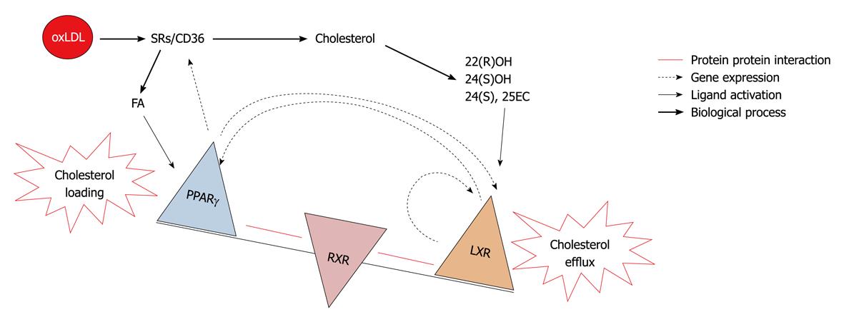
PPARγ is the closely related nuclear receptor to LXR and shares a common role in macrophage cholesterol turn over. Both receptors shares some common features, such as they form heterodimers with RXR and their endogenous activators are oxidized lipid molecules i.e. oxidized fatty acids for PPARγ and oxidized sterols for the LXR. Both are involved in the regulation of lipid metabolism in adipose tissue, macrophage and liver. Transplantation of PPARγ null bone marrow into LDLR-/- mice resulted in a significant increase in atherosclerosis[89]. Further it was reported that TZDs inhibited the development of atherosclerosis in LDLR deficient male mice[90], similar results were reported in the atherosclerotic model of apoE-/- mice[91]. All the above reported observations consider PPARγ as an antiatherogenic molecule. PPARγ expression has also been found in foam cells of atherosclerotic lesions and its expression could be increased with the oxidized LDL. PPARγ enhances uptake of oxidized LDL but not native LDL[92] by inducing the scavenger receptor CD36 leading to a vicious cycle of cholesterol loading and foam cell formation. In concordance with others, our studies suggest that PPARγ and LXR-α regulate each other. PPARγ activators have been found to enhance cholesterol efflux via the LXR-ABCA1 dependent pathway[93]. In two independent studies from our laboratory with PPARγ and LXR-α knock down cellular models using an siRNA approach it was found that both regulate expression of each other[75,94]. So the existence of this transcriptional cascade predicts that alterations in one of the elements in the cascade will affect all others and the net effect on cholesterol levels in the cell depends on how the balance between influx and efflux changes. Thus central activity of LXR-α determines if lipids are eliminated through cholesterol efflux towards HDL or accumulate and form foam cells from macrophages to induce lesion formation. This model highlights LXR-α molecule as a decision maker for atherosclerosis (Figure 5).
From in vitro to in vivo model systems, it is now clear that LXR-α have the ability to regulate key processes i.e. lipid metabolism and inflammation, which are associated with the pathogenesis of atherosclerosis, making it a candidate molecule for the therapeutic intervention of the world’s
highest death causing disease. It has been shown that treatment with a synthetic LXR agonist GW3965 can reduce atherosclerotic lesion development in two mouse models (i.e. LDLR-/- and apoE-/- mice)[95]. In addition to this, effectiveness of another synthetic agonist T-0901317 has also been reported[96]. It is evident that anti-atherosclerotic actions of synthetic LXR agonists in murine models is to a large extent independent from changes in the plasma lipid profile which indicates that this effect is predominantly a consequence of direct action of LXR activators on the vascular wall. Consistent with this notion, synthetic LXR agonists were shown to stimulate ABCA1 and ABCG1 expression in the atherosclerotic lesions of both LDL receptor- and apoE-deficient mice[95,96]. Subsequent experiments using bone marrow transplantation approaches provided direct evidence for a protective role of macrophage LXRs in atherosclerosis development. Tangirala et al[97] demonstrated that hematopoietic stem cell-specific LXRα/β deficiency aggravates atherosclerosis in both apoE and LDL receptor null mice. Another mechanism that could potentially contribute to the antiatherosclerotic action of LXR activators is their suppressing effect on macrophage inflammatory mediators production. Joseph et al[4] demonstrated that GW3965 and T0901317 inhibit expression of iNOS, cyclooxygenase-2 and interleukin-6 in macrophages subjected to bacterial infection or lipopolysaccharide stimulation. This inhibition depends on both LXRα and LXRβ and is mediated through suppression of the NF-κB signaling. Anti-inflammatory action of LXR agonists has been confirmed in vivo in a model of contact dermatitis and in the aortas of atherosclerotic mice[4].
In addition to stimulating reverse cholesterol transport and repressing inflammatory responses, LXR-α agonists may inhibit atherogenesis in several ways. For example T0901317 and GW3965 suppress platelet derived growth factor or insulin induced proliferation of vascular smooth muscle cells by inhibiting cell cycle progression from G1 to S phase[98]. The proliferation of smooth muscle cell plays an important role in growth of atherosclerotic plaques. It has been found that LXR agonist reduces the expression of cyclin D1 and cyclin A, which stimulates cyclin dependent kinases. Further TF is abundant in the lipid rich core of atherosclerotic plaques, and plaque rupture induces coagulation by exposing TF to circulating blood. It has been reported that synthetic LXR agonist attenuates LPS, TNF-α, and IL-1β induced TF expression in murine and human macrophages[71]. GW3965 has also been found to reduce cytokine induced synthesis and secretion of MMP-9, which is responsible for degrading the fibrous cap of atherosclerotic plaques and contributes to plaque rupturing[70]. A recent report shows that[99] LXR agonist attenuates the stimulatory effect of homocysteine (Hcy) on immunoglobin production by B-lymphocyte, by attenuating reactive oxygen species (ROS) and NF-κB activity. Although this study relates to a specific function of immune cells, these results are of great interest, taking into consideration the important role of the immune response in atherogenesis as well as an involvement of ROS and Hcy in cardiovascular pathology.
The immunohistochemical study indicated that LXR-α is highly expressed in macrophages present in human atherosclerotic lesions[100]. Our previous study also demonstrated that blood cellular LXR-α gene expression was higher in normolipidemic and hyperlipidemic CHD groups as compared to their corresponding groups[101], suggesting nature’s protection against the development of CHD. In concordance with others[102] findings from our laboratory also stated that the statins, which are the best drug of choice for the treatment of CHD, exert their effect via the upregulation of LXR-α gene expression[103]. Vitamin C also shares a common pathway for the upregulation of LXR-α[103]. The naturally occurring polyphenol resveratrol has been associated with the beneficial effects of red wine consumption on cardiovascular disease and has been shown to inhibit atherosclerosis in animal models. Resveratrol was shown to regulate the expression of LXR-α in human macrophages, which could be a possible molecular explanation for the beneficial effects of polyphenols[104]. However our recent study revealed the existence of deregulated LXR-α transcriptome and a paradoxical relationship between blood cellular LXR-α mRNA expression and the severity of coronary occlusion, which was explained by the presence of three critical mutations in the ligand binding domain comprising Asp324, Pro327 and Arg328, responsible for the inability of this domain to interact with its natural ligands leading thereby to a deregulated LXR-α transcriptome[35]. Keeping in view the importance of LXR-α signaling in CHD our study has raised the thrust for the search for alternative ligands for the restoration of deregulated LXR-α genomics in subjects suffering from CHD.
In summary, from cellular to animal model systems LXR activators have been found to be atheroprotective in nature by promoting reverse cholesterol transport, suppressing inflammatory processes and inhibiting vascular smooth muscle cell proliferation. Further higher expression of LXRs in the atherosclerotic lesions as well as in the peripheral blood mononuclear cells of CHD patients shows nature’s protection against the development of CHD.
LXR-α signaling pathway has an established role in atherosclerosis and possesses all the features of a candidate molecule for the treatment of CHD by its ability to modulate genes involved not only in the cellular lipid homeostasis but also in the control of inflammatory processes. The great challenge lies in the development of the alternative ligands for LXR-α having the pharmaceutical values, which can activate them and can modulate genes selectively by means of promoting reverse cholesterol transport, inhibiting inflammatory processes and avoiding its lipogenic activities. In addition to this futuristic research, safe and effective LXR-α therapeutics will make LXR-α as an extraordinary target for the treatment of CHD.
Peer reviewer: Cuihua Zhang, MD, PhD, FAHA, Associate Professor, Division of Cardiovascular Medicine, Department of Internal Medicine, Medical, Pharmacology and Physiology and Nutritional Sciences, Dalton Cardiovascular Research Center, University of Missouri-Columbia, 134 Research Park Drive, Columbia, MO 65211, United States
S- Editor Cheng JX L- Editor O’Neill M E- Editor Zheng XM
1. | Kutuk O, Basaga H. Inflammation meets oxidation: NF-kappaB as a mediator of initial lesion development in atherosclerosis. Trends Mol Med. 2003;9:549-557. [Cited in This Article: ] |
2. | Ross R. Atherosclerosis--an inflammatory disease. N Engl J Med. 1999;340:115-126. [Cited in This Article: ] |
3. | Saxena U, Goldberg IJ. Endothelial cells and atherosclerosis: lipoprotein metabolism, matrix interactions, and monocyte recruitment. Curr Opin Lipidol. 1994;5:316-322. [Cited in This Article: ] |
4. | Joseph SB, Castrillo A, Laffitte BA, Mangelsdorf DJ, Tontonoz P. Reciprocal regulation of inflammation and lipid metabolism by liver X receptors. Nat Med. 2003;9:213-219. [Cited in This Article: ] |
5. | Endemann G, Stanton LW, Madden KS, Bryant CM, White RT, Protter AA. CD36 is a receptor for oxidized low density lipoprotein. J Biol Chem. 1993;268:11811-11816. [Cited in This Article: ] |
6. | Podrez EA, Abu-Soud HM, Hazen SL. Myeloperoxidase-generated oxidants and atherosclerosis. Free Radic Biol Med. 2000;28:1717-1725. [Cited in This Article: ] |
7. | O'Brien KD, Alpers CE, Hokanson JE, Wang S, Chait A. Oxidation-specific epitopes in human coronary atherosclerosis are not limited to oxidized low-density lipoprotein. Circulation. 1996;94:1216-1225. [Cited in This Article: ] |
8. | Hulthe J, Fagerberg B. Circulating oxidized LDL is associated with subclinical atherosclerosis development and inflammatory cytokines (AIR Study). Arterioscler Thromb Vasc Biol. 2002;22:1162-1167. [Cited in This Article: ] |
9. | Ross R, Faggiotto A, Bowen-Pope D, Raines E. The role of endothelial injury and platelet and macrophage interactions in atherosclerosis. Circulation. 1984;70:III77-III82. [Cited in This Article: ] |
10. | Bonetti PO, Lerman LO, Lerman A. Endothelial dysfunction: a marker of atherosclerotic risk. Arterioscler Thromb Vasc Biol. 2003;23:168-175. [Cited in This Article: ] |
11. | Tegos TJ, Kalodiki E, Sabetai MM, Nicolaides AN. The genesis of atherosclerosis and risk factors: a review. Angiology. 2001;52:89-98. [Cited in This Article: ] |
12. | Cathcart MK, Folcik VA. Lipoxygenases and atherosclerosis: protection versus pathogenesis. Free Radic Biol Med. 2000;28:1726-1734. [Cited in This Article: ] |
13. | Hazen SL, Heinecke JW. 3-Chlorotyrosine, a specific marker of myeloperoxidase-catalyzed oxidation, is markedly elevated in low density lipoprotein isolated from human atherosclerotic intima. J Clin Invest. 1997;99:2075-2081. [Cited in This Article: ] |
14. | Chia MC. The role of adhesion molecules in atherosclerosis. Crit Rev Clin Lab Sci. 1998;35:573-602. [Cited in This Article: ] |
15. | Dong ZM, Chapman SM, Brown AA, Frenette PS, Hynes RO, Wagner DD. The combined role of P- and E-selectins in atherosclerosis. J Clin Invest. 1998;102:145-152. [Cited in This Article: ] |
16. | O'Brien KD, McDonald TO, Chait A, Allen MD, Alpers CE. Neovascular expression of E-selectin, intercellular adhesion molecule-1, and vascular cell adhesion molecule-1 in human atherosclerosis and their relation to intimal leukocyte content. Circulation. 1996;93:672-682. [Cited in This Article: ] |
17. | Harrington JR. The role of MCP-1 in atherosclerosis. Stem Cells. 2000;18:65-66. [Cited in This Article: ] |
18. | Hemmerich S, Paavola C, Bloom A, Bhakta S, Freedman R, Grunberger D, Krstenansky J, Lee S, McCarley D, Mulkins M. Identification of residues in the monocyte chemotactic protein-1 that contact the MCP-1 receptor, CCR2. Biochemistry. 1999;38:13013-13025. [Cited in This Article: ] |
19. | Krishnaswamy G, Kelley J, Yerra L, Smith JK, Chi DS. Human endothelium as a source of multifunctional cytokines: molecular regulation and possible role in human disease. J Interferon Cytokine Res. 1999;19:91-104. [Cited in This Article: ] |
20. | Sakai M, Kobori S, Miyazaki A, Horiuchi S. Macrophage proliferation in atherosclerosis. Curr Opin Lipidol. 2000;11:503-509. [Cited in This Article: ] |
21. | de Villiers WJ, Smart EJ. Macrophage scavenger receptors and foam cell formation. J Leukoc Biol. 1999;66:740-746. [Cited in This Article: ] |
22. | Brand K, Page S, Walli AK, Neumeier D, Baeuerle PA. Role of nuclear factor-kappa B in atherogenesis. Exp Physiol. 1997;82:297-304. [Cited in This Article: ] |
23. | Shin WS, Szuba A, Rockson SG. The role of chemokines in human cardiovascular pathology: enhanced biological insights. Atherosclerosis. 2002;160:91-102. [Cited in This Article: ] |
24. | Apfel R, Benbrook D, Lernhardt E, Ortiz MA, Salbert G, Pfahl M. A novel orphan receptor specific for a subset of thyroid hormone-responsive elements and its interaction with the retinoid/thyroid hormone receptor subfamily. Mol Cell Biol. 1994;14:7025-7035. [Cited in This Article: ] |
25. | Willy PJ, Umesono K, Ong ES, Evans RM, Heyman RA, Mangelsdorf DJ. LXR, a nuclear receptor that defines a distinct retinoid response pathway. Genes Dev. 1995;9:1033-1045. [Cited in This Article: ] |
26. | Repa JJ, Mangelsdorf DJ. The role of orphan nuclear receptors in the regulation of cholesterol homeostasis. Annu Rev Cell Dev Biol. 2000;16:459-481. [Cited in This Article: ] |
27. | Chawla A, Repa JJ, Evans RM, Mangelsdorf DJ. Nuclear receptors and lipid physiology: opening the X-files. Science. 2001;294:1866-1870. [Cited in This Article: ] |
28. | Wójcicka G, Jamroz-Wiśniewska A, Horoszewicz K, Bełtowski J. Liver X receptors (LXRs). Part I: structure, function, regulation of activity, and role in lipid metabolism. Postepy Hig Med Dosw (Online). 2007;61:736-759. [Cited in This Article: ] |
29. | Janowski BA, Willy PJ, Devi TR, Falck JR, Mangelsdorf DJ. An oxysterol signalling pathway mediated by the nuclear receptor LXR alpha. Nature. 1996;383:728-731. [Cited in This Article: ] |
30. | Lehmann JM, Kliewer SA, Moore LB, Smith-Oliver TA, Oliver BB, Su JL, Sundseth SS, Winegar DA, Blanchard DE, Spencer TA. Activation of the nuclear receptor LXR by oxysterols defines a new hormone response pathway. J Biol Chem. 1997;272:3137-3140. [Cited in This Article: ] |
31. | Janowski BA, Grogan MJ, Jones SA, Wisely GB, Kliewer SA, Corey EJ, Mangelsdorf DJ. Structural requirements of ligands for the oxysterol liver X receptors LXRalpha and LXRbeta. Proc Natl Acad Sci USA. 1999;96:266-271. [Cited in This Article: ] |
32. | Björkhem I, Meaney S, Diczfalusy U. Oxysterols in human circulation: which role do they have? Curr Opin Lipidol. 2002;13:247-253. [Cited in This Article: ] |
33. | Mitro N, Mak PA, Vargas L, Godio C, Hampton E, Molteni V, Kreusch A, Saez E. The nuclear receptor LXR is a glucose sensor. Nature. 2007;445:219-223. [Cited in This Article: ] |
34. | Denechaud PD, Bossard P, Lobaccaro JM, Millatt L, Staels B, Girard J, Postic C. ChREBP, but not LXRs, is required for the induction of glucose-regulated genes in mouse liver. J Clin Invest. 2008;118:956-964. [Cited in This Article: ] |
35. | Dave VP, Kaul D, Sharma Y, Bhattacharya R. Functional genomics of blood cellular LXR-alpha gene in human coronary heart disease. J Mol Cell Cardiol. 2009;46:536-544. [Cited in This Article: ] |
36. | Bramlett KS, Houck KA, Borchert KM, Dowless MS, Kulanthaivel P, Zhang Y, Beyer TP, Schmidt R, Thomas JS, Michael LF. A natural product ligand of the oxysterol receptor, liver X receptor. J Pharmacol Exp Ther. 2003;307:291-296. [Cited in This Article: ] |
37. | Tamehiro N, Sato Y, Suzuki T, Hashimoto T, Asakawa Y, Yokoyama S, Kawanishi T, Ohno Y, Inoue K, Nagao T. Riccardin C: a natural product that functions as a liver X receptor (LXR)alpha agonist and an LXRbeta antagonist. FEBS Lett. 2005;579:5299-5304. [Cited in This Article: ] |
38. | Gan X, Kaplan R, Menke JG, MacNaul K, Chen Y, Sparrow CP, Zhou G, Wright SD, Cai TQ. Dual mechanisms of ABCA1 regulation by geranylgeranyl pyrophosphate. J Biol Chem. 2001;276:48702-48708. [Cited in This Article: ] |
39. | Forman BM, Ruan B, Chen J, Schroepfer GJ Jr, Evans RM. The orphan nuclear receptor LXRalpha is positively and negatively regulated by distinct products of mevalonate metabolism. Proc Natl Acad Sci USA. 1997;94:10588-10593. [Cited in This Article: ] |
40. | Yoshikawa T, Shimano H, Yahagi N, Ide T, Amemiya-Kudo M, Matsuzaka T, Nakakuki M, Tomita S, Okazaki H, Tamura Y. Polyunsaturated fatty acids suppress sterol regulatory element-binding protein 1c promoter activity by inhibition of liver X receptor (LXR) binding to LXR response elements. J Biol Chem. 2002;277:1705-1711. [Cited in This Article: ] |
41. | Ou J, Tu H, Shan B, Luk A, DeBose-Boyd RA, Bashmakov Y, Goldstein JL, Brown MS. Unsaturated fatty acids inhibit transcription of the sterol regulatory element-binding protein-1c (SREBP-1c) gene by antagonizing ligand-dependent activation of the LXR. Proc Natl Acad Sci USA. 2001;98:6027-6032. [Cited in This Article: ] |
42. | Chiang JY, Kimmel R, Stroup D. Regulation of cholesterol 7alpha-hydroxylase gene (CYP7A1) transcription by the liver orphan receptor (LXRalpha). Gene. 2001;262:257-265. [Cited in This Article: ] |
43. | Lewis GF, Rader DJ. New insights into the regulation of HDL metabolism and reverse cholesterol transport. Circ Res. 2005;96:1221-1232. [Cited in This Article: ] |
44. | Repa JJ, Turley SD, Lobaccaro JA, Medina J, Li L, Lustig K, Shan B, Heyman RA, Dietschy JM, Mangelsdorf DJ. Regulation of absorption and ABC1-mediated efflux of cholesterol by RXR heterodimers. Science. 2000;289:1524-1529. [Cited in This Article: ] |
45. | Borst P, Elferink RO. Mammalian ABC transporters in health and disease. Annu Rev Biochem. 2002;71:537-592. [Cited in This Article: ] |
46. | Costet P, Luo Y, Wang N, Tall AR. Sterol-dependent transactivation of the ABC1 promoter by the liver X receptor/retinoid X receptor. J Biol Chem. 2000;275:28240-28245. [Cited in This Article: ] |
47. | Repa JJ, Berge KE, Pomajzl C, Richardson JA, Hobbs H, Mangelsdorf DJ. Regulation of ATP-binding cassette sterol transporters ABCG5 and ABCG8 by the liver X receptors alpha and beta. J Biol Chem. 2002;277:18793-18800. [Cited in This Article: ] |
48. | Berge KE, von Bergmann K, Lutjohann D, Guerra R, Grundy SM, Hobbs HH, Cohen JC. Heritability of plasma noncholesterol sterols and relationship to DNA sequence polymorphism in ABCG5 and ABCG8. J Lipid Res. 2002;43:486-494. [Cited in This Article: ] |
49. | Venkateswaran A, Repa JJ, Lobaccaro JM, Bronson A, Mangelsdorf DJ, Edwards PA. Human white/murine ABC8 mRNA levels are highly induced in lipid-loaded macrophages. A transcriptional role for specific oxysterols. J Biol Chem. 2000;275:14700-14707. [Cited in This Article: ] |
50. | Sabol SL, Brewer HB Jr, Santamarina-Fojo S. The human ABCG1 gene: identification of LXR response elements that modulate expression in macrophages and liver. J Lipid Res. 2005;46:2151-2167. [Cited in This Article: ] |
51. | Kennedy MA, Venkateswaran A, Tarr PT, Xenarios I, Kudoh J, Shimizu N, Edwards PA. Characterization of the human ABCG1 gene: liver X receptor activates an internal promoter that produces a novel transcript encoding an alternative form of the protein. J Biol Chem. 2001;276:39438-39447. [Cited in This Article: ] |
52. | Bodzioch M, Orsó E, Klucken J, Langmann T, Böttcher A, Diederich W, Drobnik W, Barlage S, Büchler C, Porsch-Ozcürümez M. The gene encoding ATP-binding cassette transporter 1 is mutated in Tangier disease. Nat Genet. 1999;22:347-351. [Cited in This Article: ] |
54. | Schwartz K, Lawn RM, Wade DP. ABC1 gene expression and ApoA-I-mediated cholesterol efflux are regulated by LXR. Biochem Biophys Res Commun. 2000;274:794-802. [Cited in This Article: ] |
55. | Venkateswaran A, Laffitte BA, Joseph SB, Mak PA, Wilpitz DC, Edwards PA, Tontonoz P. Control of cellular cholesterol efflux by the nuclear oxysterol receptor LXR alpha. Proc Natl Acad Sci USA. 2000;97:12097-12102. [Cited in This Article: ] |
56. | Laffitte BA, Repa JJ, Joseph SB, Wilpitz DC, Kast HR, Mangelsdorf DJ, Tontonoz P. LXRs control lipid-inducible expression of the apolipoprotein E gene in macrophages and adipocytes. Proc Natl Acad Sci USA. 2001;98:507-512. [Cited in This Article: ] |
57. | Mak PA, Laffitte BA, Desrumaux C, Joseph SB, Curtiss LK, Mangelsdorf DJ, Tontonoz P, Edwards PA. Regulated expression of the apolipoprotein E/C-I/C-IV/C-II gene cluster in murine and human macrophages. A critical role for nuclear liver X receptors alpha and beta. J Biol Chem. 2002;277:31900-31908. [Cited in This Article: ] |
58. | Hummasti S, Laffitte BA, Watson MA, Galardi C, Chao LC, Ramamurthy L, Moore JT, Tontonoz P. Liver X receptors are regulators of adipocyte gene expression but not differentiation: identification of apoD as a direct target. J Lipid Res. 2004;45:616-625. [Cited in This Article: ] |
59. | Curtiss LK, Boisvert WA. Apolipoprotein E and atherosclerosis. Curr Opin Lipidol. 2000;11:243-251. [Cited in This Article: ] |
60. | Cao G, Beyer TP, Yang XP, Schmidt RJ, Zhang Y, Bensch WR, Kauffman RF, Gao H, Ryan TP, Liang Y. Phospholipid transfer protein is regulated by liver X receptors in vivo. J Biol Chem. 2002;277:39561-39565. [Cited in This Article: ] |
61. | Laffitte BA, Joseph SB, Chen M, Castrillo A, Repa J, Wilpitz D, Mangelsdorf D, Tontonoz P. The phospholipid transfer protein gene is a liver X receptor target expressed by macrophages in atherosclerotic lesions. Mol Cell Biol. 2003;23:2182-2191. [Cited in This Article: ] |
62. | Luo Y, Tall AR. Sterol upregulation of human CETP expression in vitro and in transgenic mice by an LXR element. J Clin Invest. 2000;105:513-520. [Cited in This Article: ] |
63. | Mak PA, Kast-Woelbern HR, Anisfeld AM, Edwards PA. Identification of PLTP as an LXR target gene and apoE as an FXR target gene reveals overlapping targets for the two nuclear receptors. J Lipid Res. 2002;43:2037-2041. [Cited in This Article: ] |
64. | Schultz JR, Tu H, Luk A, Repa JJ, Medina JC, Li L, Schwendner S, Wang S, Thoolen M, Mangelsdorf DJ. Role of LXRs in control of lipogenesis. Genes Dev. 2000;14:2831-2838. [Cited in This Article: ] |
65. | Joseph SB, Laffitte BA, Patel PH, Watson MA, Matsukuma KE, Walczak R, Collins JL, Osborne TF, Tontonoz P. Direct and indirect mechanisms for regulation of fatty acid synthase gene expression by liver X receptors. J Biol Chem. 2002;277:11019-11025. [Cited in This Article: ] |
66. | Repa JJ, Liang G, Ou J, Bashmakov Y, Lobaccaro JM, Shimomura I, Shan B, Brown MS, Goldstein JL, Mangelsdorf DJ. Regulation of mouse sterol regulatory element-binding protein-1c gene (SREBP-1c) by oxysterol receptors, LXRalpha and LXRbeta. Genes Dev. 2000;14:2819-2305. [Cited in This Article: ] |
67. | Yoshikawa T, Shimano H, Amemiya-Kudo M, Yahagi N, Hasty AH, Matsuzaka T, Okazaki H, Tamura Y, Iizuka Y, Ohashi K. Identification of liver X receptor-retinoid X receptor as an activator of the sterol regulatory element-binding protein 1c gene promoter. Mol Cell Biol. 2001;21:2991-3000. [Cited in This Article: ] |
68. | Peet DJ, Turley SD, Ma W, Janowski BA, Lobaccaro JM, Hammer RE, Mangelsdorf DJ. Cholesterol and bile acid metabolism are impaired in mice lacking the nuclear oxysterol receptor LXR alpha. Cell. 1998;93:693-704. [Cited in This Article: ] |
69. | Glass CK, Witztum JL. Atherosclerosis. the road ahead. Cell. 2001;104:503-516. [Cited in This Article: ] |
70. | Castrillo A, Joseph SB, Marathe C, Mangelsdorf DJ, Tontonoz P. Liver X receptor-dependent repression of matrix metalloproteinase-9 expression in macrophages. J Biol Chem. 2003;278:10443-10449. [Cited in This Article: ] |
71. | Terasaka N, Hiroshima A, Ariga A, Honzumi S, Koieyama T, Inaba T, Fujiwara T. Liver X receptor agonists inhibit tissue factor expression in macrophages. FEBS J. 2005;272:1546-1556. [Cited in This Article: ] |
72. | De Bosscher K, Vanden Berghe W, Haegeman G. Cross-talk between nuclear receptors and nuclear factor kappaB. Oncogene. 2006;25:6868-6886. [Cited in This Article: ] |
73. | Castrillo A, Joseph SB, Vaidya SA, Haberland M, Fogelman AM, Cheng G, Tontonoz P. Crosstalk between LXR and toll-like receptor signaling mediates bacterial and viral antagonism of cholesterol metabolism. Mol Cell. 2003;12:805-816. [Cited in This Article: ] |
74. | Arai S, Shelton JM, Chen M, Bradley MN, Castrillo A, Bookout AL, Mak PA, Edwards PA, Mangelsdorf DJ, Tontonoz P. A role for the apoptosis inhibitory factor AIM/Spalpha/Api6 in atherosclerosis development. Cell Metab. 2005;1:201-213. [Cited in This Article: ] |
75. | Kaul D, Gautam A, Sikand K. Importance of LXR-alpha transcriptome in the modulation of innate immunity. Mol Cell Biochem. 2006;292:53-57. [Cited in This Article: ] |
76. | Cao G, Liang Y, Broderick CL, Oldham BA, Beyer TP, Schmidt RJ, Zhang Y, Stayrook KR, Suen C, Otto KA. Antidiabetic action of a liver x receptor agonist mediated by inhibition of hepatic gluconeogenesis. J Biol Chem. 2003;278:1131-1136. [Cited in This Article: ] |
77. | Liu Y, Yan C, Wang Y, Nakagawa Y, Nerio N, Anghel A, Lutfy K, Friedman TC. Liver X receptor agonist T0901317 inhibition of glucocorticoid receptor expression in hepatocytes may contribute to the amelioration of diabetic syndrome in db/db mice. Endocrinology. 2006;147:5061-5068. [Cited in This Article: ] |
78. | Laffitte BA, Chao LC, Li J, Walczak R, Hummasti S, Joseph SB, Castrillo A, Wilpitz DC, Mangelsdorf DJ, Collins JL. Activation of liver X receptor improves glucose tolerance through coordinate regulation of glucose metabolism in liver and adipose tissue. Proc Natl Acad Sci USA. 2003;100:5419-5424. [Cited in This Article: ] |
79. | Stulnig TM, Steffensen KR, Gao H, Reimers M, Dahlman-Wright K, Schuster GU, Gustafsson JA. Novel roles of liver X receptors exposed by gene expression profiling in liver and adipose tissue. Mol Pharmacol. 2002;62:1299-1305. [Cited in This Article: ] |
80. | Dalen KT, Ulven SM, Bamberg K, Gustafsson JA, Nebb HI. Expression of the insulin-responsive glucose transporter GLUT4 in adipocytes is dependent on liver X receptor alpha. J Biol Chem. 2003;278:48283-48291. [Cited in This Article: ] |
81. | Ulven SM, Dalen KT, Gustafsson JA, Nebb HI. Tissue-specific autoregulation of the LXRalpha gene facilitates induction of apoE in mouse adipose tissue. J Lipid Res. 2004;45:2052-2062. [Cited in This Article: ] |
82. | Grefhorst A, van Dijk TH, Hammer A, van der Sluijs FH, Havinga R, Havekes LM, Romijn JA, Groot PH, Reijngoud DJ, Kuipers F. Differential effects of pharmacological liver X receptor activation on hepatic and peripheral insulin sensitivity in lean and ob/ob mice. Am J Physiol Endocrinol Metab. 2005;289:E829-E838. [Cited in This Article: ] |
83. | Commerford SR, Vargas L, Dorfman SE, Mitro N, Rocheford EC, Mak PA, Li X, Kennedy P, Mullarkey TL, Saez E. Dissection of the insulin-sensitizing effect of liver X receptor ligands. Mol Endocrinol. 2007;21:3002-3012. [Cited in This Article: ] |
84. | Efanov AM, Sewing S, Bokvist K, Gromada J. Liver X receptor activation stimulates insulin secretion via modulation of glucose and lipid metabolism in pancreatic beta-cells. Diabetes. 2004;53 Suppl 3:S75-S78. [Cited in This Article: ] |
85. | Zitzer H, Wente W, Brenner MB, Sewing S, Buschard K, Gromada J, Efanov AM. Sterol regulatory element-binding protein 1 mediates liver X receptor-beta-induced increases in insulin secretion and insulin messenger ribonucleic acid levels. Endocrinology. 2006;147:3898-3905. [Cited in This Article: ] |
86. | Chen M, Beaven S, Tontonoz P. Identification and characterization of two alternatively spliced transcript variants of human liver X receptor alpha. J Lipid Res. 2005;46:2570-2579. [Cited in This Article: ] |
87. | Laffitte BA, Joseph SB, Walczak R, Pei L, Wilpitz DC, Collins JL, Tontonoz P. Autoregulation of the human liver X receptor alpha promoter. Mol Cell Biol. 2001;21:7558-7568. [Cited in This Article: ] |
88. | Chen M, Bradley MN, Beaven SW, Tontonoz P. Phosphorylation of the liver X receptors. FEBS Lett. 2006;580:4835-4841. [Cited in This Article: ] |
89. | Lowell BB. PPARgamma: an essential regulator of adipogenesis and modulator of fat cell function. Cell. 1999;99:239-242. [Cited in This Article: ] |
90. | Li AC, Brown KK, Silvestre MJ, Willson TM, Palinski W, Glass CK. Peroxisome proliferator-activated receptor gamma ligands inhibit development of atherosclerosis in LDL receptor-deficient mice. J Clin Invest. 2000;106:523-531. [Cited in This Article: ] |
91. | Chen Z, Ishibashi S, Perrey S, Osuga Ji, Gotoda T, Kitamine T, Tamura Y, Okazaki H, Yahagi N, Iizuka Y. Troglitazone inhibits atherosclerosis in apolipoprotein E-knockout mice: pleiotropic effects on CD36 expression and HDL. Arterioscler Thromb Vasc Biol. 2001;21:372-377. [Cited in This Article: ] |
92. | Tontonoz P, Nagy L, Alvarez JG, Thomazy VA, Evans RM. PPARgamma promotes monocyte/macrophage differentiation and uptake of oxidized LDL. Cell. 1998;93:241-252. [Cited in This Article: ] |
93. | Chawla A, Boisvert WA, Lee CH, Laffitte BA, Barak Y, Joseph SB, Liao D, Nagy L, Edwards PA, Curtiss LK. A PPAR gamma-LXR-ABCA1 pathway in macrophages is involved in cholesterol efflux and atherogenesis. Mol Cell. 2001;7:161-171. [Cited in This Article: ] |
94. | Kaul D, Anand PK, Khanna A. Functional genomics of PPAR-gamma in human immunomodulatory cells. Mol Cell Biochem. 2006;290:211-215. [Cited in This Article: ] |
95. | Joseph SB, McKilligin E, Pei L, Watson MA, Collins AR, Laffitte BA, Chen M, Noh G, Goodman J, Hagger GN. Synthetic LXR ligand inhibits the development of atherosclerosis in mice. Proc Natl Acad Sci USA. 2002;99:7604-7609. [Cited in This Article: ] |
96. | Terasaka N, Hiroshima A, Koieyama T, Ubukata N, Morikawa Y, Nakai D, Inaba T. T-0901317, a synthetic liver X receptor ligand, inhibits development of atherosclerosis in LDL receptor-deficient mice. FEBS Lett. 2003;536:6-11. [Cited in This Article: ] |
97. | Tangirala RK, Bischoff ED, Joseph SB, Wagner BL, Walczak R, Laffitte BA, Daige CL, Thomas D, Heyman RA, Mangelsdorf DJ. Identification of macrophage liver X receptors as inhibitors of atherosclerosis. Proc Natl Acad Sci USA. 2002;99:11896-11901. [Cited in This Article: ] |
98. | Blaschke F, Leppanen O, Takata Y, Caglayan E, Liu J, Fishbein MC, Kappert K, Nakayama KI, Collins AR, Fleck E. Liver X receptor agonists suppress vascular smooth muscle cell proliferation and inhibit neointima formation in balloon-injured rat carotid arteries. Circ Res. 2004;95:e110-e123. [Cited in This Article: ] |
99. | Chang L, Zhang Z, Li W, Dai J, Guan Y, Wang X. Liver-X-receptor activator prevents homocysteine-induced production of IgG antibodies from murine B lymphocytes via the ROS-NF-kappaB pathway. Biochem Biophys Res Commun. 2007;357:772-778. [Cited in This Article: ] |
100. | Watanabe Y, Jiang S, Takabe W, Ohashi R, Tanaka T, Uchiyama Y, Katsumi K, Iwanari H, Noguchi N, Naito M. Expression of the LXRalpha protein in human atherosclerotic lesions. Arterioscler Thromb Vasc Biol. 2005;25:622-627. [Cited in This Article: ] |
101. | Baba MI, Kaul D, Grover A. Importance of blood cellular genomic profile in coronary heart disease. J Biomed Sci. 2006;13:17-26. [Cited in This Article: ] |
102. | Argmann CA, Edwards JY, Sawyez CG, O'Neil CH, Hegele RA, Pickering JG, Huff MW. Regulation of macrophage cholesterol efflux through hydroxymethylglutaryl-CoA reductase inhibition: a role for RhoA in ABCA1-mediated cholesterol efflux. J Biol Chem. 2005;280:22212-22221. [Cited in This Article: ] |
103. | Kaul D, Baba MI. Genomic effect of vitamin 'C' and statins within human mononuclear cells involved in atherogenic process. Eur J Clin Nutr. 2005;59:978-981. [Cited in This Article: ] |
104. | Sevov M, Elfineh L, Cavelier LB. Resveratrol regulates the expression of LXR-alpha in human macrophages. Biochem Biophys Res Commun. 2006;348:1047-1054. [Cited in This Article: ] |