Revised: March 5, 2010
Accepted: March 12, 2010
Published online: April 26, 2010
AIM: To assess neovascularization within human carotid atherosclerotic soft plaques in patients with ischemic stroke.
METHODS: Eighty-one patients with ischemic stroke and 95 patients without stroke who had soft atherosclerotic plaques in the internal carotid artery were studied. The thickest soft plaque in each patient was examined using contrast-enhanced ultrasound. Time-intensity curves were collected from 5 s to 3 min after contrast injection. The neovascularization within the plaques in the internal carotid artery was evaluated using the ACQ software built into the scanner by 2 of the experienced investigators who were blinded to the clinical history of the patients.
RESULTS: Ischemic stroke was present in 7 of 33 patients (21%) with grade I plaque, in 14 of 51 patients (28%) with grade II plaque, in 26 of 43 patients (61%) with grade III plaque, and in 34 of 49 patients (69%) with grade IV plaque (P < 0.001 comparing grade IV plaque with grade I plaque and with grade II plaque and P = 0.001 comparing grade III plaque with grade I plaque and with grade II plaque). Analysis of the time intensity curves revealed that patients with ischemic stroke had a significantly higher intensity of enhancement (IE) than those without ischemic stroke (P < 0.01). The wash-in time (WT) of plaque was significantly shorter in stroke patients (P < 0.05). The sensitivity and specificity for IE in the plaque were 82% and 80%, respectively, and for WT were 68% and 74%, respectively. There was no significant difference in the peak intensity or time to peak between the 2 groups.
CONCLUSION: This study shows that the higher the grade of plaque enhancement, the higher the risk of ischemic stroke. The data suggest that the presence of neovascularization is a marker for unstable plaque.
- Citation: Huang PT, Chen CC, Aronow WS, Wang XT, Nair CK, Xue NY, Shen X, Li SY, Huang FG, Cosgrove D. Assessment of neovascularization within carotid plaques in patients with ischemic stroke. World J Cardiol 2010; 2(4): 89-97
- URL: https://www.wjgnet.com/1949-8462/full/v2/i4/89.htm
- DOI: https://dx.doi.org/10.4330/wjc.v2.i4.89
Carotid plaques are frequently found in patients who have suffered a stroke[1-3]. Neovascularization can be identified within the atherosclerotic plaque on endarterectomy specimens. In neovascularization, new vessels sprout from the vasa vasorum[4-6]. The development of neovascularization is considered to be an important phase in the development of plaque[7] and its vulnerability to rupture increases the risk of cerebral emboli[8,9]. Few imaging techniques can be used to detect and quantify this neovascularization[10,11]; for example, Doppler fails to do so because the neovessels are small with very slow flow and lie close to the major flow in the carotid artery lumen.
The purpose of this prospective study was to determine quantitatively whether there is a difference in the neovascular circulation within carotid plaques between patients with and without ischemic stroke using contrast-enhanced ultrasonography (CEUS).
Between September 2006 and May 2008, 86 patients with ischemic stroke were recruited in the Neurological Center of the 2nd Affiliated Hospital of Wenzhou Medical College. Ischemic stroke was defined as focal neurological symptoms lasting > 24 h with or without persisting disabilities together with a computerized tomographic scan positive for ischemic lesions, i.e. a discrete (> 10 mm) subcortical or cortical infarct in the anterior or middle cerebral artery territories, in the absence of a cardiac source of embolism (which was excluded on the basis of medical history and transesophageal echocardiography in every patient). For a patient to be included in the study, the stroke had to be recent, defined as not more than 30 d old. All patients had at least one soft plaque in the common carotid artery wall, its bifurcation, or the internal carotid artery, on the side relevant to the infarct.
Patients with ischemic infarction in the basilar artery territories were excluded as were those with transient ischemic attacks, amaurosis fugax and hemorrhagic strokes. Patients with bilateral hemispheric symptoms and those with known cardiac mural thrombus and patent foramen ovale, (as verified on transesophageal echocardiography) were also excluded from the analysis because of suspected cardioembolic origin.
Ninety-seven controls with soft carotid plaques were recruited from 1556 patients referred for thyroid ultrasound examination at the same hospital. History of stroke or other cardiovascular disease was assessed by the investigators. Individuals reporting a positive history of stroke were not eligible, whereas those reporting a positive cardiovascular history other than stroke were eligible.
Informed consent was obtained from all patients and controls prior to their examination, and the local ethics committee approved this study (No. 2006-012).
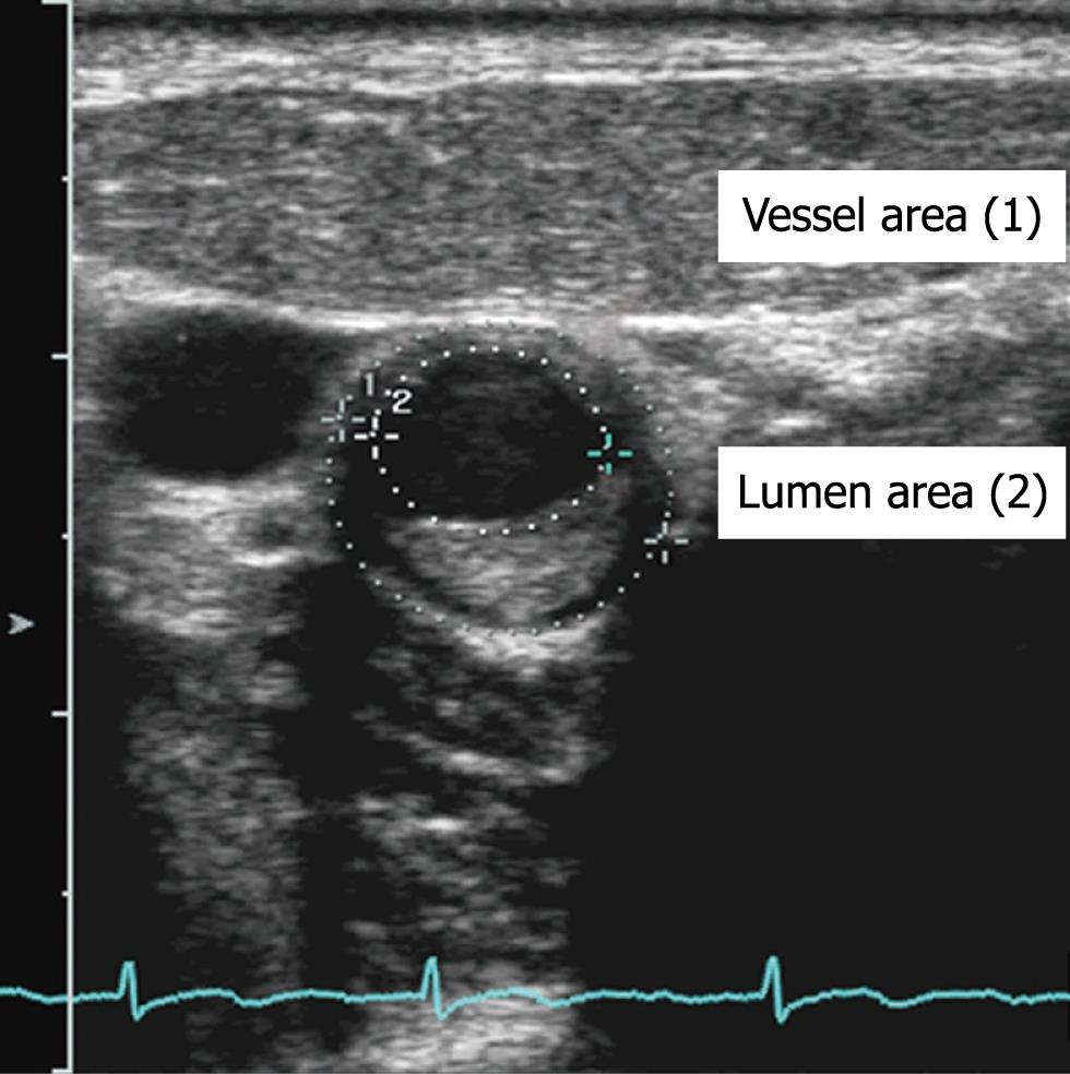
Carotid artery ultrasound was performed with an Acuson Sequoia 512 imaging system (Siemens, Mountain View, CA, USA) equipped with a 15L8 linear transducer (frequency: 8-14 MHz). With the subject supine, the extracranial carotid arteries were visualized in longitudinal and transverse sections. The common carotid arteries, carotid bifurcations, and internal carotid arteries were examined for the presence of atherosclerotic plaque which was defined as an intima-media thickness > 1.2 mm[12]. Soft plaque was defined as plaque tissue producing echogenicity less than that of the surrounding adventitia, in the absence of any calcium. The thickest soft plaque on either side (for the controls) and on the relevant side (for the stroke patients) was selected for study. In each case, the transverse view at the point of maximum plaque thickness was selected, and the corresponding cross-sectional outer vessel areas (VA) and lumen areas (LA) were manually traced (Figure 1). The VA was defined as the area within the adventitial border. Plaque area (PA) was calculated as VA-LA, and %PA (PPA) was calculated as (PA/VA) × 100 (%), as previously described by Erbel et al[13].
All subjects were examined with an Acuson Sequoia 512 imaging system equipped with Cadence™ contrast pulse sequencing technology (CPS)[14] by board-registered diagnostic radiologists with a similar amount of experience in vascular sonography (10-16 years). The thickest soft plaque located on the posterior carotid artery wall in each patient and control was examined with real-time ultrasonography using CPS, which uses phase and amplitude modulation to separate the microbubble signals from tissue echoes, at 7 MHz with a low mechanical index (0.35). The microbubble contrast agent SonoVue® (Bracco SpA, Milan, Italy) used in the study was supplied as a lyophilized powder. It was reconstituted by adding 5 mL of saline and gently shaking the vial by hand to form a homogeneous microbubble suspension. The suspension contains 8 μL/mL of sulfur hexafluoride gas stabilized by a phospholipid shell (microbubble concentration 5 mg/mL). The agent was administered intravenously as a 2 s bolus of 2.4 mL through a 19-gauge cannula in an antecubital vein. The cannula was flushed with 10 mL saline. Digital recording of the dynamic sequence was started 5 s after the injection and continued for 3 min.
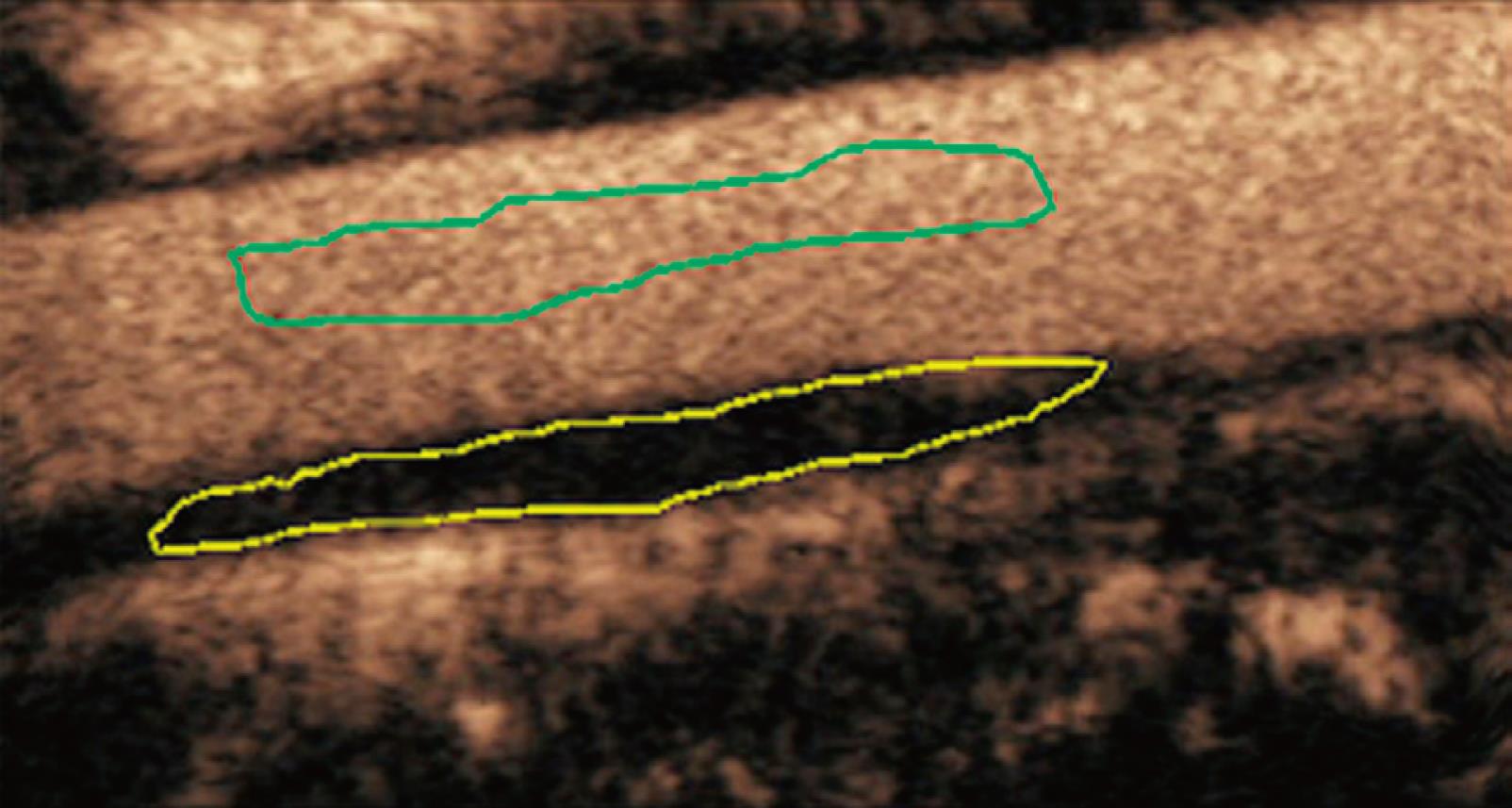
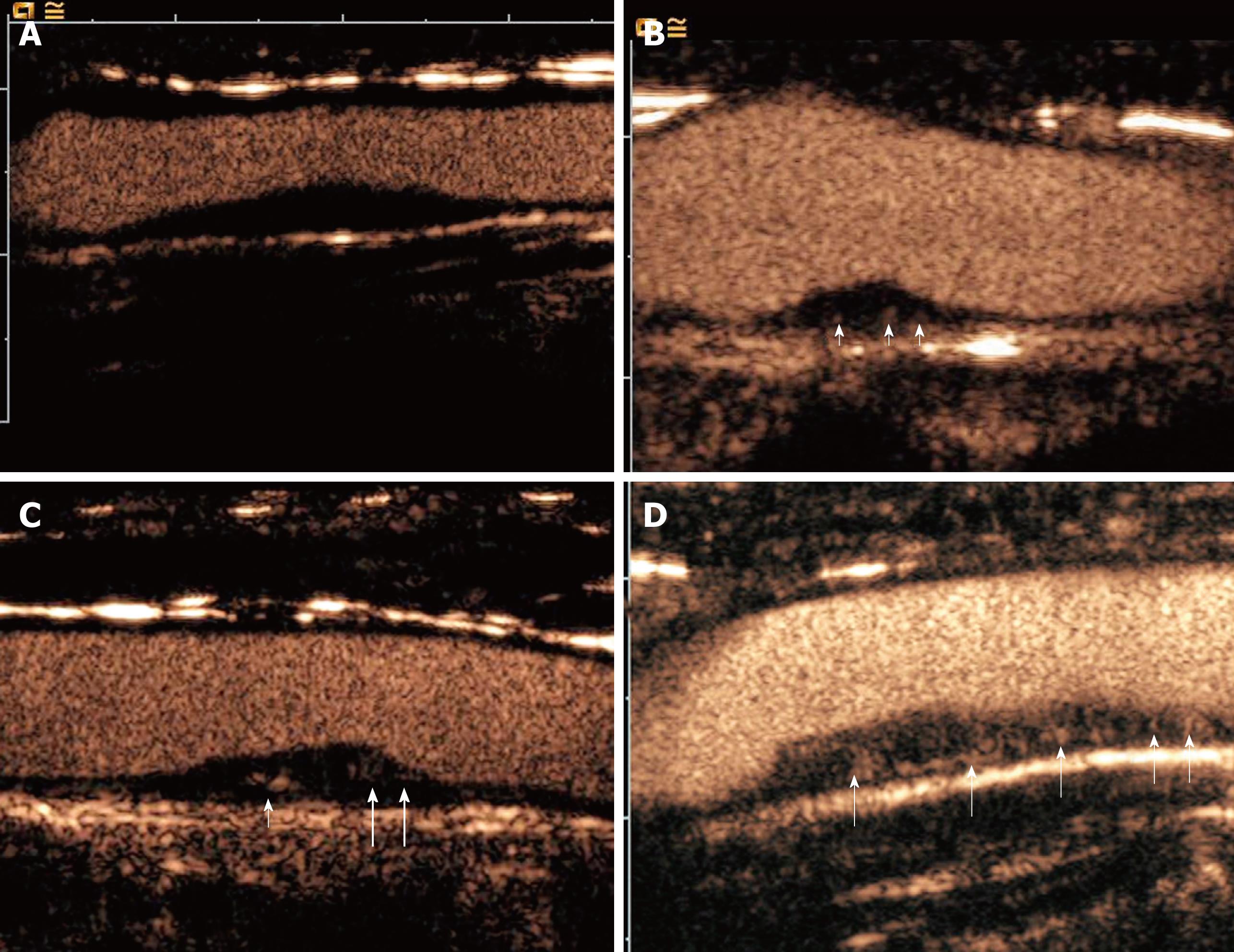
The digital cine clips were reviewed offline within 3 d of each study by 2 off-site reviewers with 5 years of experience in CEUS who had not been involved in the scanning and were blinded to the clinical data of the stroke patients and controls. The clip loop of each patient was automatically divided into 4 segments, and the first segment (during the arterial phase) was used. The neovascularization within the plaques in the posterior carotid artery was evaluated by visual interpretation and quantitative analysis using the ACQ software. Plaque CEUS enhancement was categorized: grade I: non-enhancement; grade II: the arterial wall vasa vasorum enhancement; grade III: the arterial wall vasa vasorum and plaque shoulder enhancement; and grade IV: extensive and internal plaque enhancement. For each case, a region-of interest (ROI) was then drawn freehand around the peripheral margin of the plaque using an electronic cursor. Care was taken to exclude the intralumen and periplaque tissues. To allow for the effects of movement due to the patient’s breathing, the ROI was adjusted manually frame by frame as necessary. A time-intensity curve (TIC) for the selected plaque tissue and the 4 perfusion parameters for the plaque tissue within the ROI was then derived. The mean values for the four plaque perfusion parameters for each individual patient were then computed by the built-in software (ACQ). These 4 perfusion parameters included arrival time (AT), time-to-peak (TTP), basal intensity (BI) and peak intensity (PI)[14] (Figure 2). Curves with a goodness of fit (GOF) > 0.75 were considered eligible for inclusion. The following normalized indices were calculated manually: The intensity of enhancement (IE) of the plaque was defined as PI minus BI; the wash-in time (WT) was defined as TTP minus AT. All measurements were performed 3 times, and the mean of these 3 measurements was calculated and compared for analysis.
Statistical analysis was performed with the Statistical Package for Social Sciences software (SPSS, version 9.0; SPSS, Chicago, IL, USA). Inter- and intra-observer variability for the CEUS quantitative analysis was assessed. The mean differences, standard deviation (SD), and 95% limits of agreement for each of the parameters of EI and WI for each observer and for both observers were calculated using the Bland-Altman test[15]. Intra-observer variability was determined by comparing the first and the second measurements of reviewer 1. Student’s t-test was used to determine whether there was a significant difference between the stroke patients and controls. Categorical variables including gender, smoking status, and diabetes status were compared between the 2 groups by χ2 analysis. The percent rate of stroke among different grades of plaque enhancement was also compared by chi-square analysis. A receiver-operating characteristic (ROC) curve was used to calculate the area under the ROC curve (AUC) to determine the grade of plaque enhancement, IE and WT cutoff value with the best sensitivity and specificity. A P value of less than 0.05 was considered to indicate a significant difference.
Between September 2006 and May 2008, 128 patients with strokes were referred to the Neurological Center of the 2nd Affiliated Hospital of Wenzhou Medical College. Of these patients, 42 cases were excluded for the following reasons: 26 had hemorrhagic strokes, 12 had ischemic infarction in the basilar artery territory and 4 had bilateral hemispheric symptoms and known cardiac mural thrombus. Among the remaining 86 patients and 97 controls, 5 patients with stroke were excluded because of GOF < 0.75 and 2 controls were excluded because of incomplete information on data forms. The risk factors and clinical features of the 81 patients with ischemic stroke and 95 controls are shown in Table 1.
| Variables | Ischemic stroke (n = 81) | Controls (n = 95) | P value |
| Age (yr) | 62 ± 9 | 61 ± 12 | NS |
| Men | 46 (57) | 54 (57) | NS |
| Women | 35 (43) | 41 (43) | NS |
| Systolic blood pressure (mmHg) | 146 ± 24 | 143 ± 22 | NS |
| Diastolic blood pressure (mmHg) | 80 ± 15 | 78 ± 11 | NS |
| Serum total cholesterol (mmol/L) | 6.7 ± 1.3 | 6.5 ± 1.1 | NS |
| Serum HDL cholesterol (mmol/L) | 1.4 ± 0.4 | 1.3 ± 0.4 | NS |
| Diabetes mellitus | 6 (7) | 7 (7) | NS |
| Smokers | 26 (32) | 30 (32) | NS |
| Percent carotid plaque area | 74 ± 4 | 74 ± 6 | NS |
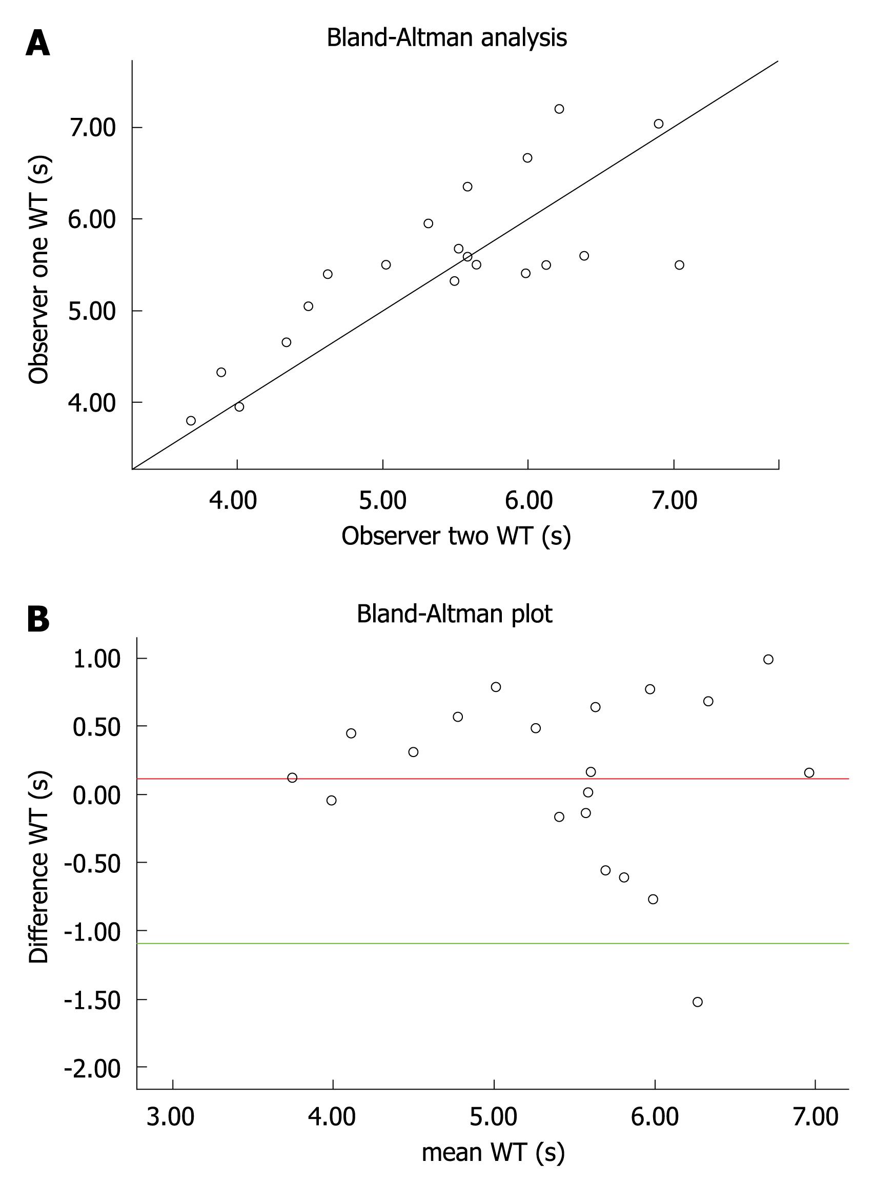
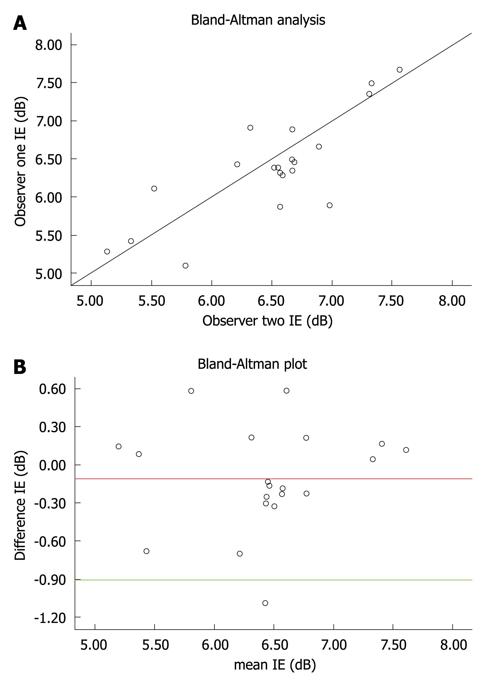
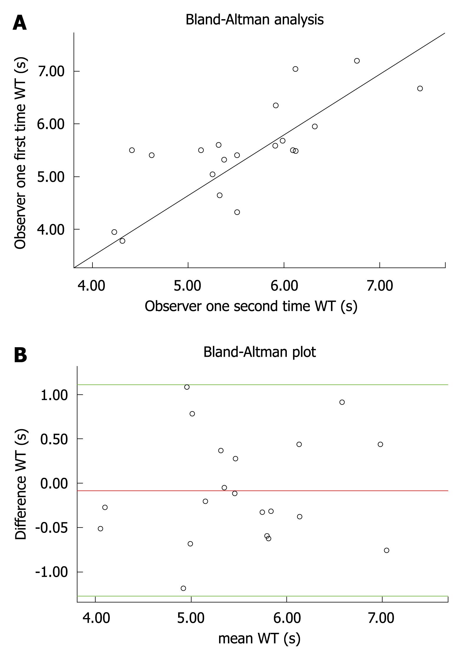
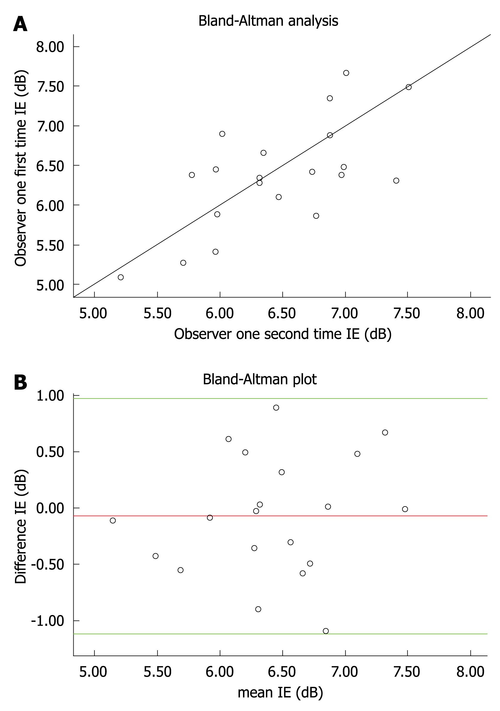
The intraclass correlation, mean difference, SD, and 95% limits of agreement for inter-observer measurements for each parameter are summarized in Table 2, with corresponding scatter and Bland-Altman agreement plots for IE and WT in Figures 3 and 4. Similar limits of agreement were obtained between the measurements from the 2 observers. The intraclass correlation ranged from 0.66 to 0.75, indicating good agreement.
| Perfusion measurement | Intraclass correlation | Mean difference | SD | Bland-Altman 95% limits of agreement |
| WT (s) | ||||
| Interobserver | 0.75 | -0.082 | 0.61 | -1.27 to 1.11 |
| Intraobserver | 0.78 | 0.110 | 0.62 | -1.11 to 1.34 |
| IE (dB) | ||||
| Interobserver | 0.66 | -0.073 | 0.53 | -1.12 to 0.97 |
| Intraobserver | 0.81 | -0.100 | 0.41 | -0.91 to 0.70 |
The intraclass correlation, mean difference, SD, and 95% limits of agreement for interobserver measurements for each parameter are summarized in Table 2, with corresponding scatter and Bland-Altman agreement plots for IE and WT in Figures 5 and 6. Similar limits of agreement were obtained between the measurements from the 2 reviewers. The intraclass correlation ranged from 0.78 to 0.81, again indicating excellent agreement. Intraobserver agreement was better than interobserver agreement for the WT and IE measurements investigated.
Plaque enhancement was grade I in 7 of 81 patients (9%) with stroke and in 26 of 95 controls (27%) (Figure 7A), grade II in 14 of 81 patients (17%) with stroke and in 37 of 95 controls (39%) (Figure 7B), grade III in 26 of 81 patients (32%) and in 17 of 95 controls (18%) (Figure 7C), and grade IV in 34 of 81 patients (42%) with stroke and in 15 of 95 controls (16%) (Figure 7D). The rate of stroke in patients with grade I plaque was 21%, in patients with grade II plaque was 28%, in patients with grade III plaque was 61%, and in patients with grade IV plaque was 69%. The rate of stroke in patients with grade IV plaque was significantly higher than that in patients with grade I or grade II plaque (P < 0.001) (Table 3). The rate of stroke in patients with grade III plaque was significantly higher than that in patients with grade I or grade II plaque (P = 0.001) (Table 3).
The sensitivity and specificity for grade of plaque enhancement (AUC = 0.721, cutoff value > grade II) were 74% and 66%, respectively (Figure 8A). Patients who had ischemic stroke had a significantly higher IE than those without ischemic stroke (P < 0.01). The WT was significantly shorter in ischemic stroke patients (P < 0.05). No other finding was significantly different between the 2 groups (Table 4). The sensitivity and specificity for IE in the plaque (AUC = 0.785, optimal cutoff value: 6.4 dB) were 82% and 80%, respectively (Figure 8B), and for WT (AUC = 0.690, optimal cutoff value: 4.15 s) were 68% and 74%, respectively (Figure 8C).
| Variable | Ischemic stroke (n = 74) | Controls (n = 69) | P value |
| Arrival time (s) | 9.6 ± 2.1 | 9.8 ± 3.4 | NS |
| Time to peak (s) | 15.1 ± 3.7 | 15.6 ± 4.1 | NS |
| WT (s) | 5.3 ± 1.4 | 5.9 ± 1.3 | < 0.05 |
| Peak intensity (dB) | 8.7 ± 2.7 | 8.6 ± 3.3 | NS |
| Basal intensity (dB) | 2.4 ± 1.0 | 2.5 ± 1.3 | NS |
| Intensity of enhancement (dB) | 6.6 ± 1.4 | 6.1 ± 1.1 | < 0.01 |
Recent reports highlight the possibility of demonstrating neovascularization within carotid plaque in real time using CEUS[16,17]. Interest centers on neovascularization as a marker of plaque progression, instability, and rupture[7-9]. This is the first in vivo human study of neovascularization within carotid plaques aimed specifically at determining whether there is a difference in neovasculature between patients with and without ischemic strokes.
Using CEUS and calculations based on the ACQ analysis software to quantify the TICs of the CPS signals from the microbubbles, our study revealed that the reproducibility of IE and WT measurements obtained by using CEUS is clinically acceptable for the evaluation of carotid plaque. The intraclass correlation of inter- and intra-observer ranged from 0.66 to 0.75 and 0.78 to 0.81, respectively.
Our study showed that the rate of ischemic stroke in patients with grade IV or grade III plaques was significantly higher than that in patients with grade I or grade II plaques. The sensitivity and specificity for grade of plaque enhancement (cutoff value > grade II) were 74% and 66%, respectively.
Our study also found that patients with ischemic stroke had a significantly greater signal intensity and shorter WT than those without ischemic stroke. The IE in carotid plaques on the relevant side was significantly higher in patients with ischemic stroke than in control patients with plaques but without ischemic stroke (P < 0.01). The WT was also significantly shorter in the ischemic stroke patients (P < 0.05). The sensitivity and specificity for IE in the plaque (cutoff value: 6.4 dB) were 82% and 80%, respectively, and for WT (cutoff value: 4.15 s) were 68% and 74%, respectively. However, we found no significant difference in the PI or TTP between the 2 groups.
The PI measures the maximum IE, and this depends on the BI, which may differ between patients, while the change in intensity after enhancement would be expected to be more robust since it is normalized by the baseline value. The temporal features, AT and TTP are affected by the factors of velocity of bolus injection, patients’ height, and cardiovascular status. Therefore, it is not surprising that they were not well correlated with the risk of stroke. This is supported by our finding that the WT, which is corrected for differences in the above factors, was shorter in the ischemic stroke patients, suggesting a more rapid flow in their plaques.
The study by Mofidi et al[18] supports our findings. These investigators reported that the presence of neovascularization in the plaque on histology correlated with occlusive clinical events (myocardial infarction and stroke). McCarthy et al[19] found a close correlation between the number of plaque neovessels and clinical manifestations in their histological study. Previous studies have reported that gender, race, older age, diabetes mellitus, smoking, and hypertension were predictors of cerebral infarction[20-23]. In our study, we compared all these risk factors and found no significant difference between the groups with and without ischemic stroke.
It is known that the presence of carotid plaques correlates with an increase in the risk of stroke and cerebral infarction[24,25], and that the degree of carotid stenosis is strongly associated with stroke risk in symptomatic patients[26,27]. The parameter of PPA was not significantly different between the 2 groups in our study. Very recently, Coli et al[28] reported that carotid plaque contrast-agent enhancement with sonographic agents correlates with the histological density of neovessels and is associated with echo poor plaques (a well-accepted marker of high risk lesions), but is unrelated to the degree of stenosis. Low echo intensity by itself does not correlate with the histological density of vasa vasorum, suggesting that contrast-enhanced ultrasound imaging may identify a subgroup of highly vascularized, potentially vulnerable plaques. Our observation of a positive relationship between contrast-agent enhancement of plaque and ischemic stroke event is in agreement with this report, as higher risk lesions are likely to have a higher value of IE and WT.
Another important finding in our study is that IE and WT were unrelated to plaque thickness. Hyperplasia of vasa vasorum and neovascularization in atherosclerosis may be driven by hypoxia[29] caused by arterial wall thickening, which may be greater in more stenotic lesions, but this does not appear to be the only mechanism. Inflammation and activation of toll-like receptors probably represent another important pathway of promoting angiogenesis in atherosclerotic lesions[30,31].
We only studied the thickest soft plaque in each patient with ischemic stroke and in each control patient, and this might not have been the most culprit plaque. The manual tracking we used to compensate for cardiorespiratory movement has an unknown inaccuracy and could not compensate for out-of-plane movement. Further study is required to evaluate the correlation between the significant dynamic contrast features and histology. Prospective clinical studies are also needed to evaluate the potential impact of contrast-enhanced ultrasound imaging of plaque neovascularization in determining the risk of cerebrovascular events and in monitoring the effect of anti-atherosclerotic therapies.
In conclusion, our study demonstrates that CEUS can be used to quantify the circulation within the neovascularization in carotid atherosclerotic plaques. The higher the grade of plaque enhancement, the higher the risk of ischemic stroke. Patients who had an ischemic stroke on the relevant side had a significantly greater IE and a shorter WT than control patients without stroke, suggesting that the presence of neovascularization is a marker for unstable plaque. The relationships between these findings and the risk of cerebral infarction need further investigation.
Carotid plaques are frequently found in patients who have suffered a stroke. Neovascularization can be identified within the atherosclerotic plaque on endarterectomy specimens. The development of neovascularization is an important phase in the development of plaque, and its vulnerability to rupture increases the risk of cerebral emboli. Few imaging techniques can be used to detect and quantify this neovascularization.
The purpose of the present prospective study was to determine quantitatively whether there is a difference in the neovascular circulation within carotid plaques between patients with and without ischemic stroke using contrast-enhanced ultrasonography (CEUS).
Our study demonstrates that CEUS can be used to quantify the circulation within the neovascularization in carotid atherosclerotic plaques. The higher the grade of plaque enhancement, the higher the risk of ischemic stroke. Patients who had an ischemic stroke on the relevant side had a significantly greater intensity of enhancement and a shorter wash-in time than control patients without stroke, suggesting that the presence of neovascularization is a marker for unstable plaque. The relationships between these findings and the risk of cerebral infarction need further investigation.
Prospective clinical studies are needed to evaluate the potential impact of CEUS of plaque neovascularization in determining the risk of cerebrovascular events and in monitoring the effect of anti-atherosclerotic therapies.
This is an interesting article dealing with the clinical importance of the assessment of neovascularization within carotid plaques in patients with ischemic stroke.
Peer reviewers: Manfredi Rizzo, MD, PhD, Assistant Professor of Internal Medicine, Department of Internal Medicine and Emerging Diseases, University of Palermo, Via del Vespro, 141, 90127 Palermo, Italy; Pil-Ki Min, MD, PhD, Cardiology Division, Heart Center, Gangnam Severance Hospital, Yonsei University College of Medicine, 712 Eonjuro, Gangnam-gu, 135-720 Seoul, South Korea; Emilio Maria G Pasanisi, MD, Echo-lab, Cardiology Department, Fondazione CNR-Regione Toscana “G. Monasterio”, via G.Moruzzi, 156124 Pisa, Italy
S- Editor Cheng JX L- Editor Webster JR E- Editor Zheng XM
| 1. | Sabetai MM, Tegos TJ, Nicolaides AN, El-Atrozy TS, Dhanjil S, Griffin M, Belcaro G, Geroulakos G. Hemispheric symptoms and carotid plaque echomorphology. J Vasc Surg. 2000;31:39-49. |
| 2. | Golledge J, Cuming R, Ellis M, Davies AH, Greenhalgh RM. Carotid plaque characteristics and presenting symptom. Br J Surg. 1997;84:1697-1701. |
| 4. | Moulton KS, Heller E, Konerding MA, Flynn E, Palinski W, Folkman J. Angiogenesis inhibitors endostatin or TNP-470 reduce intimal neovascularization and plaque growth in apolipoprotein E-deficient mice. Circulation. 1999;99:1726-1732. |
| 5. | Moulton KS, Olsen BR, Sonn S, Fukai N, Zurakowski D, Zeng X. Loss of collagen XVIII enhances neovascularization and vascular permeability in atherosclerosis. Circulation. 2004;110:1330-1336. |
| 6. | Moulton KS. Plaque angiogenesis and atherosclerosis. Curr Atheroscler Rep. 2001;3:225-233. |
| 7. | Jeziorska M, Woolley DE. Neovascularization in early atherosclerotic lesions of human carotid arteries: its potential contribution to plaque development. Hum Pathol. 1999;30:919-925. |
| 8. | Virmani R, Kolodgie FD, Burke AP, Finn AV, Gold HK, Tulenko TN, Wrenn SP, Narula J. Atherosclerotic plaque progression and vulnerability to rupture: angiogenesis as a source of intraplaque hemorrhage. Arterioscler Thromb Vasc Biol. 2005;25:2054-2061. |
| 9. | Tenaglia AN, Peters KG, Sketch MH Jr, Annex BH. Neovascularization in atherectomy specimens from patients with unstable angina: implications for pathogenesis of unstable angina. Am Heart J. 1998;135:10-14. |
| 10. | Kerwin W, Hooker A, Spilker M, Vicini P, Ferguson M, Hatsukami T, Yuan C. Quantitative magnetic resonance imaging analysis of neovasculature volume in carotid atherosclerotic plaque. Circulation. 2003;107:851-856. |
| 11. | Huang PT, Huang FG, Zou CP, Sun HY, Tian XQ, Yang Y, Tang JF, Yang PL, Wang XT. Contrast-enhanced sonographic characteristics of neovascularization in carotid atherosclerotic plaques. J Clin Ultrasound. 2008;36:346-351. |
| 12. | Salcuni M, Di Lazzaro V, Di Stasi C, Moschini M, Fiorentino P, Rollo M. [The role of Doppler US in the study of carotid system]. Rays. 1995;20:406-425. |
| 13. | Erbel R, Ge J, Görge G, Baumgart D, Haude M, Jeremias A, von Birgelen C, Jollet N, Schwedtmann J. Intravascular ultrasound classification of atherosclerotic lesions according to American Heart Association recommendation. Coron Artery Dis. 1999;10:489-499. |
| 14. | Phillips P, Gardner E. Contrast-agent detection and quantification. Eur Radiol. 2004;14 Suppl 8:P4-10. |
| 15. | Bland JM, Altman DG. Statistical methods for assessing agreement between two methods of clinical measurement. Lancet. 1986;1:307-310. |
| 16. | Neems R, Feinstein M, Goldin M, Dainauskas J, Espinoza P, Johnson M, Daniels M, Liebson PR, Macioch JE, Feinstein SB. Real-time contrast enhanced ultrasound imaging of neovascularization within the human carotid plaque. J Am Coll Cardiol. 2004;43 Suppl 2:A374. |
| 17. | Feinstein SB. Contrast ultrasound imaging of the carotid artery vasa vasorum and atherosclerotic plaque neovascularization. J Am Coll Cardiol. 2006;48:236-243. |
| 18. | Mofidi R, Crotty TB, McCarthy P, Sheehan SJ, Mehigan D, Keaveny TV. Association between plaque instability, angiogenesis and symptomatic carotid occlusive disease. Br J Surg. 2001;88:945-950. |
| 19. | McCarthy MJ, Loftus IM, Thompson MM, Jones L, London NJ, Bell PR, Naylor AR, Brindle NP. Angiogenesis and the atherosclerotic carotid plaque: an association between symptomatology and plaque morphology. J Vasc Surg. 1999;30:261-268. |
| 20. | Biller J, Thies WH. When to operate in carotid artery disease. Am Fam Physician. 2000;61:400-406. |
| 21. | Ohira T, Shahar E, Chambless LE, Rosamond WD, Mosley TH Jr, Folsom AR. Risk factors for ischemic stroke subtypes: the Atherosclerosis Risk in Communities study. Stroke. 2006;37:2493-2498. |
| 22. | Johnston SC, Gress DR, Browner WS, Sidney S. Short-term prognosis after emergency department diagnosis of TIA. JAMA. 2000;284:2901-2906. |
| 23. | Arboix A, Oliveres M, García-Eroles L, Maragall C, Massons J, Targa C. Acute cerebrovascular disease in women. Eur Neurol. 2001;45:199-205. |
| 24. | Hollander M, Bots ML, Del Sol AI, Koudstaal PJ, Witteman JC, Grobbee DE, Hofman A, Breteler MM. Carotid plaques increase the risk of stroke and subtypes of cerebral infarction in asymptomatic elderly: the Rotterdam study. Circulation. 2002;105:2872-2877. |
| 25. | Moody AR, Murphy RE, Morgan PS, Martel AL, Delay GS, Allder S, MacSweeney ST, Tennant WG, Gladman J, Lowe J. Characterization of complicated carotid plaque with magnetic resonance direct thrombus imaging in patients with cerebral ischemia. Circulation. 2003;107:3047-3052. |
| 26. | Barnett HJ, Taylor DW, Eliasziw M, Fox AJ, Ferguson GG, Haynes RB, Rankin RN, Clagett GP, Hachinski VC, Sackett DL. Benefit of carotid endarterectomy in patients with symptomatic moderate or severe stenosis. North American Symptomatic Carotid Endarterectomy Trial Collaborators. N Engl J Med. 1998;339:1415-1425. |
| 27. | Rothwell PM, Gutnikov SA, Warlow CP. Reanalysis of the final results of the European Carotid Surgery Trial. Stroke. 2003;34:514-523. |
| 28. | Coli S, Magnoni M, Sangiorgi G, Marrocco-Trischitta MM, Melisurgo G, Mauriello A, Spagnoli L, Chiesa R, Cianflone D, Maseri A. Contrast-enhanced ultrasound imaging of intraplaque neovascularization in carotid arteries: correlation with histology and plaque echogenicity. J Am Coll Cardiol. 2008;52:223-230. |
| 30. | Moulton KS, Vakili K, Zurakowski D, Soliman M, Butterfield C, Sylvin E, Lo KM, Gillies S, Javaherian K, Folkman J. Inhibition of plaque neovascularization reduces macrophage accumulation and progression of advanced atherosclerosis. Proc Natl Acad Sci USA. 2003;100:4736-4741. |


