Editorial Open Access
Copyright ©The Author(s) 2025. Published by Baishideng Publishing Group Inc. All rights reserved.
World J Cardiol. Jan 26, 2025; 17(1): 100203
Published online Jan 26, 2025. doi: 10.4330/wjc.v17.i1.100203
Known yet underdiagnosed: Invasive assessment of coronary microvascular disease and its implications
Sukhdeep Bhogal, Department of Cardiology, Sovah Health, Martinsville, VA 24112, United States
Akash Batta, Bishav Mohan, Department of Cardiology, Dayanand Medical College and Hospital, Ludhiana 141001, Punjab, India
ORCID number: Sukhdeep Bhogal (0000-0001-9212-557X); Akash Batta (0000-0002-7606-5826); Bishav Mohan (0000-0002-4337-3603).
Author contributions: Bhogal S performed the literature review and data collection; Bhogal S and Batta A designed the editorial, analysed the data and wrote the manuscript and subsequently revised it; Bhogal S and Mohan B supervised the manuscript and provided key feedback and suggestions; all authors have read and approved the final manuscript.
Conflict-of-interest statement: All authors declare no conflict of interest in publishing the manuscript.
Open Access: This article is an open-access article that was selected by an in-house editor and fully peer-reviewed by external reviewers. It is distributed in accordance with the Creative Commons Attribution NonCommercial (CC BY-NC 4.0) license, which permits others to distribute, remix, adapt, build upon this work non-commercially, and license their derivative works on different terms, provided the original work is properly cited and the use is non-commercial. See: https://creativecommons.org/Licenses/by-nc/4.0/
Corresponding author: Akash Batta, Associate Professor, MD, Department of Cardiology, Dayanand Medical College and Hospital, Tagore Nagar, Civil Lines, Ludhiana 141001, Punjab, India. akashbatta02@gmail.com
Received: August 9, 2024
Revised: December 22, 2024
Accepted: January 3, 2025
Published online: January 26, 2025
Processing time: 164 Days and 13 Hours

Abstract

Coronary microvascular disease (CMD) is one of the commonest causes of cardiac chest pain. The condition is more prevalent in women, and incidence is known to increase with age, hypertension, and diabetes. The pathophysiological pathways are heterogenous and related to intrinsic vascular and endothelial dysfunction. Furthermore, this entity is known to be associated with adverse cardiovascular outcomes. Despite this, there is inertia amongst cardiologists to further evaluate patients with non-critical coronary artery disease and suspected CMD. With refinement in technology, we have now better understanding of CMD and invasive testing in the catheterization laboratory is a viable option for confirming the diagnosis of CMD. However, despite advances in diagnosing and stratifying this entity, therapeutic options remain limited and poorly defined. In this editorial, we will briefly focus on the pathophysiology and invasive assessment and therapeutic options available for CMD.

Key Words: Coronary microvascular disease; Endothelial dysfunction; Invasive assessment; Coronary flow reserve; Hyperemic myocardial velocity resistance; Pressurewire X

Core Tip: Coronary microvascular disease (CMD) is one of the commonest causes of chest pain in outpatient clinics and yet is grossly underappreciated and undertreated. Despite the increasing awareness in cardiology community about CMD, there is still a huge lacuna in our approach to identifying and managing this entity. In this editorial, we review the most recent literature surrounding CMD and in particular we focus on its invasive assessment and diagnosis in the cardiac catheterization laboratory. Further, we discuss how this knowledge will ultimately translate into clinical benefit for the patients and why knowledge about CMD is an absolute necessity in today’s world.


INTRODUCTION

Coronary artery disease is a leading cause of mortality worldwide and associated with significant morbidity. Nonetheless, studies have shown that up to two thirds of patients who exhibit anginal symptoms have non-obstructive coronary arteries during coronary angiography but have demonstratable ischemia, an entity known as ischemia with non-obstructive coronary arteries[1]. Of these, half of the patients have coronary microvascular disease (CMD) or coronary vasospasm[2]. CMD refers to a spectrum of functional and structural changes at the level of coronary microcirculation, impairing the coronary blood flow leading to myocardial ischemia[3]. It is one of the common causes of chest pain, is more prevalent in women and is a predictor for increased major adverse cardiac events[4,5]. The invasive diagnosis requires special tools or equipment, which is not universally available in catheterization laboratory or hospitals and so the entity is infrequently diagnosed, and no tailored therapy is offered to these patients. The methods include CMD assessment via Doppler technique (Combowire XT or Flowire) or thermodilution method using a pressure-temperature sensor guidewire (Pressurewire X). Invasive assessment of CMD is indicated for the patients with symptoms suggestive of angina and signs of myocardial ischemia at noninvasive testing in the absence of significant epicardial stenosis on coronary or computed tomographic angiography. These patients continue to have recurrent angina with impaired quality of life, unnecessary cardiac testing in multiple settings, and adverse outcomes regardless of the presence of coronary artery stenosis[2,6]. Lately, there is data emerging that the entity may be associated with cancer therapy-related cardiovascular toxicities[7]. The recently updated 2024 European Society of Cardiology (ESC) chronic coronary syndrome guidelines have upgraded the recommendation to class I [level of evidence (LOE): B] from class IIa (LOE: B) in 2019 for invasive assessment of CMD in patients with persistent symptoms, who have either normal or hemodynamically non-significant moderate stenosis of coronary arteries[8,9]. Also, the updated 2023 American College of Cardiology (ACC) and American Heart Association (AHA) guidelines for chronic coronary disease provide class IIa (LOE: B) recommendation for invasive coronary physiologic guided medical therapy to improve angina severity and quality of life[10].

PATHOPHYSIOLOGY

The endothelium plays an important role in the physiological tone of coronary vasculature through release of several vasodilators including nitric oxide (NO) and endothelium-derived hyperpolarizing factor. The underlying pathophysiology of CMD is complex and heterogenous and related to intrinsic vascular and endothelial dysfunction. The molecular mechanism include alteration in the cellular milieu, and overproduction of reactive oxygen species (ROS) resulting in inflammatory response. The changes at the mitochondrial level are believed to be initiated by a proapoptotic protein(p66shc) that stimulates nicotinamide adenine dinucleotide phosphate oxidases, and activates the vicious cycle of ROS production[11,12]. This in turn promotes uncoupling of endothelial NO synthetase, altering its activity of NO-mediated vasodilation with enhancement of vasoconstricting thromboxane and endothelin-1 activity via RhoA/Rho-kinase pathway[13]. Furthermore, in presence of endothelial dysfunction, acetylcholine binds to the muscarinic receptor on vascular smooth muscle cell and, leading to unopposed vasoconstriction[14]. The hypercontraction of vascular smooth muscle cell via impairment of calcium sensitivity modulation, phosphorylation of contractile myofilaments and increased ROS synthesis, plays a crucial role in increasing the susceptibility of coronary vessels to spasm[15].

The endothelium independent mechanisms are comparatively less studied and include diminished vasodilator response of vascular smooth muscle cell to adenosine or papaverine. Besides this, other variables such as blood pressure, heart rate, diastolic time also influence functional responsiveness of coronary microcirculation[15]. Moreover, structural alterations associated with CMD include luminal narrowing of arterioles and capillaries, perivascular fibrosis, and capillary diminution in setting of left ventricular hypertrophy[3].

INVASIVE ASSESSMENT OF CMD

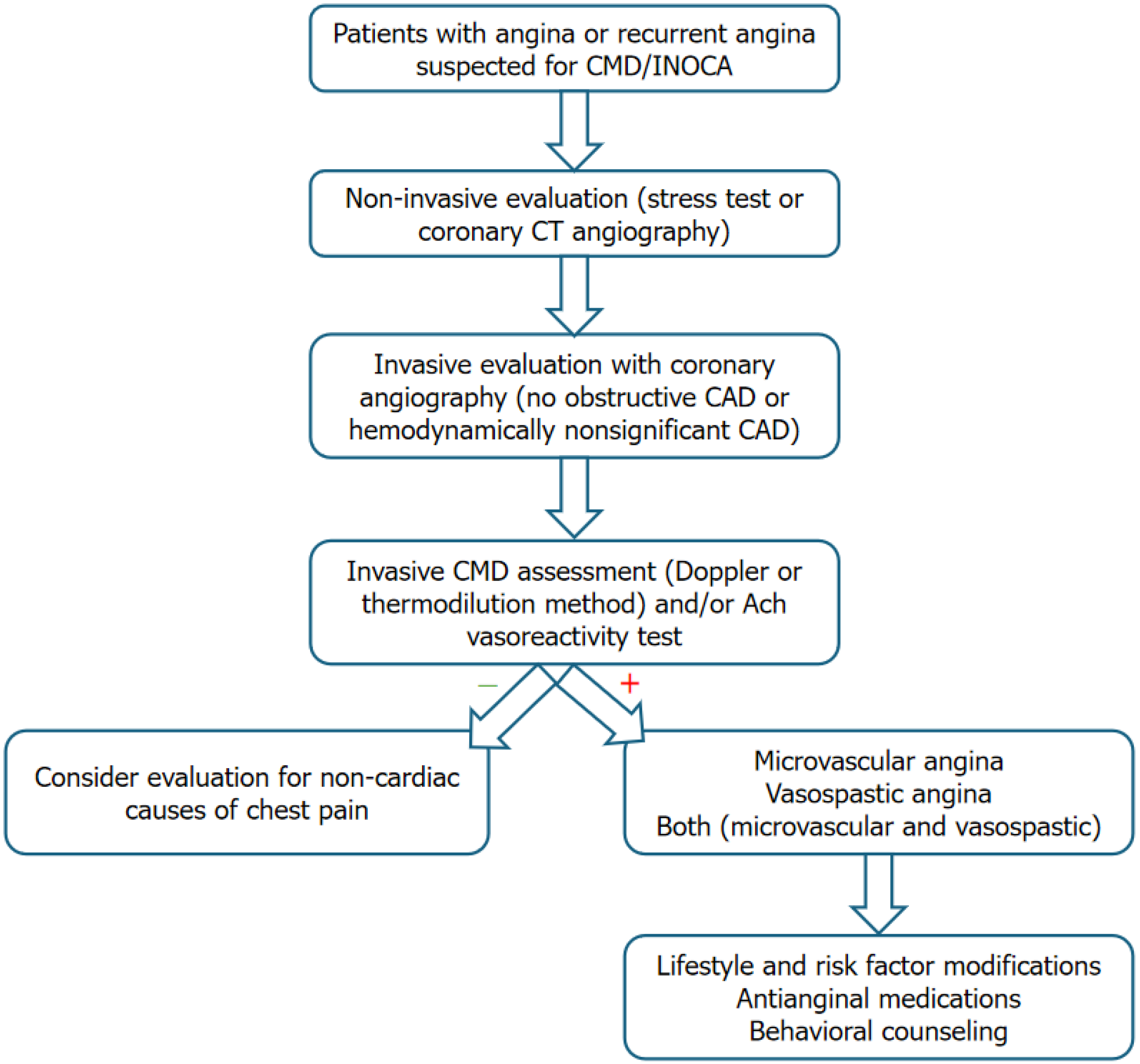
Invasive assessment of CMD is indicated for the patients with symptoms suggestive of angina and signs of myocardial ischemia at noninvasive testing in the absence of significant epicardial stenosis on coronary or computed tomographic angiography[6]. The options include CMD assessment via Doppler technique (Combowire XT or Flowire, Philips Volcano corporation, San Diego, CA) or thermodilution method using a pressure-temperature sensor guidewire (Pressurewire X, Abbott, Santa Clara, CA, United States). An algorithm highlighting the recommended approach to a patient with suspected CMD is depicted in Figure 1.

Figure 1
Figure 1 An algorithm for coronary microvascular disease assessment. CAD: Coronary artery disease; CMD: Coronary microvascular disease; CT: Computed tomography; INOCA: Ischemia with non-obstructive coronary arteries.

Using the combowire XT, doppler flow velocity, aortic pressure, and distal coronary pressure and the electrocardiographic signals are recorded using the ComboMap system (Philips, Eindhoven)[16]. Then steady state hyperemia is induced using adenosine (140 μg/kg/minute) for endothelium-independent vasodilation[17]. Coronary flow reserve (CFR) is calculated using doppler flow velocity (hyperemic flow velocity divided by resting flow velocity) and hyperemic myocardial velocity resistance (HMR) (HMR by dividing intracoronary pressure by hyperemic flow velocity)[16]. The doppler value of CFR ≤ 2.5 is considered prognostically significant[6]. Prior studies have shown HMR > 1.9 was an independent predictor of recurrence of symptoms and HMR ≥ value of 2.5 mmHg/cm/second provided adequate sensitivity and specificity for predicting CMD[18].

The thermodilution test is based on the dilution theory “Stewart-Hamilton equation” principle, which implies that the flow is inversely proportional to the temperature of the saline solution mixed with blood in the distal part of the vessel[19]. The CMD assessment can either be done by injecting saline at room temperature (colder than body temperature) as a bolus (bolus method) or continuous infusion at a fixed rate (continuous method). In a study of 102 patients comparing bolus and continuous methods of thermodilution, continuous thermodilution demonstrated higher reproducibility than bolus method[20].

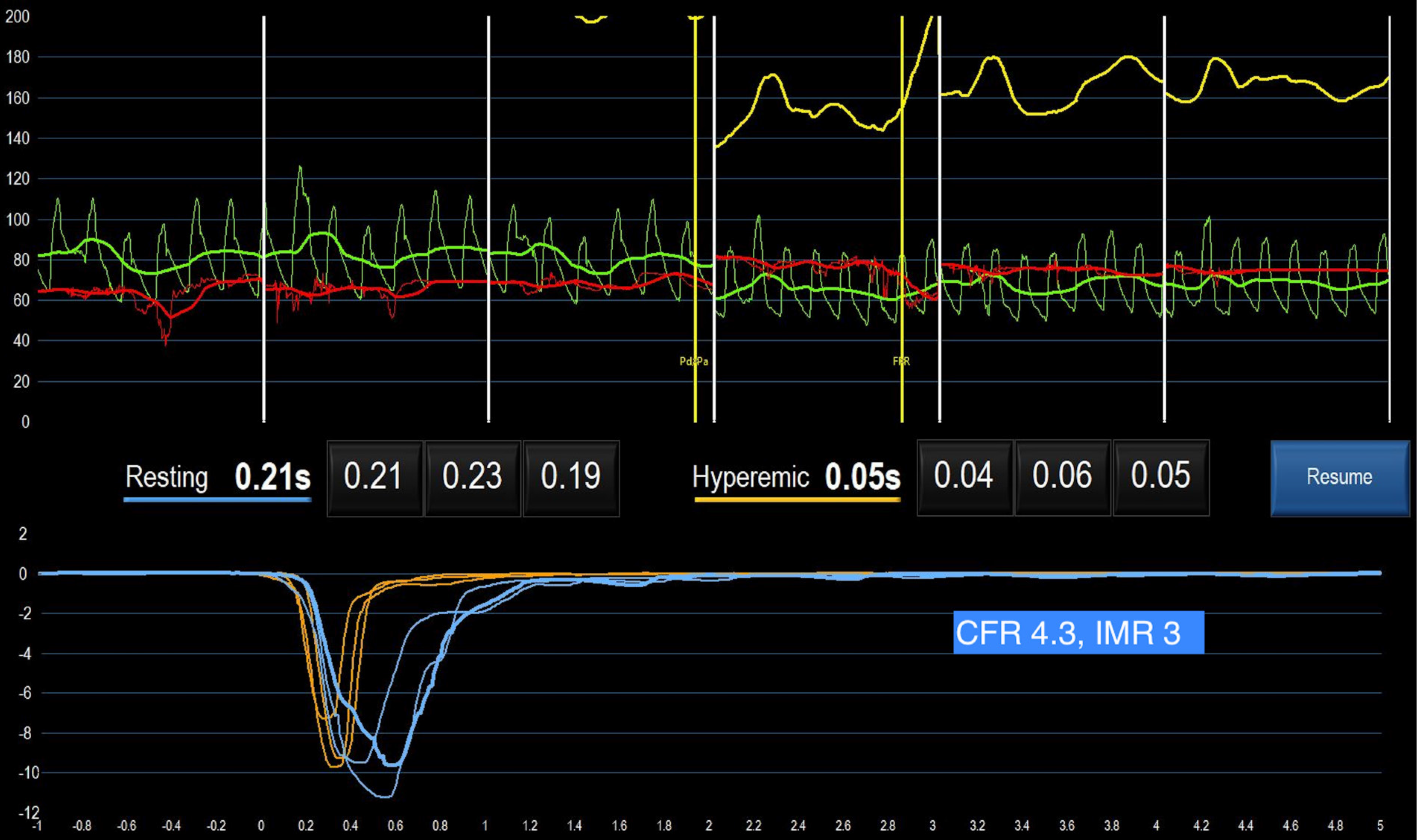
In bolus method, standard practice to inject bolus of 3 mL to 4 mL of saline from a 6 French guiding catheter and a V-shaped temperature change curve (fast descending limb and slow ascending limb) is recorded in the distal part of coronary artery using pressure wire X, and readings are repeated in setting of hyperemia using intravenous adenosine (Figure 2) and intracoronary papaverine[21]. The flow (Q) in mL/second is calculated using equation, Q = I (quantity of indicator) divided by ∫0c(t)dtwhich is time-dependent surface area under the thermodilution. If the exact volume of saline is not known (possibility of spillover into the aorta), Q can be estimated by dividing the vascular volume (in mL) by the mean transit time of the saline (Tmn; in secs). CFR via this method is calculated as the ratio between resting and hyperemic Tmn[22]. CFR interrogates the flow status in both epicardial vessels and microcirculation, while index of microcirculatory resistance (IMR) calculation is based on the analogy of Ohm’s law for electric resistance, that evaluates microvascular resistance, independent of epicardial stenoses. It is defined as distal coronary pressure (Pd) divided by the inverse of the Tmn during maximal hyperemia and thus IMR = Pd x Tmn at hyperemia[23]. According to thermodilution methods, CFR < 2.0 is considered prognostically significant and IMR value of ≥ 25 is representative of microvascular dysfunction[5].

Figure 2
Figure 2 A case study of coronary microvascular disease using bolus thermodilution method with normal coronary flow reserve. CFR: Coronary flow reserve; IMR: Index of microcirculatory resistance.

Continuous thermodilution method directly measures absolute coronary blood flow (Q; mL/minute) as well as absolute microvascular resistance [Rμ; wood units (WU)]. In addition to pressurewire X, this method requires a monorail infusion microcatheter with four external side holes and two internal side holes (RayFlow, Hexacath) that enables instantaneous mixing of saline solution and blood in the very proximal part of the vessel and a regular power injector to infuse saline solution at a steady rate. Ideally, the monorail infusion microcatheter and pressurewire X are parked in the proximal and distal segments of the vessel to be examined, respectively[21]. The power injector is automated to infuse at the rate of 10 mL/minute for resting phase and 20 mL/minute for the hyperemic measurements. The absolute coronary blood flow is calculated as Q (mL/minute) = 1.08 × (T/Ti) x Qi, where Tiis the temperature of the saline when it enters the coronary artery (°C), T is temperature of mixed saline blood measured in the distal segment of the artery (°C), 1.08 is constant for the densities and specific heat of blood and normal saline and Qi is saline infusion rate (mL/minute). First, T is measured after complete mixing of blood with saline and once steady state for T is achieved, then temperature sensor of wire is pulled back to the tip of infusion catheter for measuring Ti. Absolute microvascular resistance is computed as Rμ (WU) as a ratio of distal coronary pressure (Pd) and absolute coronary blood flow (Q). CFRabsolute is calculated by dividing Qhyperemia by Qrest and value < 2.0 is abnormal. Microvascular resistance reserve (MRR) is calculated as: MRR = CFR/FFR × Parest/Pahyperemia, where Pa is aortic pressure and FFR is fractional flow reserve. MRR > 2.7 essentially excludes the presence of CMD, while MRR < 2.1 confirms CMD[24].

Lastly, vasoreactivity testing by intracoronary infusion of acetylcholine is recommended. A standardized method involves successive infusion of acetylcholine at concentrations of 10−6 moL/L, 10−5 moL/L, and 10−4 moL/L, respectively[6,25]. Vasospastic angina is diagnosed when there is ≥ 90% diameter reduction of coronary vessel, plus presence of ischemic electrocardiographic changes and angina[6].

MANAGEMENT

A patient-centered multidisciplinary approach should be considered for the management of CMD. Currently there is no evidence-based treatment available for CMD as most of prior studies were small and heterogeneous[26]. Lifestyle modifications including directing at addressing risk factors (hypertension, dyslipidemia, and diabetes), reduce symptoms and behavioral interventions including smoking cessation are warranted[10]. Blood pressure control can reduce the frequency and intensity of anginal symptoms[27]. Patients with microvascular angina with reduced CFR and increased IMR can be benefited from beta-blockers (carvedilol, nebivolol, bisoprolol), calcium channel blockers (CCB) and or angiotensin-converting enzyme inhibitors (ACEis). In patients with epicardial or microvascular spasm with acetylcholine testing, CCBs are first line medications of choice. In patients with hypertensive microvascular angina, ACEis have shown improve hyperemic myocardial blood flow and decrease angina frequency[28,29]. The recent 2023 ACC/AHA guidelines recommend beta-blockers as a first line agent, followed by CCBs as a second line therapy if beta-blockers are ineffective or not well tolerated[10]. CORonary MICrovascular Angina trial demonstrated that a stratified medical therapy approach of an interventional diagnostic procedure with linked therapy was associated with reduction in angina frequency and better quality of life at 6 months and 1 year[30,31]. Nicorandil, a potassium channel activator with coronary microvascular dilatory effect may be an effective alternative for some patients[32]. Ranolazine, an inhibitor of late sodium current in cardiomyocytes, have shown mixed results in various studies[33,34]. Trimetazidine, which increases cellular tolerance to ischemia is recommended by ESC in patients who are intolerant to medications for angina[8]. Few patients remain refractory to medications and with the gaps in the knowledge and lack of evidence-based treatment, large future studies are warranted. Women's Ischemia Trial to Reduce Events in Non-Obstructive CAD trial is a multicenter, prospective, randomized, blinded outcome trial (NCT03417388) currently in phase 4, is investigating intensive medical treatment versus usual care in 4422 symptomatic women patients with signs and symptoms of ischemia but no obstructive CAD.

CONCLUSION

CMD is a common cause of angina, often overlooked and underdiagnosed resulting in recurrent angina, multiple unnecessary cardiac testing and associated with adverse outcomes. This editorial intends to increase the awareness of CMD among the primary physicians and cardiologist to keep low threshold for referral or considering further testing in suspected patients having recurrent angina or angina with no obstructive coronary artery. Undoubtedly, future research is warranted to investigate it further including availability of evidence-based treatment options and most importantly, to improve diagnostic awareness and relieve patient sufferings.

Footnotes

Provenance and peer review: Invited article; Externally peer reviewed.

Peer-review model: Single blind

Specialty type: Cardiac and cardiovascular systems

Country of origin: India

Peer-review report’s classification

Scientific Quality: Grade B, Grade B

Novelty: Grade B, Grade B

Creativity or Innovation: Grade B, Grade C

Scientific Significance: Grade B, Grade B

P-Reviewer: Xue HM S-Editor: Luo ML L-Editor: A P-Editor: Zhao YQ

References
1.  Patel MR, Peterson ED, Dai D, Brennan JM, Redberg RF, Anderson HV, Brindis RG, Douglas PS. Low diagnostic yield of elective coronary angiography. N Engl J Med. 2010;362:886-895.  [PubMed]  [DOI]  [Cited in This Article: ]  [Cited by in Crossref: 1067]  [Cited by in F6Publishing: 1192]  [Article Influence: 79.5]  [Reference Citation Analysis (0)]
2.  Mileva N, Nagumo S, Mizukami T, Sonck J, Berry C, Gallinoro E, Monizzi G, Candreva A, Munhoz D, Vassilev D, Penicka M, Barbato E, De Bruyne B, Collet C. Prevalence of Coronary Microvascular Disease and Coronary Vasospasm in Patients With Nonobstructive Coronary Artery Disease: Systematic Review and Meta-Analysis. J Am Heart Assoc. 2022;11:e023207.  [PubMed]  [DOI]  [Cited in This Article: ]  [Cited by in Crossref: 4]  [Cited by in F6Publishing: 83]  [Article Influence: 27.7]  [Reference Citation Analysis (0)]
3.  Camici PG, Crea F. Coronary microvascular dysfunction. N Engl J Med. 2007;356:830-840.  [PubMed]  [DOI]  [Cited in This Article: ]  [Cited by in Crossref: 1191]  [Cited by in F6Publishing: 1229]  [Article Influence: 68.3]  [Reference Citation Analysis (0)]
4.  AlBadri A, Bairey Merz CN, Johnson BD, Wei J, Mehta PK, Cook-Wiens G, Reis SE, Kelsey SF, Bittner V, Sopko G, Shaw LJ, Pepine CJ, Ahmed B. Impact of Abnormal Coronary Reactivity on Long-Term Clinical Outcomes in Women. J Am Coll Cardiol. 2019;73:684-693.  [PubMed]  [DOI]  [Cited in This Article: ]  [Cited by in Crossref: 163]  [Cited by in F6Publishing: 156]  [Article Influence: 26.0]  [Reference Citation Analysis (0)]
5.  Lee JM, Jung JH, Hwang D, Park J, Fan Y, Na SH, Doh JH, Nam CW, Shin ES, Koo BK. Coronary Flow Reserve and Microcirculatory Resistance in Patients With Intermediate Coronary Stenosis. J Am Coll Cardiol. 2016;67:1158-1169.  [PubMed]  [DOI]  [Cited in This Article: ]  [Cited by in Crossref: 184]  [Cited by in F6Publishing: 198]  [Article Influence: 22.0]  [Reference Citation Analysis (0)]
6.  Kunadian V, Chieffo A, Camici PG, Berry C, Escaned J, Maas AHEM, Prescott E, Karam N, Appelman Y, Fraccaro C, Louise Buchanan G, Manzo-Silberman S, Al-Lamee R, Regar E, Lansky A, Abbott JD, Badimon L, Duncker DJ, Mehran R, Capodanno D, Baumbach A. An EAPCI Expert Consensus Document on Ischaemia with Non-Obstructive Coronary Arteries in Collaboration with European Society of Cardiology Working Group on Coronary Pathophysiology & Microcirculation Endorsed by Coronary Vasomotor Disorders International Study Group. Eur Heart J. 2020;41:3504-3520.  [PubMed]  [DOI]  [Cited in This Article: ]  [Cited by in Crossref: 162]  [Cited by in F6Publishing: 449]  [Article Influence: 112.3]  [Reference Citation Analysis (0)]
7.  Chitturi KR, Bhogal S, Kassaian SE, Merdler I, Abusnina W, Chaturvedi A, Ben-Dor I, Waksman R, Case BC, Barac A, Hashim HD. Coronary microvascular dysfunction and cancer therapy-related cardiovascular toxicity. Cardiovasc Revasc Med. 2024;68:45-49.  [PubMed]  [DOI]  [Cited in This Article: ]  [Cited by in Crossref: 1]  [Reference Citation Analysis (0)]
8.  Knuuti J, Wijns W, Saraste A, Capodanno D, Barbato E, Funck-Brentano C, Prescott E, Storey RF, Deaton C, Cuisset T, Agewall S, Dickstein K, Edvardsen T, Escaned J, Gersh BJ, Svitil P, Gilard M, Hasdai D, Hatala R, Mahfoud F, Masip J, Muneretto C, Valgimigli M, Achenbach S, Bax JJ; ESC Scientific Document Group. 2019 ESC Guidelines for the diagnosis and management of chronic coronary syndromes. Eur Heart J. 2020;41:407-477.  [PubMed]  [DOI]  [Cited in This Article: ]  [Cited by in Crossref: 2791]  [Cited by in F6Publishing: 4219]  [Article Influence: 843.8]  [Reference Citation Analysis (0)]
9.  Vrints C, Andreotti F, Koskinas KC, Rossello X, Adamo M, Ainslie J, Banning AP, Budaj A, Buechel RR, Chiariello GA, Chieffo A, Christodorescu RM, Deaton C, Doenst T, Jones HW, Kunadian V, Mehilli J, Milojevic M, Piek JJ, Pugliese F, Rubboli A, Semb AG, Senior R, Ten Berg JM, Van Belle E, Van Craenenbroeck EM, Vidal-Perez R, Winther S; ESC Scientific Document Group. 2024 ESC Guidelines for the management of chronic coronary syndromes. Eur Heart J. 2024;45:3415-3537.  [PubMed]  [DOI]  [Cited in This Article: ]  [Cited by in Crossref: 64]  [Cited by in F6Publishing: 120]  [Article Influence: 120.0]  [Reference Citation Analysis (0)]
10.  Writing Committee Members; Virani SS, Newby LK, Arnold SV, Bittner V, Brewer LC, Demeter SH, Dixon DL, Fearon WF, Hess B, Johnson HM, Kazi DS, Kolte D, Kumbhani DJ, LoFaso J, Mahtta D, Mark DB, Minissian M, Navar AM, Patel AR, Piano MR, Rodriguez F, Talbot AW, Taqueti VR, Thomas RJ, van Diepen S, Wiggins B, Williams MS. 2023 AHA/ACC/ACCP/ASPC/NLA/PCNA Guideline for the Management of Patients With Chronic Coronary Disease: A Report of the American Heart Association/American College of Cardiology Joint Committee on Clinical Practice Guidelines. J Am Coll Cardiol. 2023;82:833-955.  [PubMed]  [DOI]  [Cited in This Article: ]  [Cited by in Crossref: 52]  [Cited by in F6Publishing: 117]  [Article Influence: 58.5]  [Reference Citation Analysis (0)]
11.  Li Y, Pagano PJ. Microvascular NADPH oxidase in health and disease. Free Radic Biol Med. 2017;109:33-47.  [PubMed]  [DOI]  [Cited in This Article: ]  [Cited by in Crossref: 59]  [Cited by in F6Publishing: 61]  [Article Influence: 7.6]  [Reference Citation Analysis (0)]
12.  Prabhahar A, Batta A, Hatwal J, Kumar V, Ramachandran R, Batta A. Endothelial dysfunction in the kidney transplant population: Current evidence and management strategies. World J Transplant. 2025;15.  [PubMed]  [DOI]  [Cited in This Article: ]  [Reference Citation Analysis (2)]
13.  Tsai SH, Lu G, Xu X, Ren Y, Hein TW, Kuo L. Enhanced endothelin-1/Rho-kinase signalling and coronary microvascular dysfunction in hypertensive myocardial hypertrophy. Cardiovasc Res. 2017;113:1329-1337.  [PubMed]  [DOI]  [Cited in This Article: ]  [Cited by in Crossref: 55]  [Cited by in F6Publishing: 51]  [Article Influence: 6.4]  [Reference Citation Analysis (0)]
14.  Lanza GA, Careri G, Crea F. Mechanisms of coronary artery spasm. Circulation. 2011;124:1774-1782.  [PubMed]  [DOI]  [Cited in This Article: ]  [Cited by in Crossref: 222]  [Cited by in F6Publishing: 229]  [Article Influence: 16.4]  [Reference Citation Analysis (0)]
15.  Pries AR, Badimon L, Bugiardini R, Camici PG, Dorobantu M, Duncker DJ, Escaned J, Koller A, Piek JJ, de Wit C. Coronary vascular regulation, remodelling, and collateralization: mechanisms and clinical implications on behalf of the working group on coronary pathophysiology and microcirculation. Eur Heart J. 2015;36:3134-3146.  [PubMed]  [DOI]  [Cited in This Article: ]  [Cited by in Crossref: 128]  [Cited by in F6Publishing: 133]  [Article Influence: 13.3]  [Reference Citation Analysis (0)]
16.  Everaars H, de Waard GA, Driessen RS, Danad I, van de Ven PM, Raijmakers PG, Lammertsma AA, van Rossum AC, Knaapen P, van Royen N. Doppler Flow Velocity and Thermodilution to Assess Coronary Flow Reserve: A Head-to-Head Comparison With [15O]H2O PET. JACC Cardiovasc Interv. 2018;11:2044-2054.  [PubMed]  [DOI]  [Cited in This Article: ]  [Cited by in Crossref: 52]  [Cited by in F6Publishing: 110]  [Article Influence: 15.7]  [Reference Citation Analysis (0)]
17.  Layland J, Carrick D, Lee M, Oldroyd K, Berry C. Adenosine: physiology, pharmacology, and clinical applications. JACC Cardiovasc Interv. 2014;7:581-591.  [PubMed]  [DOI]  [Cited in This Article: ]  [Cited by in Crossref: 157]  [Cited by in F6Publishing: 172]  [Article Influence: 15.6]  [Reference Citation Analysis (0)]
18.  Williams RP, de Waard GA, De Silva K, Lumley M, Asrress K, Arri S, Ellis H, Mir A, Clapp B, Chiribiri A, Plein S, Teunissen PF, Hollander MR, Marber M, Redwood S, van Royen N, Perera D. Doppler Versus Thermodilution-Derived Coronary Microvascular Resistance to Predict Coronary Microvascular Dysfunction in Patients With Acute Myocardial Infarction or Stable Angina Pectoris. Am J Cardiol. 2018;121:1-8.  [PubMed]  [DOI]  [Cited in This Article: ]  [Cited by in Crossref: 69]  [Cited by in F6Publishing: 66]  [Article Influence: 9.4]  [Reference Citation Analysis (0)]
19.  Hamilton WF, Moore JW, Kinsman JM, Spurling RG. Studies on the circulation. AJP. 1932;99:534-551.  [PubMed]  [DOI]  [Cited in This Article: ]
20.  Gallinoro E, Bertolone DT, Fernandez-Peregrina E, Paolisso P, Bermpeis K, Esposito G, Gomez-Lopez A, Candreva A, Mileva N, Belmonte M, Mizukami T, Fournier S, Vanderheyden M, Wyffels E, Bartunek J, Sonck J, Barbato E, Collet C, De Bruyne B. Reproducibility of bolus versus continuous thermodilution for assessment of coronary microvascular function in patients with ANOCA. EuroIntervention. 2023;19:e155-e166.  [PubMed]  [DOI]  [Cited in This Article: ]  [Cited by in Crossref: 25]  [Cited by in F6Publishing: 39]  [Article Influence: 19.5]  [Reference Citation Analysis (0)]
21.  Candreva A, Gallinoro E, van 't Veer M, Sonck J, Collet C, Di Gioia G, Kodeboina M, Mizukami T, Nagumo S, Keulards D, Fournier S, Pijls NHJ, De Bruyne B. Basics of Coronary Thermodilution. JACC Cardiovasc Interv. 2021;14:595-605.  [PubMed]  [DOI]  [Cited in This Article: ]  [Cited by in Crossref: 11]  [Cited by in F6Publishing: 40]  [Article Influence: 10.0]  [Reference Citation Analysis (0)]
22.  De Bruyne B, Pijls NH, Smith L, Wievegg M, Heyndrickx GR. Coronary thermodilution to assess flow reserve: experimental validation. Circulation. 2001;104:2003-2006.  [PubMed]  [DOI]  [Cited in This Article: ]  [Cited by in Crossref: 175]  [Cited by in F6Publishing: 202]  [Article Influence: 8.4]  [Reference Citation Analysis (0)]
23.  Fearon WF, Balsam LB, Farouque HM, Caffarelli AD, Robbins RC, Fitzgerald PJ, Yock PG, Yeung AC. Novel index for invasively assessing the coronary microcirculation. Circulation. 2003;107:3129-3132.  [PubMed]  [DOI]  [Cited in This Article: ]  [Cited by in Crossref: 418]  [Cited by in F6Publishing: 483]  [Article Influence: 22.0]  [Reference Citation Analysis (0)]
24.  de Vos A, Jansen TPJ, van 't Veer M, Dimitriu-Leen A, Konst RE, Elias-Smale S, Paradies V, Rodwell L, van den Oord S, Smits P, van Royen N, Pijls N, Damman P. Microvascular Resistance Reserve to Assess Microvascular Dysfunction in ANOCA Patients. JACC Cardiovasc Interv. 2023;16:470-481.  [PubMed]  [DOI]  [Cited in This Article: ]  [Cited by in Crossref: 1]  [Cited by in F6Publishing: 30]  [Article Influence: 15.0]  [Reference Citation Analysis (0)]
25.  Okumura K, Yasue H, Matsuyama K, Goto K, Miyagi H, Ogawa H, Matsuyama K. Sensitivity and specificity of intracoronary injection of acetylcholine for the induction of coronary artery spasm. J Am Coll Cardiol. 1988;12:883-888.  [PubMed]  [DOI]  [Cited in This Article: ]  [Cited by in Crossref: 236]  [Cited by in F6Publishing: 229]  [Article Influence: 6.2]  [Reference Citation Analysis (0)]
26.  Suhrs HE, Michelsen MM, Prescott E. Treatment strategies in coronary microvascular dysfunction: A systematic review of interventional studies. Microcirculation. 2019;26:e12430.  [PubMed]  [DOI]  [Cited in This Article: ]  [Cited by in Crossref: 12]  [Cited by in F6Publishing: 14]  [Article Influence: 2.3]  [Reference Citation Analysis (0)]
27.  Williams B, Mancia G, Spiering W, Agabiti Rosei E, Azizi M, Burnier M, Clement DL, Coca A, de Simone G, Dominiczak A, Kahan T, Mahfoud F, Redon J, Ruilope L, Zanchetti A, Kerins M, Kjeldsen SE, Kreutz R, Laurent S, Lip GYH, McManus R, Narkiewicz K, Ruschitzka F, Schmieder RE, Shlyakhto E, Tsioufis C, Aboyans V, Desormais I; ESC Scientific Document Group. 2018 ESC/ESH Guidelines for the management of arterial hypertension. Eur Heart J. 2018;39:3021-3104.  [PubMed]  [DOI]  [Cited in This Article: ]  [Cited by in Crossref: 4314]  [Cited by in F6Publishing: 6042]  [Article Influence: 863.1]  [Reference Citation Analysis (0)]
28.  Neglia D, Fommei E, Varela-Carver A, Mancini M, Ghione S, Lombardi M, Pisani P, Parker H, D'amati G, Donato L, Camici PG. Perindopril and indapamide reverse coronary microvascular remodelling and improve flow in arterial hypertension. J Hypertens. 2011;29:364-372.  [PubMed]  [DOI]  [Cited in This Article: ]  [Cited by in Crossref: 63]  [Cited by in F6Publishing: 66]  [Article Influence: 4.7]  [Reference Citation Analysis (0)]
29.  Pauly DF, Johnson BD, Anderson RD, Handberg EM, Smith KM, Cooper-DeHoff RM, Sopko G, Sharaf BM, Kelsey SF, Merz CN, Pepine CJ. In women with symptoms of cardiac ischemia, nonobstructive coronary arteries, and microvascular dysfunction, angiotensin-converting enzyme inhibition is associated with improved microvascular function: A double-blind randomized study from the National Heart, Lung and Blood Institute Women's Ischemia Syndrome Evaluation (WISE). Am Heart J. 2011;162:678-684.  [PubMed]  [DOI]  [Cited in This Article: ]  [Cited by in Crossref: 176]  [Cited by in F6Publishing: 167]  [Article Influence: 11.9]  [Reference Citation Analysis (0)]
30.  Ford TJ, Stanley B, Sidik N, Good R, Rocchiccioli P, McEntegart M, Watkins S, Eteiba H, Shaukat A, Lindsay M, Robertson K, Hood S, McGeoch R, McDade R, Yii E, McCartney P, Corcoran D, Collison D, Rush C, Sattar N, McConnachie A, Touyz RM, Oldroyd KG, Berry C. 1-Year Outcomes of Angina Management Guided by Invasive Coronary Function Testing (CorMicA). JACC Cardiovasc Interv. 2020;13:33-45.  [PubMed]  [DOI]  [Cited in This Article: ]  [Cited by in Crossref: 76]  [Cited by in F6Publishing: 159]  [Article Influence: 26.5]  [Reference Citation Analysis (0)]
31.  Ford TJ, Stanley B, Good R, Rocchiccioli P, McEntegart M, Watkins S, Eteiba H, Shaukat A, Lindsay M, Robertson K, Hood S, McGeoch R, McDade R, Yii E, Sidik N, McCartney P, Corcoran D, Collison D, Rush C, McConnachie A, Touyz RM, Oldroyd KG, Berry C. Stratified Medical Therapy Using Invasive Coronary Function Testing in Angina: The CorMicA Trial. J Am Coll Cardiol. 2018;72:2841-2855.  [PubMed]  [DOI]  [Cited in This Article: ]  [Cited by in Crossref: 315]  [Cited by in F6Publishing: 482]  [Article Influence: 68.9]  [Reference Citation Analysis (0)]
32.  Guarini G, Huqi A, Morrone D, Capozza P, Todiere G, Marzilli M. Pharmacological approaches to coronary microvascular dysfunction. Pharmacol Ther. 2014;144:283-302.  [PubMed]  [DOI]  [Cited in This Article: ]  [Cited by in Crossref: 20]  [Cited by in F6Publishing: 21]  [Article Influence: 1.9]  [Reference Citation Analysis (0)]
33.  Bairey Merz CN, Handberg EM, Shufelt CL, Mehta PK, Minissian MB, Wei J, Thomson LE, Berman DS, Shaw LJ, Petersen JW, Brown GH, Anderson RD, Shuster JJ, Cook-Wiens G, Rogatko A, Pepine CJ. A randomized, placebo-controlled trial of late Na current inhibition (ranolazine) in coronary microvascular dysfunction (CMD): impact on angina and myocardial perfusion reserve. Eur Heart J. 2016;37:1504-1513.  [PubMed]  [DOI]  [Cited in This Article: ]  [Cited by in Crossref: 121]  [Cited by in F6Publishing: 142]  [Article Influence: 14.2]  [Reference Citation Analysis (0)]
34.  Mehta PK, Sharma S, Minissian M, Harsch MR, Martinson M, Nyman JA, Shaw LJ, Bairey Merz CN, Wenger NK. Ranolazine Reduces Angina in Women with Ischemic Heart Disease: Results of an Open-Label, Multicenter Trial. J Womens Health (Larchmt). 2019;28:573-582.  [PubMed]  [DOI]  [Cited in This Article: ]  [Cited by in Crossref: 11]  [Cited by in F6Publishing: 10]  [Article Influence: 1.7]  [Reference Citation Analysis (0)]