Copyright
©The Author(s) 2018.
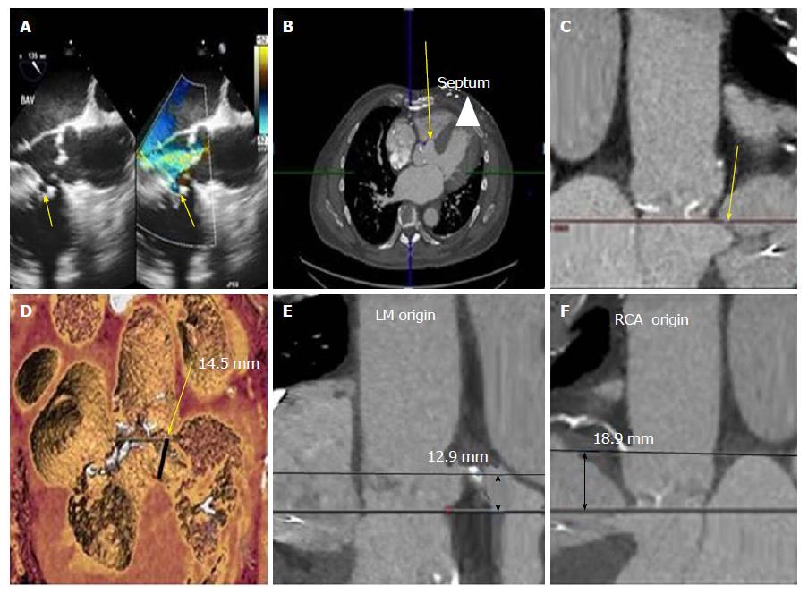
Figure 1 Assessment of the calcific aortic valve and interventricular septal aneurysm prior to transcatheter aortic valve replacement.
A: Transesophageal echocardiography shows a perimembranous ventricular septal aneurysm (arrow) and severe calcific stenotic aortic valve with moderate aortic regurgitation; B: Cardiac computer tomography (CT) findings confirmed the focal interventricular septal aneurysm (arrow) below the aortic annulus in the transverse view along the membranous septum with left ventricular outflow tract extension (arrowhead); C: Cardiac CT in coronal view showing interventricular septal aneurysm (arrow) below the annulus; D: The dimension at the neck of septal aneurysm (arrow) was 14.5 mm on 3-D reconstructed CT image; E and F: The aortic annulus to left main ostium distance was 12.9 mm and annulus to right coronary ostium distance was 18.9 mm.
- Citation: Banga S, Barzallo MA, Nighswonger CL, Mungee S. Transcatheter aortic valve replacement in membranous interventricular septum aneurysm with left ventricular outflow tract extension. World J Cardiol 2018; 10(1): 1-5
- URL: https://www.wjgnet.com/1949-8462/full/v10/i1/1.htm
- DOI: https://dx.doi.org/10.4330/wjc.v10.i1.1


