Copyright
©The Author(s) 2017.
World J Cardiol. Sep 26, 2017; 9(9): 715-722
Published online Sep 26, 2017. doi: 10.4330/wjc.v9.i9.715
Published online Sep 26, 2017. doi: 10.4330/wjc.v9.i9.715
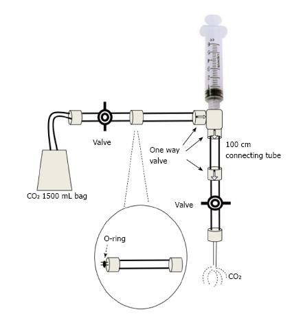
Figure 1 The modified plastic bag system with O-ring.
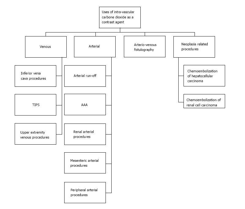
Figure 2 Potential uses of carbon dioxide angiography.
AAA: Abdominal aortic aneurysm.
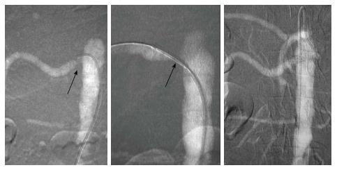
Figure 3 A carbon dioxide renal arteriogram showing renal artery orifice stenosis with subsequent stent placement and resolution of the stenosis with good flow.
The carbon dioxide contrast is injected through the sheath. Adapted with permission from Dr. Kyung Cho.
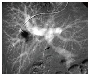
Figure 4 Carbon dioxide wedged hepatic protogram showing portal vein stenosis (arrow).
Adapted with permission from Dr. Kyung Cho.
- Citation: Ali F, Mangi MA, Rehman H, Kaluski E. Use of carbon dioxide as an intravascular contrast agent: A review of current literature. World J Cardiol 2017; 9(9): 715-722
- URL: https://www.wjgnet.com/1949-8462/full/v9/i9/715.htm
- DOI: https://dx.doi.org/10.4330/wjc.v9.i9.715


