Copyright
©The Author(s) 2017.
World J Cardiol. Aug 26, 2017; 9(8): 702-709
Published online Aug 26, 2017. doi: 10.4330/wjc.v9.i8.702
Published online Aug 26, 2017. doi: 10.4330/wjc.v9.i8.702
Figure 1 Patient flowchart.
CA: Coronary angiography; CS: Cardiogenic shock; ECG: Electrocardiogram; LBBB: Left bundle branch block; SCA: Sudden cardiac arrest.
Figure 2 Kaplan Meier curves for all cause 5-year survival depending on the finding of an acute coronary lesion.
ACL: Acute coronary lesion.
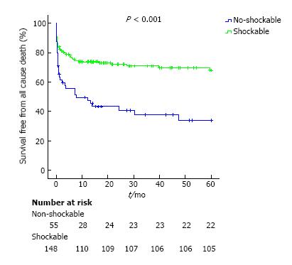
Figure 3 Kaplan-Meier curves for 5 years survival.
- Citation: Martínez-Losas P, Salinas P, Ferrera C, Nogales-Romo MT, Noriega F, Del Trigo M, Núñez-Gil IJ, Nombela-Franco L, Gonzalo N, Jiménez-Quevedo P, Escaned J, Fernández-Ortiz A, Macaya C, Viana-Tejedor A. Coronary angiography findings in cardiac arrest patients with non-diagnostic post-resuscitation electrocardiogram: A comparison of shockable and non-shockable initial rhythms. World J Cardiol 2017; 9(8): 702-709
- URL: https://www.wjgnet.com/1949-8462/full/v9/i8/702.htm
- DOI: https://dx.doi.org/10.4330/wjc.v9.i8.702


