Copyright
©The Author(s) 2017.
World J Cardiol. Jun 26, 2017; 9(6): 539-546
Published online Jun 26, 2017. doi: 10.4330/wjc.v9.i6.539
Published online Jun 26, 2017. doi: 10.4330/wjc.v9.i6.539
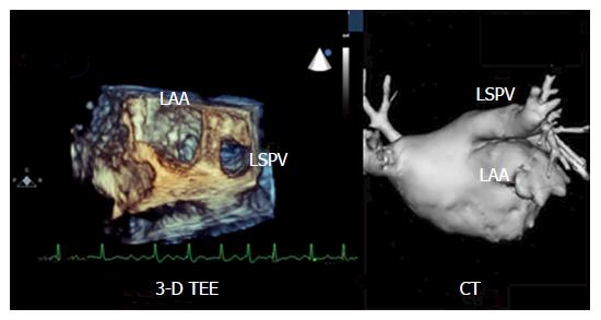
Figure 1 Three-dimensional transesophageal echocardiography-reconstruction providing an overview over the left atrial anatomy.
LSPV: Left superior pulmonary vein; LAA: Left atrial appendage; CT: Computed tomography; 3-D TEE: Three-dimensional transesophageal echocardiography.
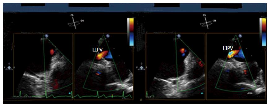
Figure 2 Three-dimensional transesophageal echocardiography performed 3 years after catheter ablation of atrial fibrillation.
Slightly increased flow velocity in the left inferior pulmonary vein (LIPV) ostium indicating a minor pulmonary vein stenosis [pulmonary vein diameter at 3-year follow-up: 2.1 mm (compared to 2.6 mm at baseline)].
- Citation: Kettering K, Gramley F, von Bardeleben S. Catheter ablation of atrial fibrillation facilitated by preprocedural three-dimensional transesophageal echocardiography: Long-term outcome. World J Cardiol 2017; 9(6): 539-546
- URL: https://www.wjgnet.com/1949-8462/full/v9/i6/539.htm
- DOI: https://dx.doi.org/10.4330/wjc.v9.i6.539


