Copyright
©The Author(s) 2017.
World J Cardiol. Mar 26, 2017; 9(3): 283-288
Published online Mar 26, 2017. doi: 10.4330/wjc.v9.i3.283
Published online Mar 26, 2017. doi: 10.4330/wjc.v9.i3.283
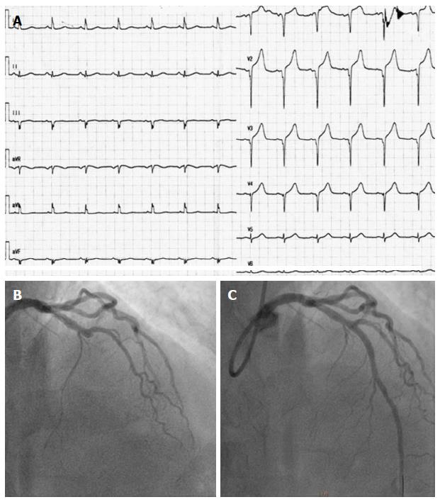
Figure 1 First presentation with acute coronary syndrome.
A: Electrocardiograph upon admission; B: Coronary angiography showing critical stenosis in left anterior descending; C: After implantation of resolute integrity drug-eluting stent.
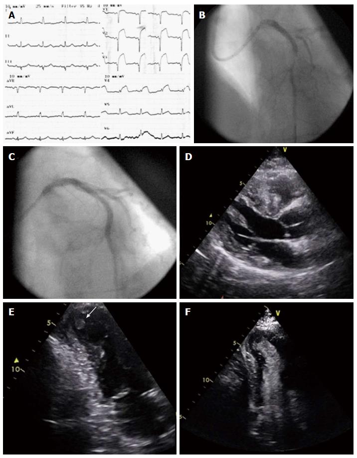
Figure 2 Kounis syndrome following rice pudding consumption.
A: Electrocardiograph upon admission; B: Coronary angiography showing stent thrombosis; C: After implantation of drug-eluting stent (stent in stent); D: Parasternal long axis view showing excessive hypertrophy of the septum; E: Thrombus in aneurysmatic apex, apical 2-chamber view; F: Contrast derived image, with thrombus in the apex, apical 2-chamber view.
- Citation: Tzanis G, Bonou M, Mikos N, Biliou S, Koniari I, Kounis NG, Barbetseas J. Early stent thrombosis secondary to food allergic reaction: Kounis syndrome following rice pudding ingestion. World J Cardiol 2017; 9(3): 283-288
- URL: https://www.wjgnet.com/1949-8462/full/v9/i3/283.htm
- DOI: https://dx.doi.org/10.4330/wjc.v9.i3.283


