Copyright
©The Author(s) 2017.
World J Cardiol. Mar 26, 2017; 9(3): 212-229
Published online Mar 26, 2017. doi: 10.4330/wjc.v9.i3.212
Published online Mar 26, 2017. doi: 10.4330/wjc.v9.i3.212
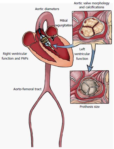
Figure 1 Main morphologic and functional parameters to assess by Multi-Imaging approach in the setting of pre-interventional evaluation and planning of Transcatheter Aortic Valve Replacement procedures (illustrator by Germano Massenzio).
PAPs: Pulmonary arterial systolic pressure.
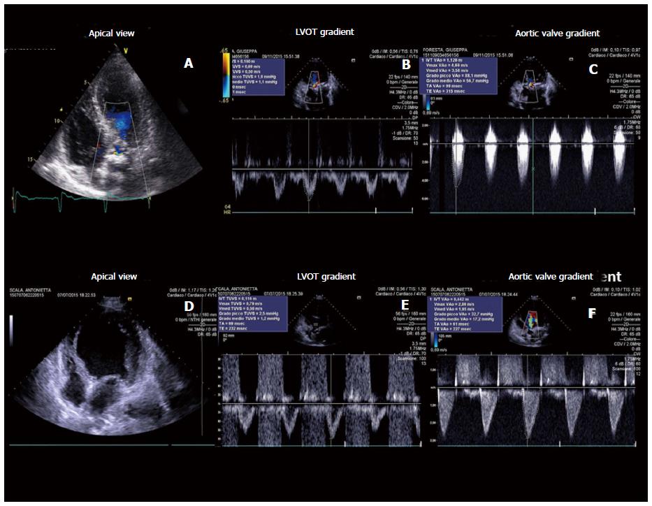
Figure 2 Transthoracic echocardiography gives detailed anatomic description of aortic valve complex and allows to estimate with enough reliability the haemodynamic entity of valvular stenosis by assessment of functional aortic valve area, derived using continuity equation.
Two examples of severe aortic stenosis with normal ejection fraction and gradients (A-C), and with classical “low flow-low gradient” pattern (D-F). LVOT: Left ventricular outflow tract.
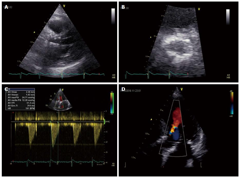
Figure 3 Post-implantation echocardiographic transcatheter aortic valve replacement assessment in long-axis (A) and short-axis (B) parasternal views; Normal trans-prothesis flow gradient by Doppler analysis (C); Mild paravalvular aortic regurgitation in this apical 5-chamber view of the same patient (D).
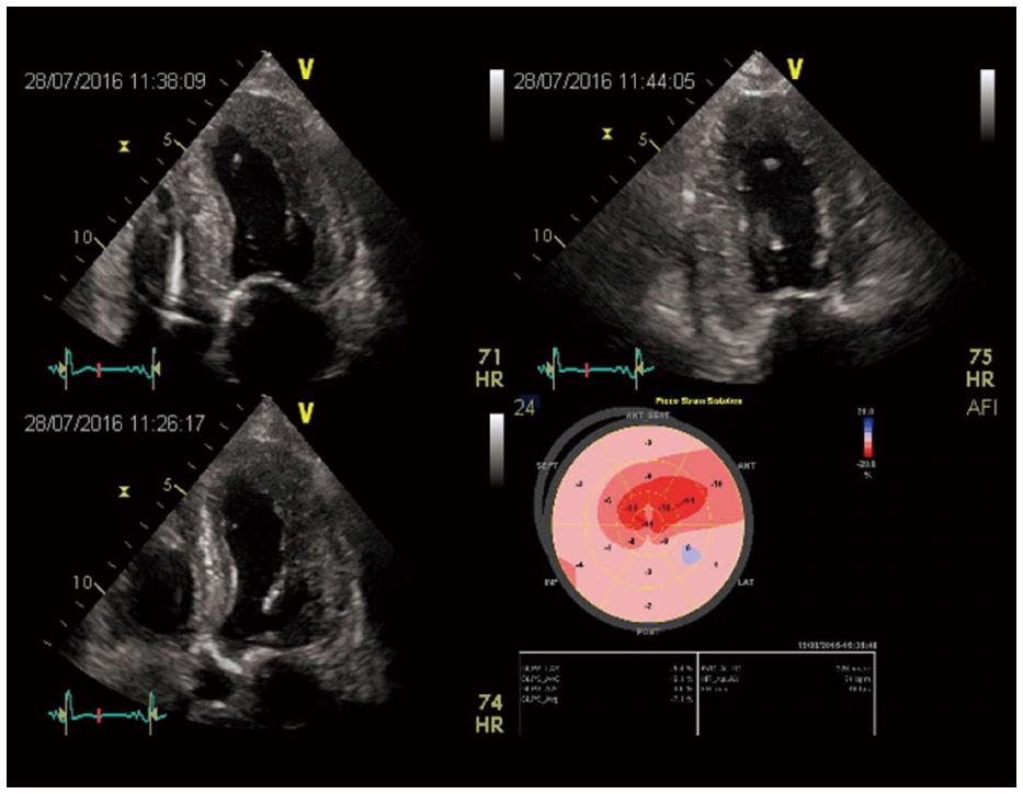
Figure 4 Two-dimensional LV strain in a patient with low flow-low gradient aortic stenosis, showing a severe and diffused impairment of myocardial deformation.
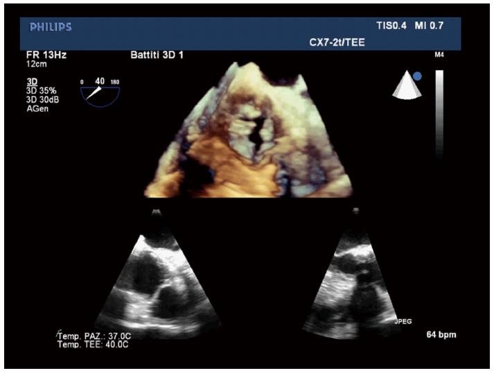
Figure 5 Three dimensional transesophageal echocardiography allows to visualize the real shape of left ventricular outflow tract, and has proved more effective in providing optimal annular measurement and was more useful in predicting paravalvular aortic regurgitation compared to 2D-transesophageal echocardiography.
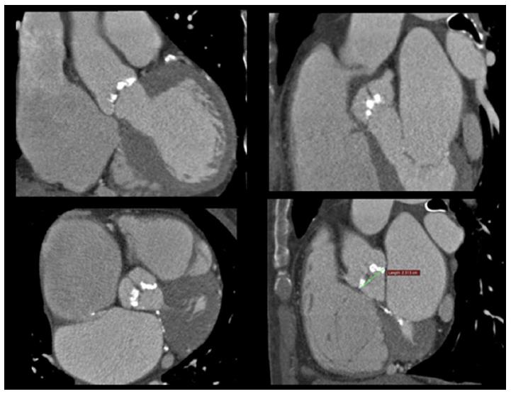
Figure 6 Multi-slice enhanced computed tomography images showing the aortic valve cusps and the first tract of the ascending aorta, with associated presence of extensive valvular calcifications.
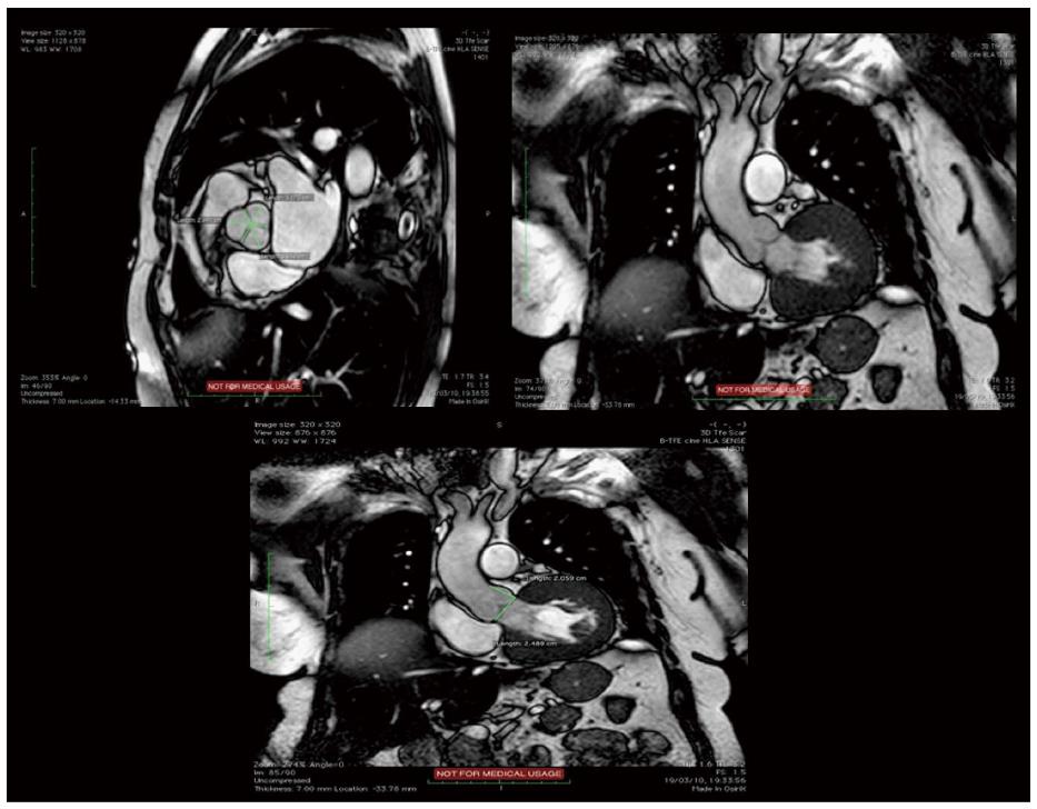
Figure 7 Balanced fast-field echo unenhanced magnetic resonance images showing the normal tricuspid aortic valve, with the typical “Mercedes-Benz Sign” and the first tract of the ascending aorta.
- Citation: Cocchia R, D’Andrea A, Conte M, Cavallaro M, Riegler L, Citro R, Sirignano C, Imbriaco M, Cappelli M, Gregorio G, Calabrò R, Bossone E. Patient selection for transcatheter aortic valve replacement: A combined clinical and multimodality imaging approach. World J Cardiol 2017; 9(3): 212-229
- URL: https://www.wjgnet.com/1949-8462/full/v9/i3/212.htm
- DOI: https://dx.doi.org/10.4330/wjc.v9.i3.212


