Copyright
©The Author(s) 2017.
World J Cardiol. Oct 26, 2017; 9(10): 773-786
Published online Oct 26, 2017. doi: 10.4330/wjc.v9.i10.773
Published online Oct 26, 2017. doi: 10.4330/wjc.v9.i10.773
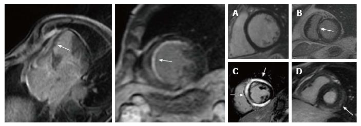
Figure 1 Late gadolinium-enhanced cardiac magnetic resonance images of ischemic (left panel) and non-ischemic (right panel) dilated cardiomyopathy.
In ischemic cardiomyopathy LGE has a subendocardial or transmural distribution in myocardial segments following a coronary artery. In non-ischemic etiology, it shows a “midwall” pattern (panel B, C) or subepicardial distribution (panel D). LGE: Late gadolinium enhancement.
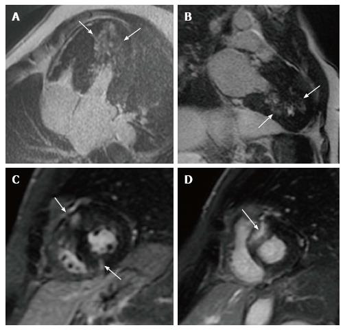
Figure 2 Late gadolinium enhancement patterns in hypertrophic cardiomyopathy patients.
A: LGE in the lateral wall (small arrows) and in the interventricular ventricular septum; B: LGE in the LV apex and inferior wall; C: LGE localized to the insertion area of the RV wall into the anterior and posterior ventricular septum; D: Transmural LGE involving the ventricular septum. LGE: Late gadolinium enhancement.
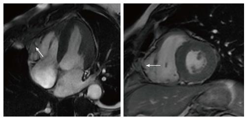
Figure 3 Subtricuspid involvement in arrhythmogenic right ventricular cardiomyopathy.
Dilated right ventricle with bulging of the subtricuspid region (arrow). The right ventricular apex is relatively spared.
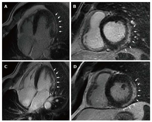
Figure 4 Left dominant arrhythmogenic cardiomyopathy.
Long-axis (A and C) and short-axis (B and D) postcontrast CMR views of two 34-year-old identical twin brothers showing a subepicardial/midmyocardial stria of LGE involving the lateral and inferolateral left ventricular wall (white arrows). CMR: Cardiac magnetic resonance; LGE: Late gadolinium enhancement.
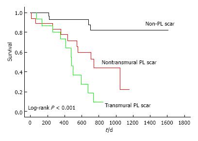
Figure 5 Left ventricular lead position, transmurality of scar, and outcome after cardiac resynchronization therapy.
PL: Posterolateral.
- Citation: De Maria E, Aldrovandi A, Borghi A, Modonesi L, Cappelli S. Cardiac magnetic resonance imaging: Which information is useful for the arrhythmologist? World J Cardiol 2017; 9(10): 773-786
- URL: https://www.wjgnet.com/1949-8462/full/v9/i10/773.htm
- DOI: https://dx.doi.org/10.4330/wjc.v9.i10.773


