Copyright
©The Author(s) 2017.
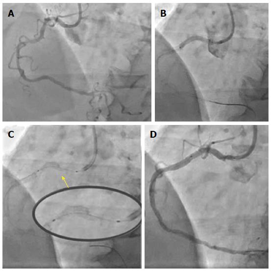
Figure 1 Longitudinal deformation of a stent implanted at the ostium of the right coronary artery.
A: Initial angiogram showing severe calcific lesions in proximal and mid right coronary artery; B: Proximal lesion stented all the way to cover the ostium with a Resolute Onyx 3.0 mm × 22 mm stent; C: Longitudinal deformation of proximal stent treated with 3.0 non-compliant balloon and another 3.0 mm × 18 mm Resolute Onyx stent; D: Final angiographic result.
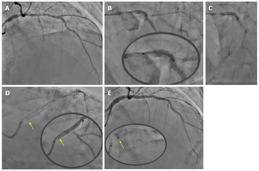
Figure 2 Longitudinal deformation of an ostial left main stem stent.
A: Initial angiogram showing significant calcific ostial and distal left main lesions with further significant proximal LAD calcific disease; B: After 1.5 burr rotablation, LMS into proximal LAD was stented with a 3.5 mm × 34 mm Resolute Onyx (B) to 14atm covering the LMS ostium (C). The LMS segment of the stent was post-dilated with a 4.5 NC balloon and the jailed LCx wire removed; D: Longitudinal deformation of the ostial LMS stent (yellow arrow pointing at the proximal deformed edge of the stent); E: Final angiographic result after covering the ostial LMS with a 4.0 mm × 8 mm Resolute Onyx stent. LAD: Left anterior descending; LMS: Left main stem.
- Citation: Panoulas VF, Demir OM, Ruparelia N, Malik I. Longitudinal deformation of a third generation zotarolimus eluting stent: “The concertina returns!”. World J Cardiol 2017; 9(1): 60-64
- URL: https://www.wjgnet.com/1949-8462/full/v9/i1/60.htm
- DOI: https://dx.doi.org/10.4330/wjc.v9.i1.60


